INTRODUCTION
With improved survival following transplantation, increas- ing importance is now being placed upon the long-term effects of therapy including osteoporosis. Several reviews regarding bone complications in transplanted patients have appeared in the literature (1-5). Most of these works refer to renal, hepatic, and cardiac transplants, whereas references to bone marrow transplantation (BMT) remain scarce, especially about the long-term changes in bone mineral metabolism. Bone diseases following BMT show several differences from those following other organ transplants. BMT recipients are relatively young and the duration of immunosuppressive therapy is usually short. The duration from diagnosis to BMT is relatively short, and thus pre-transplant bone mineral density (BMD) in BMT is higher than those in other solid organ transplantation (6).
However, loss of bone mineral density is usually detected following BMT in short-term studies (6-9).
We previously reported one-year change of bone mineral metabolism in which early bone loss is significant after BMT (7). However, data on the long-term change of BMD following BMT are rare. We observed the changes of yearly BMD during the post-BMT 3 yr and found a unique pattern in the bone loss and recovery according to the skeletal sites.
PATIENTS AND METHODS
We prospectively investigated 11 patients (5 females and 6 males) undergoing allogeneic BMT for hematologic diseases (7 with leukemia, 3 with severe aplastic anemia, and 1 with myelodysplastic syndrome). The mean age of patients was 28.7
±8.6 yr at the time of BMT (Table 1). Each patient received high dose cyclophosphamide (60 mg/kg/day) for two to four days. Six patients also received total body irradiation (TBI, 10- 13.2 Gy) as a conditioning regimen. Intravenous cyclosporin A was administered at a dose of 5 mg/kg/day one day before BMT and at 3 mg/kg/day until the 20th day after BMT to the patients in order to prevent graft-versus-host disease (GVHD).
Thereafter, oral cyclosporin A at 6 mg/kg/day was begun and continued for 6 to 12 months. Chronic GVHD developed in 3 patients. Established GVHD was treated with a combina- tion of intravenous methylprednisolone or oral prednisolone and cyclosporin A. Doses were tapered when GVHD was con- trolled clinically.
During the 3-yr study period, pre-BMT and yearly BMD were measured. BMDs of the lumbar spine (lumbar vertebrae L2-L4) and of the total proximal femur were measured by dual-energy radiography absorptiometry (DEXA) using a Lunar Expert (Lunar, Madison, WI, U.S.A.). The precision
Won Young Lee, Moo Il Kang�, Ki Hyun Baek�, Eun Sook Oh*, Ki Won Oh*, Kwang Woo Lee�, Sun Woo Kim, Choon Choo Kim�
Department of Internal Medicine, Kangbuk Samsung Hospital, Sungkyunkwan University School of Medicine, Seoul; Mizmedi Hospital*, Seoul; Department of Internal Medicine, The Catholic University of Korea�, Seoul, Korea
Address for correspondence Won-Young Lee, M.D.
Division of Endocrinology and Metabolism Department of Internal Medicine, Kangbuk Samsung Hospital, Sungkyunkwan University School of Medicine,108 Pyung-dong, Jongro-gu, Seoul 110-746, Korea
Tel : +82-2-2001-2075, Fax : +82-2-2001-2049 E-mail : [email protected]
749
The Skeletal Site-Differential Changes in Bone Mineral Density Following Bone Marrow Transplantation: 3-Year Prospective Study
Loss of bone mass is usually detected after bone marrow transplantation (BMT) during the early post-transplant period. However, little is known about the long-term effects of BMT on bone metabolism. We have prospectively investigated 11 patients undergoing BMT. Bone mineral density (BMD) was measured before BMT, and 1, 2, and 3 yr after BMT. Serum markers of bone turnover were serially measured before BMT and 1, 2, 3, 4, and 12 weeks, 6 months, and 1 yr after BMT. The mean change in the lumbar spine (L2-4) BMD, calculated as the percent change from the baseline to the level at 1, 2, and 3 yr was -4.7% (NS), -1.1% (NS), and +6.4%
(p<0.05), respectively. The mean change in the total proximal femur BMD from the baseline to the level at 1, 2, and 3 yr was -8.5% (p<0.01), -8.7% (p<0.05) and -5.6% (p<0.05), respectively. In summary, there was little decline in lumbar BMD at 1 yr following BMT and gradual recovery until 3 yr. In contrast, femoral BMD decreased much more than the lumbar area at 1 yr and did not recover until 3 yr.
The mechanism of skeletal site-selective differences in the changes of BMD needs to be elucidated.
Key Words : Bone Marrow Transplantation; Bone Density
Received : 31 May 2002 Accepted : 11 September 2002
of the method (coefficient of variation) was 1.0% at both locations.
Serum osteocalcin and carboxy-terminal cross-linked telopep- tide of type I collagen (ICTP) were measured serially before BMT and at 1, 2, 3, 4, and 12 wk, 6 months, and 1 yr after BMT. Serum osteocalcin (N-tact� osteo SP, Incstar, U.S.A.) and ICTP (Telopeptide ICTP, Orion Diagnostica, Finland) were determined by radioimmunoassay. The maximum inter- and intra-assay coefficients of variation for the range of concentrations evaluated were 7.7% and 5.4% for osteo- calcin, and 10.7% and 3.6% for ICTP, respectively.
All women went into a menopausal state immediately after BMT and remained amenorrhea. In these female patients, hor- mone replacement therapy was administered from 1 yr after BMT. This study was approved by the Institutional Review Board of St. Mary’s Hospital and informed consent was obtained from all the participants.
Study design
Wilcoxon’s signed rank test was used to analyze the thyroid hormone parameters and cytokine profiles in matched pairs dur- ing the pre- and post-transplant period. The Mann-Whitney U test was used to compare the thyroid hormone levels according to TBI or steroid doses. Correlation between thyroid hormone and cytokine levels were assessed using the Spearman rank test.
A p value of <0.05 was considered to be statistically significant.
RESULTS
Three-year changes of bone mineral density
The mean changes in the lumbar spine (L2-4) BMD, calcu- lated as the percent change from the baseline to the level at 1, 2, and 3 yr were -4.7% (statistically not significant, NS), -1.1% (NS), and +6.4% (p<0.05), respectively. The mean changes in the total proximal femur BMD from baseline to the level at 1, 2, and 3 yr were -8.5% (p<0.01), -8.7% (p<0.05), and -5.6% (p<0.05), respectively (Fig. 1).
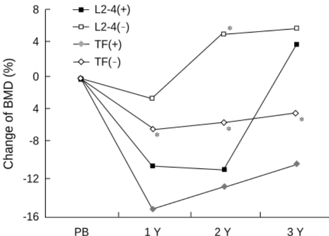
According to the GVHD occurrence, the GVHD group (n=3) had significantly more bone loss in femoral bone than the non- GVHD group (n=8), but not in lumbar spine. The mean changes of total femoral BMD in GVHD vs non-GVHD patients were -15.0% vs -5.9% at 1 yr (p<0.05), -12.4% vs -5.1% at 2 yr (NS), and -9.8% vs -3.8% at 3 yr (p<0.05), respectively. The mean changes of lumbar BMD in GVHD vs non-GVHD patients were -10.0% vs -2.0% at 1 yr (NS), -10.3% vs 5.3% at 2 yr (p<0.05), and +3.9% vs +5.9% at 3 yr (NS), respectively (Fig. 2).
According to TBI, patients with TBI (n=6) tended to have more bone loss than those without TBI (n=5), but without statis- tical significance. The mean changes of total femoral BMD in
Age at the time of BMT (yr)
Mean 28.7±8.6 (16~47)
Age stratification
< 20 2
20-29 3
30-39 5
≥40 1
Total Body Irradiation Yes 6
No 5
GVHD Yes 3
No 8 Baseline Z score
L2-4 -1.32±0.99 (-2.7~0)
Total femur 0.18±1.15 (-1.2~2.2)
Table 1. Patient characteristics
Bone Mneral Density (g/cm2) 1.15
1.1
1.05
1
0.95
0.9
0.85
PB 1 Y 2 Y 3 Y L2-4
TF
* * *
*
Percent Change (%)
8
4
0
-4
-8
-12
L2-4 TF
* * *
*
*
Fig. 1. The mean changes of bone mineral density before and after BMT (A) and percent changes of BMD from the baseline of pre-BMT (B). *, p<0.05; **, p< 0.01 against baseline. PB, Pre-BMT. TF, proximal total femur.
PB 1 Y 2 Y 3 Y
A B
patients with TBI vs non-TBI patients were -11.0% vs -5.3%
at 1 yr (NS), -9.6% vs -5.5% at 2 yr (NS), and -7.4% vs -3.2%
at 3 yr (NS), respectively. The mean changes of lumbar BMD in patients with TBI vs non-TBI patients were -8.5% vs +0.6% at 1 yr (NS), -6.3% vs +6.5% at 2 yr (NS), and +2.2% vs +9.1%
at 3 yr (NS), respectively (Fig. 3).
According to sex, female patients (n=5) tended to have more bone loss in the lumbar spine than male patients (n=6), but not in femoral bone. The mean changes of total femoral BMD in female vs male patients were -8.6% vs -8.2% at 1 yr (NS), -8.3%
vs -6.1% at 2 yr (NS), and -5.8% vs -5.2% at 3 yr (NS), respec- tively. The mean changes of lumbar BMD in female vs male
patients were -8.1% vs -1.2% at 1 yr (NS), -2.0% vs +4.2%
at 2 yr (NS), and +2.6% vs +7.7% at 3 yr (NS), respectively (Fig. 4).
Changes of biochemical markers
The serum ICTP increased progressively until four weeks after BMT. Thereafter, it began to decrease and reached basal values after one year (pre-BMT 7.4±1.6 ng/mL, post-BMT 4 wk 11.1±3.2 ng/mL, post-BMT 1 yr 8.3±0.8 ng/mL). Serum osteocalcin decreased progressively until three weeks after BMT. Thereafter, it recovered back to its initial basal level by 12 months (pre-BMT 3.6±1.9 ng/mL, post-BMT 3 wk 2.0
±0.8 ng/mL, post-BMT 1 yr 4.6±0.9 ng/mL) (Fig. 5).
Change of BMD (%)
8
4
0
4
-8
-12
-16
L2-4(+) L2-4(-) TF(+) TF(-)
Fig. 2.The differences in bone mineral density between patients with (n=3) and without (n=8) graft-versus-host disease (GVHD) before and after BMT, in view of percent change of BMD from baseline. There are no significant differences in the age, sex and TBI treatment between two groups. *, p<0.05 between patients with GVHD and without GVHD. +, with GVHD; -, without GVHD.
* *
*
*
Change of BMD(%)
12
8
4
0
-4
-8
-12
L2-4(+) L2-4(-) TF(+) TF(-)
Fig. 3. The differences in bone mineral density between patients with (n=6) and without (n=5) total body irradiation (TBI) before and after BMT, in view of percent change of BMD from baseline. There are no significant differences in the age, sex and GVHD frequency between two groups. *, p<0.05; **, p<0.01 against baseline. +, with TBI; -, without TBI. PB, Pre-BMT.
* ** **
**
Change of BMD (%)
8
4
0
4
-8
-12
-16
L2-4(M) L2-4(F) TF(M) TF(F)
Fig. 4. The differences in bone mineral density between male (n=6) and female (n=5) patients before and after BMT, in view of percent change of BMD from baseline. There are no significant differences in the age, TBI and GVHD frequency between two groups. *, p<0.05, **, p<0.01 against baseline. M, male patients;
F, Female patient. PB, Pre-BMT.
**
*
OC, ICTIP (ng/mL), Cr (mg/dL)
16 14 12 10 8 6 4 2 0
OC ICTP Cr
Fig. 5. The changes of serum bone turnover markers and serum creatinine before and after BMT. Data are given as mean values (±SEM). *, p<0.05 against baseline. PB, Pre-BMT.
* * *
*
PB 1w 2w 3w 4w 3M 6M 1Y PB 1 Y 2 Y 3 Y
PB 1 Y 2 Y 3 Y
PB 1 Y 2 Y 3 Y
Changes of estradiol, testosterone, and gonadotropin levels
After 6 months after BMT, in female patients, serum LH and FSH increased significantly and serum estradiol tended to decrease, but without statistical significance (LH; from 6.6
±4.9 IU/L to 38.7±9.3 IU/L, FSH; from 10.0±6.9 IU/L to 74.9±6.3 IU/L, estradiol; from 62.3±7.0 pmol/L to 24.9
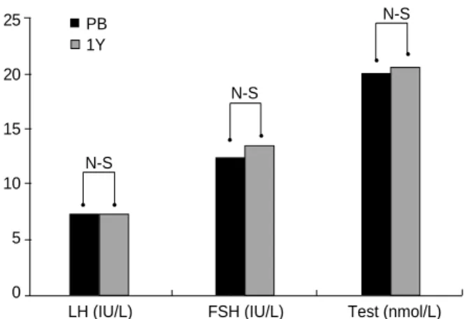
±0.3 pmol/L) (Fig. 6). In male patients, serum LH, FSH, and testosterone levels did not change significantly after BMT (Fig. 7).
DISCUSSION
Previous studies have demonstrated a 30 to 35% increase of the incidence of lumbar fracture following heart and liver transplantation and an 8 to 17% loss of BMD within 1 yr fol- lowing kidney transplantation (1-5). However, little is known about the effect of BMT on the skeletal system. In their cross- sectional studies, Kelly et al. (8) and Bhatia et al. (11) reported a significant decrease in bone density at the lumbar spine and femoral neck in a group of subjects following allogeneic BMT Recently, Ebeling et al. also found that post-allogeneic BMT patients lost 11.7% of femoral neck BMD and 3.9% of lum- bar BMD during the post-BMT 1 yr (9). We also previously reported significant bone loss following BMT and found that femoral bone loss was greater than vertebral bone loss in 67 patients during the post-BMT 1 yr (10).
In this study, we observed the serial BMD change during the post-BMT 3 yr and found that the change was different between vertebral and femoral bone. The initial femoral bone loss was more prominent than vertebral bone loss during the post-BMT 1 yr. Furthermore, most of vertebral bone loss fol- lowing BMT was recovered during post-BMT 3 yr, but the femoral bone loss was not recovered. Similar results were shown in other short-term studies on bone mineral metabolism after
BMT (11, 12). Schulte et al. reported that initial 6 months were important period in post-BMT bone loss and they ob- served much lower rate in bone loss after 1 yr in the 2-yr prospective study (13). In their study, partial recovery of spine BMD was also demonstrated, but not femoral compartments, which was consistent with our study. However, the study for longer period in a prospective manner following BMT has not been reported, to our knowledge. Although the number of patients in this study was small, this was the first study to observe the serial changes of BMD during the post-BMT 3-yr period. Exact mechanisms for this site-selectivity need to be elucidated, but it may be partly due to the differences in the tissue expression of several proteins related to bone metabolism including certain growth factors and BMP-2 according to the skeletal site (14, 15). The BMD response to therapeutic agents (e.g. bisphosphonate and hormone replacement therapy) is also different between lumbar and femoral bone, but the clear mechanism for this is currently unknown (16-18).
In this study, patients with GVHD or TBI tended to expe- rience more bone loss than those without GVHD or TBI. If more subjects were included in the study, the differences accord- ing to these risk factors could be greater to reach statistical significance. Patients suffering from GVHD receive larger amounts of immunosuppressive agents and thus have higher risk of bone loss. GVHD itself might have negative effect on bone marrow microenvironment in which bone turnover actually takes place (19). Because osteoblasts and osteoclasts are present in bone marrow, transplant-related stromal injury and cytokine changes in addition to GVHD itself might be related to post-BMT bone loss (20). We previously reported that the increase of bone marrow interleukin-6 was significant- ly associated with bone resorption after BMT (20). TBI can induce hypogonadism especially in women and may cause growth hormone deficiency, both of which are well known risk factors of osteoporosis.
The cause of significant increase in lumbar BMD at post- BMT 3 yr compared to basal values is uncertain, but gradual
90 80 70 60 50 40 30 20 10 0
Fig. 6. The changes in serum LH, FSH, and estradiol in female patients (n=5) before and after BMT. Data are given as the mean values (±SEM). PB, Pre-BMT.
NS
p=0.09
p<0.01
LH (IU/L) FSH (IU/L) E2 (pmol/L)
25
20
15
10
5
0
Fig. 7. The changes in serum LH, FSH, and testosterone in male patients (n=6) before and after BMT. Data are given as the mean values (±SEM). PB, Pre-BMT.
N-S
N-S
N-S
LH (IU/L) FSH (IU/L) Test (nmol/L) PB
1Y PB
6M
recovery of stromal cell function might partly contribute to it. Gradual reduction in the doses of immunosuppressants could be one of the main causes. It seems that continuous estrogen replacement might also contribute to this increase in lumbar BMD, but this is probably not the dominant cause for it because male subjects also experienced the similar rates of increase in lumbar BMD without hormone replacement therapy.
The women had a tendency to experience more bone loss than men, especially in vertebral bone. This may be related to hypogonadism induced by BMT in women, but not in men.
Thus, female patients received cyclic estrogen and progesterone replacement therapy since 1 yr after BMT and experienced gradual recovery in vertebral BMD until 3 yr. Without the hormone replacement therapy, the bone loss in the female patients might have been greater. It is well known that gonadal dysfunction in women occurs frequently after combined radi- ation treatment and cytotoxic chemotherapy (21, 22). Since hypogonadism is one of the main causes of post-BMT bone loss, evaluation of estrogen and testosterone status after BMT is needed and hormone replacement should follow when indi- cated (23). Growth hormone deficiency should be evaluated among long-term survivors. The decrease in bone formation marker and increase in bone resorption marker, observed in the immediate post-BMT period, might explain the initial post-BMT bone loss.
In conclusion, although there was a decline in lumbar BMD during the first year following BMT, gradual recovery occurred until post-BMT 3 yr. Femoral BMD decreased much more than lumbar BMD at 1 yr and did not recover until 3 yr. The clear mechanism for this skeletal site-selective difference needs to be elucidated through further studies. Evaluation of gonadal function and measurement of BMD in long-term survivors following BMT are recommended, especially in patients with GVHD or TBI.
ACKNOWLEDGMENTS
The results of the present study have been published in an abstract form in the Endocrine Society 83th annual meeting, 2001. This work was supported by the grant from Kangbuk Samsung Hospital and the grant from the Korean Ministry of Health and Welfare (01-PJ1-PG1-01CH08-0001).
REFERENCES
1. Arnold JC, Hauser D, Ziegler R, Kommerell B, Otto G, Theilmann L, Wuster C. Bone disease after liver transplantation. Transplant Proc 1992; 24: 2709-10.
2. Boot AM, Nauta J, Hokken-Koelega AC, Pols HA, de Ridder MA, Keizer-Schrama SM. Renal transplantation and osteoporosis. Arch Dis Child 1995; 72: 502-6.
3. Grotz WH, Mundinger FA, Gugel B, Exner VM, Kirste G, Schollmeyer PJ. Bone mineral density after kidney transplantation. Transplanta- tion 1995; 59: 982-6.
4. Julian BA, Laskow DA, Dubovsky J, Dubovsky EV, Curtis JJ, Quar- les LD. Rapid loss of vertebral mineral density after renal transplanta- tion. N Engl J Med 1991; 325: 544-50.
5. Lee AH, Mull RL, Keenan GF, Callegari PE, Dalinka MK, Eisen HJ, Mancini DM, Disesa VJ, Attie MF. Osteoporosis and bone morbidity in cardiac transplant recipients. Am J Med 1994; 96: 35-41.
6. Castaneda S, Carmona L, Carvajal I, Arranz R, Diaz A, Garcia-Vadillo A. Reduction of bone mass in women after bone marrow transplanta- tion. Calcif Tissue Int 1997; 60: 343-7.
7. Kang MI, Lee WY, Oh KW, Han JH, Song KH, Cha BY, Lee KW, Son HY, Kang SK, Kim CC. The short-term changes of bone mineral metabolism following bone marrow transplantation. Bone 2000; 26:
275-9.
8. Kelly PJ, Atkinson K, Ward RL, Sambrook PN, Biggs JC, Eisman JA. Reduced bone mineral density in men and women with allogeneic bone marrow transplantation. Transplantation 1990; 50: 881-3.
9. Ebeling PR, Thomas DM, Erbas B, Hopper JL, Szer J, Grigg AP. Mech- anisms of bone loss following allogeneic and autologous hemopoietic stem cell transplantation. J Bone Miner Res 1999; 14: 342-50.
10. Lee WY, Cho SW, Oh ES, Oh KW, Lee JM, Yoon KH, Kang MI, Cha BY, Lee KW, Son HY, Kang SK, Kim CC. The effect of bone marrow transplantation on the osteoblastic differentiation of human bone marrow stromal cells. J Clin Endocrinol Metab 2002; 87: 329- 35.
11. Bhatia S, Ramsay NKC, Weisdorf D, Griffiths H, Robison LL. Bone mineral density in patients undergoing bone marrow transplantation for myeloid malignancies. Bone Marrow Transplant 1998; 22: 87-90.
12. Valimaki MJ, Kinnunen K, Volin L, Tahtela R, Loyttyniemi E, Laitinen K, Makela P, Keto P, Ruutu T. A prospective study of bone loss and turnover after allogeneic bone marrow transplantation: effect of cal- cium supplementation with or without calcitonin. Bone Marrow Transplant 1999; 23: 355-61.
13. Schulte C, Beelen DW, Schaefer UW, Mann K. Bone loss in long-term survivors after transplantation of hematopoietic stem cells: A prospec- tive study. Osteoporosis Int 2000; 11: 344-53.
14. Milne M, Quail JM, Rosen CJ, Baran DT. Insulin-like growth factor binding proteins in femoral and vertebral bone marrow stromal cells:
expression and regulation by thyroid hormone and dexamethasone. J Cell Biochem 2001; 81: 229-40.
15. Milne M, Quail JM, Baran DT. Dexamethasone stimulates osteogenic differentiation in vertebral and femoral bone marrow cell cultures:
comparison of IGF-I gene expression. J Cell Biochem 1998; 71: 382- 91.
16. Pols HA, Felsenberg D, Hanley DA, Stepan J, Munoz-Torres M, Wilkin TJ, Qin-sheng G, Galich AM, Vandormael K, Yates AJ, Stych B.
Multinational, placebo-controlled, randomized trial of the effects of alendronate on bone density and fracture risk in postmenopausal women with low bone mass: results of the FOSIT study. Foxamax International Trial Study Group. Osteoporosis Int 1999; 9: 461-8.
17. Orwoll E, Ettinger M, Weiss S, Miller P, Kendler D, Graham J, Adami S, Weber K, Lorenc R, Pietschmann P, Vandormael K, Lombardi A.
Alendronate for the treatment of osteoporosis in men. N Engl J Med 2000; 343: 604-10.
18. Kohrt WM, Birge SJ Jr. Differential effects of estrogen treatment on bone mineral density of the spine, hip, wrist and total body in late postmenopausal women. Osteoporosis Int 1995; 5: 150-5.
19. Stern JM, Chesnut CH III, Bruemmer B, Sullivan KM, Lenssen PS, Aker SN, Sanders J. Bone density loss during treatment of chronic GVHD. Bone Marrow Transplant 1996; 17: 395-400.
20. Lee WY, Kang MI, Oh ES, Oh KW, Han JH, Cha BY, Lee KW, Son HY, Kang SK, Kim CC. The role of cytokines in the changes in bone turnover following bone marrow transplantation. Osteoporosis Int
2002; 13: 62-8.
21. Schimmer AD, Quatermain M, Imrie K, Ali V, McCrae J, Stewart AK, Crump M, Derzko C, Keating A. Ovarian function after autologous bone marrow transplantation. J Clin Oncol 1998; 16: 2359-63.
22. Mertens AC, Ramsay NK, Kouris S, Neglia JP. Patterns of gonadal dysfunction following bone marrow transplantation. Bone Marrow Transplant 1998; 22: 345-50.
23. Schimmer AD, Minden MD, Keating A. Osteoporosis after blood and marrow transplantation: clinical aspects. Biol Blood Marrow Trans- plant 2000; 6: 175-81.