root apex and the other is the route from the deep periodontal pocket. Infection sometimes expands into the fascial space directly and causes a fascial space infection. The fascial space is a potential space that can be expanded by purulent exudate. The fascial spaces that may be directly affected by odontogenic infections are called ‘primary spaces’, such as the canine, buccal, infratemporal, submental, sublingual, or submandibular spaces. Failure to control primary spaces in- fection may cause them to spread to ‘secondary spaces’, such as the submasseteric, pterygomandibular, and temporal spac- es. Infections that are not treated properly can spread beyond secondary spaces to deep neck fascial spaces such as the lateral pharyngeal, retropharyngeal, and prevertebral spaces.
It is difficult to treat patients who have these space infections without drainage of the purulent exudates, since those areas are surrounded by connective tissues that have poor blood supply1.
The term “diabetes mellitus (DM)” describes a metabolic disorder of multiple etiology characterized by chronic hy-
I. Introduction
Risk of infection depends on several factors, including host defense mechanisms, functional or anatomical abnormalities of the host, and virulence of the infecting microorganism. It is not only the host defense that determines the outcome of infection, but the timing and appropriateness of antimicrobial treatment as well. There are two main routes of infection in oral and maxillofacial infections; one is the route from the
Chul-Hwan Kim
Department of Oral and Maxillofacial Surgery, College of Dentistry, Dankook University, 119 Dandae-ro, Dongnam-gu, Cheonan 31116, Korea
TEL: +82-41-550-0271 FAX: +82-41-551-8988 E-mail: [email protected]
ORCID: http://orcid.org/0000-0002-5199-2420
This is an open-access article distributed under the terms of the Creative Commons Attribution Non-Commercial License (http://creativecommons.org/licenses/by-nc/4.0/), which permits unrestricted non-commercial use, distribution, and reproduction in any medium, provided the original work is properly cited.
CC
Analysis of glycosylated hemoglobin (HbA1c) level on maxillofacial fascial space infection in diabetic patients
Jong-Won Jang, Chul-Hwan Kim, Moon-Young Kim
Department of Oral and Maxillofacial Surgery, College of Dentistry, Dankook University, Cheonan, Korea
Abstract(J Korean Assoc Oral Maxillofac Surg 2015;41:251-258)
Objectives: This study was performed to evaluate the impact of glycosylated hemoglobin (HbA1c) level on characteristics and prognosis of maxil- lofacial fascial infection in diabetic patients.
Materials and Methods: We reviewed the medical records of 72 patients (35 patients with HbA1c lower than 7.0% and 37 patients with HbA1c higher than 7.0%) diagnosed with maxillofacial fascial space infection and hospitalized for treatment at the Department of Oral and Maxillofacial Sur- gery in Dankook University Hospital (Cheonan, Korea) from January 2005 to February 2014. We compared demographics, parameters of glucoregula- tion (HbA1c), laboratory parameters of inflammation (white blood cell [WBC], C-reactive protein [CRP] count), type and number of involved spaces, type and number of antibiotics, period of hospitalization, number of surgical operations, need for tracheostomy, complications, computed tomography (CT), and microorganisms between the two groups.
Results: Compared with the well-controlled diabetes mellitus (DM) group (HbA1c <7.0%), patients in the poorly-controlled (HbA1c ≥7.0%) DM group had the following characteristics: longer hospitalization periods, higher values of laboratory parameters of inflammation (WBC, CRP count) at the time of admission, higher number of antibiotics prescribed, more frequent complications, frequent deep neck space involvement, and distinctive main causative microorganisms. As the HbA1c level increases, hospitalization periods and incidence of complications increase gradually.
Conclusion: This retrospective study suggests that regulation of DM significantly impacts maxillofacial fascial infection. Poorly controlled DM with high HbA1c level negatively influences the prognosis of infection.
Key words: Glycosylated hemoglobin A, Dental infection control, Diabetes mellitus
[paper submitted 2015. 5. 18 / revised 2015. 6. 30 / accepted 2015. 7. 12]
Copyright Ⓒ 2015 The Korean Association of Oral and Maxillofacial Surgeons. All rights reserved.
during admission after consultation with the Department of Endocrinology. Patients diagnosed with secondary infec- tions from fractures, osteomyelitis, or cyst formation, patients hospitalized only for dentoalveolar abscess, and patients who were administered antibiotics before admission were ex- cluded from this study. A total of 72 patients were included in this study. The HbA1c levels of all patients at the time of admission were evaluated. We divided these patients into two groups according to HbA1c level.
• Group 1: Thirty-five patients with DM with HbA1c level under 7.0% (HbA1c <7.0%) and included 18 male and 17 female patients.
• Group 2: Thirty-seven patients with DM with HbA1c level over 7.0% (HbA1c ≥7.0%) and included 20 male and 17 female patients.
Investigations between the two groups regarding patient age, parameters of glucoregulation (HbA1c), laboratory parameters of inflammation (white blood cell [WBC], C- reactive protein [CRP] count), type and number of involved spaces, type and number of antibiotics, period of hospitaliza- tion, number of surgical operations, need for tracheostomy, complications, computed tomography (CT), and type of mi- croorganism were performed.
2. Laboratory examination
Laboratory values included indicators such as WBC count on admission and at discharge and CRP at admission and at discharge to evaluate severity and subsidence of infection.
3. Diagnosis of maxillofacial fascial infection
Three-dimensional neck CT with enhancement was used to identify which fascial space was involved with infection for all patients. The same examiner read all CT images and diag- nosed the infection.
4. Surgical procedures and bacterial culture test
Immediately after admission, we administered intravenous antibiotics and fluid therapy for all patients and obtained a CT image as soon as possible in accordance with nil per os time.
Amoxicillin/clavulanic acid (augmentin) was used as the first drug of choice. Clindamycin and a cephalosporin were subsequently used empirically according to the progress of laboratory tests. We performed emergency operations for pa- tients complaining of dysphagia, dyspnea, or reduced oxygen perglycemia with disturbances of carbohydrate, fat, and pro-
tein metabolism, resulting from defects in insulin secretion, insulin action, or both. Diabetes is usually diagnosed based on plasma glucose criteria, either by fasting plasma glucose (FPG) or the 2-hour plasma glucose (2-h PG) value after a 75-g oral glucose tolerance test (OGTT). Recently, an Inter- national Expert Committee added glycosylated hemoglobin (HbA1c) as a third option to diagnose diabetes2,3. Patients with DM have higher risks of infection due to abnormal phagocytosis, persistent reduction of blood flow, and cell- mediated immune abnormalities typical of diabetic patients4-6. HbA1c is a form of hemoglobin that is measured primar- ily to identify the average plasma glucose concentration over the previous two to three months prior to the measurement.
Because HbA1c testing can be performed at any time of day and without special patient preparation, it is more convenient for patients and health care providers compared to the oral glucose tolerance test or measuring FPG7.
There are many studies comparing diabetics and nondia- betics with infections in the maxillofacial region via blood glucose level at the time of admission or with a past medical history of DM8-12. However, admission glucose levels or fast- ing glucose levels are subject to changes of daily activities, such as diet content, amount of exercise, and physical or emotional stress before examination, even in well-controlled DM before admission13. On the other hand, HbA1c reflects long-term glycemic status and is a more stable measurement than FPG7. Therefore, HbA1c might be a better indicator for glucose control in diabetes than fasting blood sugar14. How- ever, scarce studies exist on infection of DM patients with different HbA1c levels in the oral and maxillofacial surgery fields. Therefore, this study was conducted to evaluate the clinical impact of HbA1c level and regulation of DM on the characteristics and prognosis of oral and maxillofacial fascial infection in patients with DM.
II. Materials and Methods
1. Patients
We reviewed the medical records of patients diagnosed with maxillofacial fascial space infection and hospitalized for treatment at the Department of Oral and Maxillofacial Surgery in Dankook University Hospital (Cheonan, Korea) from January 2005 to February 2014. Among these patients, we selected patients with DM from records of previous medi- cal history of DM or patients who newly diagnosed with DM
III. Results
1. Demographics and HbA1c level
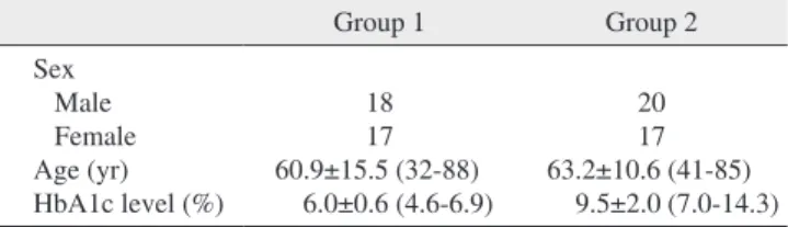
The average patient age in group 1 and group 2 was 60.9 years (range, 32-88 years) and 63.2 years (range, 41-85 years), respectively. The age showed no statistically signifi- cant difference between the two groups (P>0.05). The aver- age level of HbA1c level was 6.0% (range, 4.6%-6.9%) in group 1 and 9.5% (range, 7.0%-14.3%) in group 2.(Table 1)
2. Duration of hospitalization
The mean hospitalization days of group 1 and group 2 was 9.6 days (range, 4-36 days) and 15.1 days (range, 4-66 days), respectively. Patients in group 2 had longer hospitalizations than those in group 1.(Table 2) This showed a statistically significant difference between the two groups. In the linear saturation immediately after obtaining CT imaging. In cases
of non-emergency patients, we performed elective operations after localization of the abscess. We performed incision and drainage with insertion of silastic drain under general anes- thesia or local anesthesia in accordance with the status of the patient. Tracheostomy was performed on any patient with questionable postoperative airway patency. Pus samples from the site of infection were collected during incision and drain- age (I&D), using sterile agar gel transport swabs. Sixteen pa- tients among group 1 and 23 patients among group 2 yielded identifiable pathogens. Susceptibility to various antibiotics was evaluated and antibiotics were tailored to culture results.
We performed daily dressing changes for the operation site and treatment was terminated when clinical and radiographic signs improved and after CRP and WBC were normalized.
5. Data analysis
For statistical analysis between the two study groups, we performed a t-test, Mann-Whitney test, logistic regression test, chi-square test, and linear regression analysis (IBM SPSS Statistics version 21.0; IBM Co., Armonk, NY, USA).
Data were presented as the mean±standard deviation (SD). A P-value less than 0.05 was considered to be significant.
Table 1. Demographics and HbA1c levels for group 1 and group 2
Group 1 Group 2
Sex Male Female Age (yr) HbA1c level (%)
18 17 60.9±15.5 (32-88)
6.0±0.6 (4.6-6.9)
20 17 63.2±10.6 (41-85)
9.5±2.0 (7.0-14.3) (HbA1c: glycosylated hemoglobin)
Group 1: 35 patients with diabetes mellitus (DM) with HbA1c <7.0%, Group 2: 37 patients with DM with HbA1c ≥7.0%.
Values are presented as number or mean±standard deviation (range).
Jong-Won Jang et al: Analysis of glycosylated hemoglobin (HbA1c) level on maxillo- facial fascial space infection in diabetic patients. J Korean Assoc Oral Maxillofac Surg 2015
Table 2. Duration of hospitalization of group 1 and group 2
Group 1 Group 2 P-value
Duration of
hospitalization (day) 9.6±6.5 (4-36) 15.1±13.7 (4-66) 0.032 Group 1: 35 patients with diabetes mellitus (DM) with glycosylated hemoglobin (HbA1c) <7.0%, Group 2: 37 patients with DM with HbA1c ≥7.0%.
Jong-Won Jang et al: Analysis of glycosylated hemoglobin (HbA1c) level on maxillo- facial fascial space infection in diabetic patients. J Korean Assoc Oral Maxillofac Surg 2015
Table 3. Linear regression analysis between HbA1c level and hospitalization period
Independent variable
Unstandardized
coefficient Standardized
coefficient t P-value
B SD β
HbA1c 2.436 0.496 0.506 4.908 0.000
(HbA1c: glycosylated hemoglobin, SD: standard deviation)
Jong-Won Jang et al: Analysis of glycosylated hemoglobin (HbA1c) level on maxillo- facial fascial space infection in diabetic patients. J Korean Assoc Oral Maxillofac Surg 2015
Table 4. Parameters of inflammation in group 1 and group 2
Parameter Group 1 Group 2
Admission Discharge Admission Discharge
WBC (/mm3)
CRP (mg/dL) 12,580 (6,920-22,620)
11.51 (1.50-43.67) 6,689 (1,510-12,900)
1.04 (0.26-0.88) 15,719 (4,440-24,930)
17.97 (1.68-56.20) 8,108 (3,890-19,430) 1.35 (0.3-5.00) (WBC: white blood cell, CRP: C-reactive protein)
Group 1: 35 patients with diabetes mellitus (DM) with glycosylated hemoglobin (HbA1c) <7.0%, Group 2: 37 patients with DM with HbA1c
≥7.0%.
Values are presented as mean (range).
Jong-Won Jang et al: Analysis of glycosylated hemoglobin (HbA1c) level on maxillofacial fascial space infection in diabetic patients. J Korean Assoc Oral Maxillofac Surg 2015
5. Tracheostomy, number of antibiotics, and operation
The tracheostomy ratio was 5.7% (2 patients) and 13.5% (5 patients) in groups 1 and 2, respectively (P>0.05). Patients in group 2 needed a higher number of antibiotics for treatment.
The mean number of prescribed antibiotics was 2.0 (range, 2-3) in group 1 and 2.4 (range, 1-6) in group 2, which was a statistically significant difference (P=0.039). However, the mean number of surgical procedures was not significantly different between the two groups (P=0.56).
6. Complications
The ratio of patients with complications was 17.1% (6 patients) in group 1 and 51.4% (19 patients) in group 2. The most commonly encountered complications were airway obstruction and skin defects such as necrotic skin, pitting edema, and fistula formation. There was one patient in group 2 that expired due to septic shock.(Table 6) Chi-square analy- sis on the prevalence of complications between the groups re- vealed a significantly higher prevalence in group 2 compared to the group 1 (relative risk=3.005, P=0.002).(Table 7) Logis- tic regression analysis showed that as HbA1c levels increase by 1, the prevalence of complication increases by a factor of 1.404, which was found to be significant.(Table 8) Patients were categorized by 1% of HbA1c level and linear by linear analysis was performed.(Fig. 1) The prevalence of complica- tions was low, even with an HbA1c level lower than 7% to 8%; however, complications increased drastically in groups with an HbA1c level greater than 8% to 9%.
regression analysis, hospitalization days increased by 2.436 days as HbA1c levels increased by 1%.(Table 3)
3. Laboratory examination
WBC count and CRP of both groups was increased at the time of admission.(Table 4) This pre-operative WBC count and CRP was higher in group 2 than in group 1 and showed statistical significance (P<0.05). At the time of discharge, these two laboratory parameters showed almost normal val- ues and there were no statistically significant differences between the two groups in terms of these two parameters (P>0.05).
4. Distribution of the involved fascial space
The submandibular, buccal, and submental spaces were the most commonly involved spaces.(Table 5) The range of the number of involved spaces was one through five in group 1 and one through nine in group 2. The mean±SD number of involved spaces was 1.62±1.0 and 2.16±1.7 in group 1 and group 2, respectively. However, this did not show statisti- cally significant differences between the two groups (P=0.10).
There were 14 patients (40.0%) in group 1 and 21 patients (56.8%) in group 2 with multiple-space infection (more than 2 spaces). Note that deep neck space infections such as the ret- ropharyngeal or prevertebral space were seen only in group 2.
Table 5. Distribution of the involved fascial space
Fascial space Group 1 Group 2 Total
Submandibular Buccal Submental Sublingual Submasseteric Pterygomandibular Lateral pharyngeal Temporal Retropharyngeal Prevertebral Canine Infratemporal Total
15 13 9 8 3 4 1 3- - 1 - 57
19 18 7 4 8 5 7 5 3 2 1 1 80
34 (24.8) 31 (22.6) 16 (11.7) 12 (8.8) 11 (8.0) 9 (6.6) 8 (5.8) 8 (5.8) 3 (2.2) 2 (1.5) 2 (1.5) 1 (0.7) 137 (100) Group 1: 35 patients with diabetes mellitus (DM) with glycosylated hemoglobin (HbA1c) <7.0%, Group 2: 37 patients with DM with HbA1c ≥7.0%.
Values are presented as number or number (%).
Jong-Won Jang et al: Analysis of glycosylated hemoglobin (HbA1c) level on maxillo- facial fascial space infection in diabetic patients. J Korean Assoc Oral Maxillofac Surg 2015
Table 6. Complications of maxillofacial fascial space infection
Group 1 Group 2 Total
Airway obstruction Skin defect Sepsis Pleural effusion Acute renal failure Trismus
Emphysema Pneumonia
Descending mediastinitis Death
Total
3 2 - - - 1 - 1 - - 7
5 4 4 3 3 1 2 1 1 1 25
8 6 4 3 3 2 2 2 1 1 32 Group 1: 35 patients with diabetes mellitus (DM) with glycosylated hemoglobin (HbA1c) <7.0%, Group 2: 37 patients with DM with HbA1c ≥7.0%.
Values are presented as number.
Jong-Won Jang et al: Analysis of glycosylated hemoglobin (HbA1c) level on maxillo- facial fascial space infection in diabetic patients. J Korean Assoc Oral Maxillofac Surg 2015
IV. Discussion
DM is not only a predisposing factor for common infections, but also responds poorly to infections once they have devel- oped, especially when glucose levels are uncontrolled8,15,16. The mechanisms in which diabetes predisposes to infection may be attributable to hyperglycemia. Many factors increase susceptibility of hyperglycemia to infections. Hyperglycemia has adverse effects on the immune system, causing impaired chemotaxis, adherence of microorganisms to polymor- phonuclear leukocytes and lymphocytes, and disruption of phagocytosis. Hyperglycemia reduces the ability of WBCs to break down phagocited microorganisms. Since the process of phagocytosis is a basic defense against bacteria and fungi, the disruption of this process is thought to be responsible for a higher incidence of infections in diabetics17. Geerlings and Hoepelman18 suggested that the function of neutrophils, such as chemotaxis or production of cytokines, is reduced under high blood sugar levels. These defects of the immune system along with vascular abnormalities render diabetic patients at higher risk for a variety of invasive infections19. Chronic dis- eases such as diabetes occur more often in older patients. In 7. Main causative microorganisms
In total, there were 16 patients in group 1 and 23 patients in group 2 that yielded identifiable pathogens.(Table 9) In group 1, the most common microorganism cultured was Strepto- coccus viridans (33.3%, 8/24), followed by Staphylococcus epidermidis (12.5%, 3/24). In group 2, the most common microorganism cultured was Klebsiella pneumonia (34.3%, 12/35) followed by S. viridans (8.6%, 3/35) and Streptococ- cus constellatus (8.6%, 3/35).
Fig. 1. Glycosylated hemoglobin (HbA1c) level and rate of compli- cation.
Jong-Won Jang et al: Analysis of glycosylated hemoglobin (HbA1c) level on maxillo- facial fascial space infection in diabetic patients. J Korean Assoc Oral Maxillofac Surg 2015
Complicationrate(%)
4-5 100
90 80 70 60 50 40 30 20 10 0
HbA1c level (%)
5-6 6-7 7-8 8-9 9-10 10-11 >11
Table 9. Distribution of causative microorganisms Group 1 Group 2 Aerobic bacteria
Acinetobacter baumannii Psuedomonas aeruginosa Facultative anaerobic bacteria Aeromonas salmonicida Enterobacter cloacae Klebsiella pneumonia Staphylococcus aureus Staphylococcus epidermidis Staphylococcus hominis Streptococcus anginosus Streptococcus constellatus Streptococcus gordonii Streptococcus intermedius Streptococcus mitis Streptococcus oralis Streptococcus salivarius Streptococcus sanguinis Streptococcus viridans Anaerobic bacteria Bacteroides
Fusobacterium mortiferum Gemella morbillorum Prevotella buccae
1 - 1 - - - 3 - 2 2 - - 2 1 1 - 8 1 1 1 -
1 1 - 1 12
2 1 1 - 3 1 1 1 - 1 1 3 1 1 2 1 Group 1: 35 patients with diabetes mellitus (DM) with glycosylated hemoglobin (HbA1c) <7.0%, Group 2: 37 patients with DM with HbA1c ≥7.0%.
Jong-Won Jang et al: Analysis of glycosylated hemoglobin (HbA1c) level on maxillo- facial fascial space infection in diabetic patients. J Korean Assoc Oral Maxillofac Surg 2015
Table 7. Chi-square analysis of HbA1c level and complication be- tween the two groups
Complication Group 1 Group 2 Relative risk P-value No
Yes 29 (82.9)
6 (17.1) 18 (48.6)
19 (51.4) 3.005 0.002
Group 1: 35 patients with diabetes mellitus (DM) with glycosylated hemoglobin (HbA1c) <7.0%, Group 2: 37 patients with DM with HbA1c ≥7.0%.
Values are presented as number (%).
Jong-Won Jang et al: Analysis of glycosylated hemoglobin (HbA1c) level on maxillo- facial fascial space infection in diabetic patients. J Korean Assoc Oral Maxillofac Surg 2015
Table 8. Logistic regression analysis between HbA1c level and complication
Complication prevalence
Exp(B) 95% CI P-value
HbA1c level (%) 1.404 1.112-1.772 0.004
(HbA1c: glycosylated hemoglobin, CI: confidence interval)
Jong-Won Jang et al: Analysis of glycosylated hemoglobin (HbA1c) level on maxillo- facial fascial space infection in diabetic patients. J Korean Assoc Oral Maxillofac Surg 2015
rial fibrillation occurred significantly more in patients with HbA1c levels higher than 7.0%. Dronge et al.26 reported that after performing urologic, vascular, or orthopedic surgery in diabetic patients, the prevalence of postoperative infection was higher in patients with higher HbA1c levels. However, studies on the prognosis of fascial infection in diabetic pa- tients according to the HbA1c levels in maxillofacial surgery field are rare.
Lee et al.27 reported that 81.6% of odontogenic infection showed single fascial space involvement. In contrast, Rega et al.28 reported that multiple-space infections were more com- mon than single-space infections in odontogenic infection.
In our study, 34 patients (48.6%) had more than two fascial space involvements in both groups. The most commonly involved fascial space in this study was the submandibular space followed by the buccal space in both groups. This result was similar to other previously reported studies19,21. Deep neck infections, such as lateral, retropharyngeal, or pre- vertebral spaces may result in life-threatening complications including upper airway obstruction, descending mediastinitis, jugular vein thrombosis, venous septic emboli, carotid artery rupture, septic shock, and disseminated intravascular coagu- lopathy29,30. Thus, these infections require early diagnosis and aggressive treatment. Note that the retropharyngeal (3/80) and prevertebral (2/80) spaces were involved only in the poorly-controlled DM group. This result of our study is simi- lar to that of a previous study of Huang et al.10, who reported that hyperclycemic patients are vulnerable to deep neck in- fections.
There are many studies reported that periods of hospi- talization were longer in DM patients, and WBC and CRP levels were higher in DM patients compared to non-DM patients20,21,31. Similarly, in this study, patients in the poorly- controlled DM group (group 1) had a comparatively longer hospital stay compared to the well-controlled DM group (group 2) (P<0.05). Regardless of the group, HbA1c level and hospitalization period were in a linear relationship, with the latter increasing as the former increases. As the HbA1c level increased by 1%, hospitalization period increased by 2.4 days.(Table 3) WBC count and CRP levels at time of admis- sion were significantly higher in the poorly-controlled DM group compared to the well-controlled DM group (P<0.05).
These results can be said to be a reflection of persistent in- flammation and delayed healing, which could be due to a prolonged inflammatory response to cytokine dysregulation and enhanced fibroblast apoptosis by hyperglycemia32,33.
Amoxicillin/clavulanic acid (augmentin) was used first this study, the average age of the well-controlled DM group
and the poorly-controlled DM group was 60.9 years and 63.2 years, respectively.
There have been many studies comparing diabetics and nondiabetics with infections in the maxillofacial region.
Many of them divided the groups by blood glucose level on admission or past medical history of DM. They concluded that diabetic patients are more likely to develop complica- tions, greater incidence rates of involved spaces, and abnor- mal hematologic findings19-21. Sim et al.13 reported that physi- cal and emotional stress increases blood glucose level via activation of both the adrenergic and glucocorticoid systems.
Koraćević et al.22 reported that increased glucose levels dur- ing the stress might be a result of sympathetic nervous sys- tem activation, which raises the production of catecholamines that stimulate processes of glyconeogenesis, glycogenolysis, and liposysis. In addition, infection itself can be a cause of hyperglycemia. One of the most important metabolic features of an infection is catecholaminemia, and it may disrupt the regulation of blood glucose in four ways: a) increased gluco- neogenesis, b) reduction of the intrinsic secretion of insulin, c) increased resistance to intrinsic insulin, and d) increased uti- lization of glucagon23. Therefore, blood glucose levels on ad- mission may change at the time of admission even if glucose metabolism was previously well controlled before admission in diabetic patients.
On the other hand, the HbA1c level appears to be a more reliable marker for average glycaemia in the last two to three months and seems to be a better measurement for the evalua- tion of glucose regulation on infection.
The American Diabetes Association recommends that HbA1c should be below 7.0% for glycemic targets for most patients with diabetes. Lowering HbA1c below or around 7.0% has been shown to reduce microvascular complications and macrovascular disease. Therefore, we set the borderline level of HbA1c between the two groups at 7.0%. However, this standard only considers complications of DM itself, and lacks concerns regarding prognosis after surgical procedures or certain other diseases. There are some reports on diseases and postoperative complications according to the HbA1c lev- els in diabetic patients. Jupiter et al.24 reported a significant difference of prevalence of postoperative complications after ankle and foot surgery between well-controlled diabetic pa- tients (HbA1c <7.0%) and poorly-controlled diabetic patients (HbA1c >7.0%)8. Halkos et al.25 reported that after coronary artery bypass surgery of diabetic patients, postoperative com- plications such as infection, myocardial infarction, and arte-
clinicians should take into account the preponderance of this microorganism when choosing empirical antibiotics.
Phagocytic function has been shown to be compromised when glucose levels range from 198.6 to 270.8 mg/dL (11 to 15 mmol/L). Therefore, glucose control plays an important role in the treatment strategy of hyperglycemic patients with infection15. There is a report that patients with high HbA1c levels tend to have more acute infections, not only in maxil- lofacial regions but in general as well23. Moreover, in a study of influence of glycemic control on outcomes of common infections, Leibovici et al.36 reported that in patients with- out a fatal underlying disease, a higher fatality rate could be demonstrated in patients with higher HbA1c levels. Diabetic patients with poor glycaemic control were at high risk for a fatal outcome. It has been postulated that a poorly controlled diabetic state predisposes the patient to sepsis, and once this disease is established further, adverse carbohydrate me- tabolism may result. A recent report on diabetes control and infections showed that impaired neutrophil bactericidal func- tion is associated with poor blood glucose control and that it is likely that neutrophil bactericidal function will improve as blood glucose control improves37. In addition to the aggres- sive treatment that is needed in diabetic patients compared to non-diabetic patients when infection occurs, we also found that regulation of blood glucose (HbA1c within 7.0%) is critical to the prognosis of infection. HbA1c levels of 7.0%, which are suggested by the ADA, were confirmed to be a re- liable marker for prognosis in oral and maxillofacial surgery patients with infections.
V. Conclusion
In conclusion, this study confirmed that good preoperative glycemic control, as measured by HbA1c levels less than 7%, is associated with a significantly better prognosis in oral and maxillofacial fascial infection. This simple laboratory test drawn preoperatively may provide the clinician with a more accurate risk profile and provide additional prognostic in- formation when discussing morbidity with patients and their families.
Conflict of Interest
No potential conflict of interest relevant to this article was reported.
as the drug of choice. Clindamycin and cephalosporin were subsequently used empirically according to the progress of laboratory tests. In patients who underwent a pus culture test, we tailored antibiotics according to the sensitivity results.
The mean number of antibiotics prescribed was higher in the poorly-controlled DM group compared to the well-controlled DM group. Number of I&D or the tracheostomy ratio were not significantly higher in the poorly-controlled group. The poorly-controlled DM group had more frequent and severe complications. In addition, the prevalence of complications increased consistently as HbA1c levels increased; at a 1%
increase of HbA1c level, the prevalence of complications increased by a factor of 1.4.(Table 8) The major complication was airway obstruction followed by skin defects such as skin necrosis, fistula formation, exfoliative dermatitis, and pitting edema. Skin defects might be, to a larger extent, related to the operation site and delayed healing, due to poorly-controlled glucose levels. Severe complications such as sepsis and de- scending mediastinitis were seen in the poorly controlled DM group only and one patient in this group died due to septic shock and pneumonia. These results are consistent with the findings of previous studies12,20,34. As seen in Fig. 1, the prev- alence of complications increased by a small extent until the 7% to 8% range, but increased drastically for the 8% to 9%
range and continued. Therefore, an HbA1c level of 8% could be considered as a reference point for predicting the preva- lence of complications in maxillofacial infection patients.
The bacteriologic patterns of maxillofacial infection are usually polymicrobial, including aerobes, microaerophilics, and anaerobes. Many studies reported that K. pneumonia and Streptococcus spp. were the most commonly isolated organisms among diabetic patients. Streptococcus spp. and Staphylococcus spp. are the most commonly isolated organ- isms in non-diabetic patients1,10,12,27,28
. In our study, Strepto- coccus spp. was most commonly isolated in group 1. This causative microorganisms of the well-controlled DM group were similar to those of the non-diabetic patients. On the oth- er hand, in group 2, K. pneumonia was the most commonly isolated. This K. pneumonia plays a role in deep neck infec- tion10. Possible contributing factors of the preponderance of K. pneumonia include increased oropharyngeal colonization by gram-negative bacilli, and defects of host defenses, espe- cially phagocytic function impairment in hyperglcaemia35. Clindamycin and other antibiotics are often used to treat head and neck infections, but should not be administered alone for the therapy of infection in poorly controlled DM patients be- cause of their limitation in controlling K. pneumoniea. Thus,
Wkly 2008;138:512-9.
18. Geerlings SE, Hoepelman AI. Immune dysfunction in patients with diabetes mellitus (DM). FEMS Immunol Med Microbiol 1999;26:
259-65.
19. Rao DD, Desai A, Kulkarni RD, Gopalkrishnan K, Rao CB. Com- parison of maxillofacial space infection in diabetic and nondiabetic patients. Oral Surg Oral Med Oral Pathol Oral Radiol Endod 2010;
110:e7-12.
20. Chang JS, Yoo KH, Yoon SH, Ha J, Jung S, Kook MS, et al. Odon- togenic infection involving the secondary fascial space in diabetic and non-diabetic patients: a clinical comparative study. J Korean Assoc Oral Maxillofac Surg 2013;39:175-81.
21. Zheng L, Yang C, Zhang W, Cai X, Kim E, Jiang B, et al. Is there association between severe multispace infections of the oral maxil- lofacial region and diabetes mellitus? J Oral Maxillofac Surg 2012;
70:1565-72.
22. Koraćević G, Vasiljević S, Velicković-Radovanović R, Sakac D, Obradović S, Damjanović M, et al. Stress hyperglycemia in acute myocardial infarction. Vojnosanit Pregl 2014;71:858-69.
23. Burekovic A, Dizdarevic-Bostandzic A, Godinjak A. Poorly regu- lated blood glucose in diabetic patients-predictor of acute infec- tions. Med Arch 2014;68:163-6.
24. Jupiter DC, Humphers JM, Shibuya N. Trends in postoperative infection rates and their relationship to glycosylated hemoglobin levels in diabetic patients undergoing foot and ankle surgery. J Foot Ankle Surg 2014;53:307-11.
25. Halkos ME, Puskas JD, Lattouf OM, Kilgo P, Kerendi F, Song HK, et al. Elevated preoperative hemoglobin A1c level is predictive of adverse events after coronary artery bypass surgery. J Thorac Car- diovasc Surg 2008;136:631-40.
26. Dronge AS, Perkal MF, Kancir S, Concato J, Aslan M, Rosenthal RA. Long-term glycemic control and postoperative infectious com- plications. Arch Surg 2006;141:375-80.
27. Lee JK, Kim HD, Lim SC. Predisposing factors of complicated deep neck infection: an analysis of 158 cases. Yonsei Med J 2007;
48:55-62.
28. Rega AJ, Aziz SR, Ziccardi VB. Microbiology and antibiotic sen- sitivities of head and neck space infections of odontogenic origin. J Oral Maxillofac Surg 2006;64:1377-80.
29. Wills PI, Vernon RP Jr. Complications of space infections of the head and neck. Laryngoscope 1981;91:1129-36.
30. Beck HJ, Salassa JR, McCaffrey TV, Hermans PE. Life-threatening soft-tissue infections of the neck. Laryngoscope 1984;94:354-62.
31. Zheng L, Yang C, Kim E, Zhang W, Cai X, Jiang B, et al. The clinical features of severe multi-space infections of the head and neck in patients with diabetes mellitus compared to non-diabetic patients. Br J Oral Maxillofac Surg 2012;50:757-61.
32. Liu R, Desta T, He H, Graves DT. Diabetes alters the response to bacteria by enhancing fibroblast apoptosis. Endocrinology 2004;
145:2997-3003.
33. Naguib G, Al-Mashat H, Desta T, Graves DT. Diabetes prolongs the inflammatory response to a bacterial stimulus through cytokine dysregulation. J Invest Dermatol 2004;123:87-92.
34. Huang TT, Liu TC, Chen PR, Tseng FY, Yeh TH, Chen YS. Deep neck infection: analysis of 185 cases. Head Neck 2004;26:854-60.
35. Sahly H, Podschun R, Ullmann U. Klebsiella infections in the im- munocompromised host. Adv Exp Med Biol 2000;479:237-49.
36. Leibovici L, Yehezkelli Y, Porter A, Regev A, Krauze I, Harell D.
Influence of diabetes mellitus and glycaemic control on the charac- teristics and outcome of common infections. Diabet Med 1996;13:
457-63.
37. Gallacher SJ, Thomson G, Fraser WD, Fisher BM, Gemmell CG, MacCuish AC. Neutrophil bactericidal function in diabetes mel- litus: evidence for association with blood glucose control. Diabet Med 1995;12:916-20.
ORCID
Jong-Won Jang, http://orcid.org/0000-0003-1515-032X Chul-Hwan Kim, http://orcid.org/0000-0002-5199-2420 Moon-Young Kim, http://orcid.org/0000-0001-9596-7481
References
1. Chang CM, Lu FH, Guo HR, Ko WC. Klebsiella pneumoniae fas- cial space infections of the head and neck in Taiwan: emphasis on diabetic patients and repetitive infections. J Infect 2005;50:34-40.
2. American Diabetes Association. Diagnosis and classification of diabetes mellitus. Diabetes Care 2014;37 Suppl 1:S81-90.
3. International Expert Committee. International Expert Committee report on the role of the A1C assay in the diagnosis of diabetes.
Diabetes Care 2009;32:1327-34.
4. Delamaire M, Maugendre D, Moreno M, Le Goff MC, Allannic H, Genetet B. Impaired leucocyte functions in diabetic patients. Dia- bet Med 1997;14:29-34.
5. Muller LM, Gorter KJ, Hak E, Goudzwaard WL, Schellevis FG, Hoepelman AI, et al. Increased risk of common infections in patients with type 1 and type 2 diabetes mellitus. Clin Infect Dis 2005;41:281-8.
6. Tanaka Y. Immunosuppressive mechanisms in diabetes mellitus.
Nihon Rinsho 2008;66:2233-7.
7. Rohlfing CL, Little RR, Wiedmeyer HM, England JD, Madsen R, Harris MI, et al. Use of GHb (HbA1c) in screening for undiag- nosed diabetes in the U.S. population. Diabetes Care 2000;23:187- 8. Shah BR, Hux JE. Quantifying the risk of infectious diseases for 91.
people with diabetes. Diabetes Care 2003;26:510-3.
9. Infante-Cossío P, Fernández-Hinojosa E, Mangas-Cruz MA, González-Pérez LM. Ludwig's angina and ketoacidosis as a first manifestation of diabetes mellitus. Med Oral Patol Oral Cir Bucal 2010;15:e624-7.
10. Huang TT, Tseng FY, Yeh TH, Hsu CJ, Chen YS. Factors affecting the bacteriology of deep neck infection: a retrospective study of 128 patients. Acta Otolaryngol 2006;126:396-401.
11. Chandu A, Macisaac RJ, Smith AC, Bach LA. Diabetic ketoaci- dosis secondary to dento-alveolar infection. Int J Oral Maxillofac Surg 2002;31:57-9.
12. Huang TT, Tseng FY, Liu TC, Hsu CJ, Chen YS. Deep neck in- fection in diabetic patients: comparison of clinical picture and outcomes with nondiabetic patients. Otolaryngol Head Neck Surg 2005;132:943-7.
13. Sim YB, Park SH, Kang YJ, Kim SM, Lee JK, Jung JS, et al. The regulation of blood glucose level in physical and emotional stress models: possible involvement of adrenergic and glucocorticoid systems. Arch Pharm Res 2010;33:1679-83.
14. Lippi G, Mattiuzzi C, Targher G. Glycated hemoglobin, diabetes, and cardiovascular risk in nondiabetic adults. N Engl J Med 2010;
362:2030; author reply 2031.
15. Alexander M, Krishnan B, Shenoy N. Diabetes mellitus and odon- togenic infections: an exaggerated risk? Oral Maxillofac Surg 2008;12:129-30.
16. Carton JA, Maradona JA, Nuño FJ, Fernandez-Alvarez R, Pérez- Gonzalez F, Asensi V. Diabetes mellitus and bacteraemia: a com- parative study between diabetic and non-diabetic patients. Eur J Med 1992;1:281-7.
17. Stoeckle M, Kaech C, Trampuz A, Zimmerli W. The role of dia- betes mellitus in patients with bloodstream infections. Swiss Med