CASE REPORT
십이지장 궤양 천공 단순 봉합수술 후 완전 피막형 자가확장 금속 스텐트 삽입술로 치료된 봉합 부위 누출
유영진, 이용강1, 이중호2, 이형순2
연세대학교 의과대학 외과학교실, 국민건강보험 일산병원 소화기내과1, 외과2
Covered Self-expandable Metallic Stent Insertion as a Rescue Procedure for Postoperative Leakage after Primary Repair of Perforated Duodenal Ulcer
Young Jin Yoo, Yong Kang Lee1, Joong Ho Lee2 and Hyung Soon Lee2
Department of Surgery, Yonsei University College of Medicine, Seoul; Division of Gastroenterology, Departments of Internal Medicine1 and Surgery2, National Health Insurance Service Ilsan Hospital, Goyang, Korea
Surgery has been the standard treatment for perforated duodenal ulcers, with mostly good results. However, the resolution of post- operative leakage after primary repair of perforated duodenal ulcer remains challenging. There are several choices for re-operation required in persistent leakage from perforated duodenal ulcers. However, many of these choices are complicated surgical procedures requiring prolonged general anesthesia that may increase the chances of morbidity and mortality. Several recent reports have demon- strated postoperative leakage after primary repair of a perforated duodenal ulcer treated with endoscopic insertion using a covered self-expandable metallic stent, with good clinical results. We report a case with postoperative leakage after primary repair of a perfo- rated duodenal ulcer treated using a covered self-expandable metallic stent. (Korean J Gastroenterol 2018;72:262-266) Key Words: Duodenal ulcer; Peptic ulcer perforation; Self expandable metallic stents
Received April 11, 2018. Revised May 1, 2018. Accepted May 23, 2018.
CC This is an open access article distributed under the terms of the Creative Commons Attribution Non-Commercial License (http://creativecommons.org/licenses/
by-nc/4.0) which permits unrestricted non-commercial use, distribution, and reproduction in any medium, provided the original work is properly cited.
Copyright © 2018. Korean Society of Gastroenterology.
교신저자: 이형순, 10444, 경기도 고양시 일산동구 일산로 100, 국민건강보험 일산병원 외과
Correspondence to: Hyung Soon Lee, Department of Surgery, National Health Insurance Service Ilsan Hospital, 100 Ilsan-ro, Ilsandong-gu, Goyang 10444, Korea. Tel:
+82-31-900-0975, Fax: +82-31-900-0138, E-mail: [email protected], ORCID: https://orcid.org/0000-0001-9825-8648 Financial support: None. Conflict of interest: None.
INTRODUCTION
The incidence of duodenal ulcer perforation has been re- duced since the distribution of commercialized histamine-2 receptor blockers and proton pump inhibitors; however, ul- cer-related mortality remains to be an issue. Duodenal ulcer perforation requires emergency surgery and is usually well controlled when initially treated with surgical intervention.
However, postoperative leakage has been associated with morbidity as it requires re-operation with prolonged general anesthesia time and severe inflammation, which leads to a
challenging problem for surgeons.1,2
Therefore, presently, surgeons are seeking alternative methods to treating postoperative leakage. One such effort is covered self-expandable metallic stent (SEMS) insertion us- ing endoscopy. Recently, covered SEMS insertion as a treat- ment for postoperative leakage after duodenal ulcer perfo- ration surgery or other gastrointestinal tract surgery has been studied.3-7 We report a case of endoscopic insertion of a cov- ered SEMS as a rescue procedure for postoperative leakage after primary repair of perforated duodenal ulcer.
Fig. 1. Preoperative endoscopic examination demonstrating a 3-cm-sized ulcer perforation at the first portion of the duodenum covered by yellowish debris tissue.
Fig. 2. Abdomen X-ray findings and esophagogastroduodenoscopy before SEMS insertion. (A) The diatrizoate sodium and diatrizoate meglumine solution procedure demonstrating leakage in the primary repair site of the duodenal ulcer perforation. (B) Esophagogastroduodenoscopy showing a huge ulcer in the first portion of the duodenum with suture material and absorbable polyglycolic acid sheet. SEMS, self-expandable metallic stent.
CASE REPORT
A 69-year-old man presented to the emergency department with abdominal pain and distention for three days. He only had a history of hypertension and no specified abdominal operation history. He was admitted to a local hospital three
days prior; but only symptom control was performed. At admis- sion, he was in septic shock, and physical examination revealed direct tenderness and rebound tenderness of the whole abdomen. Laboratory data showed the following: white blood cell count of 8,440/µL with an elevated neutrophil segment of 81.7%; erythrocyte sedimentation rate of 110 mm/hr; and C-reactive protein level of 45.07 mg/dL. Abdominopelvic CT showed ulcer perforation at the anterior wall of the duodenal first portion with peritonitis. Esophagogastroduodenoscopy performed at a previous local hospital demonstrated a huge ulcer with perforation at the first portion of the duodenum (Fig. 1). The patient was initially treated with antibiotics and fluid resuscitation.
We immediately performed emergency surgery. In the oper- ation room, a 3-cm-sized ulcer perforation with severe in- flammation was found. Primary repair of the duodenal ulcer perforation was performed. However, the surrounding tissue of the perforation area was friable, making it difficult to suture. Additionally, we found severe intra-abdominal in- flammation with gastric content spillage and massive irriga- tion with normal saline. However, omentopexy could not be performed due to omental cake causing severe inflammation.
Thus, the fibrin sealant (Tisseel, Baxter International Inc., Westlake Village, CA, USA) and absorbable polyglycolic acid A
A BB
Fig. 4. Change in the negative suction drain amount after SEMS insertion. SEMS, self-expandable metallic stent.
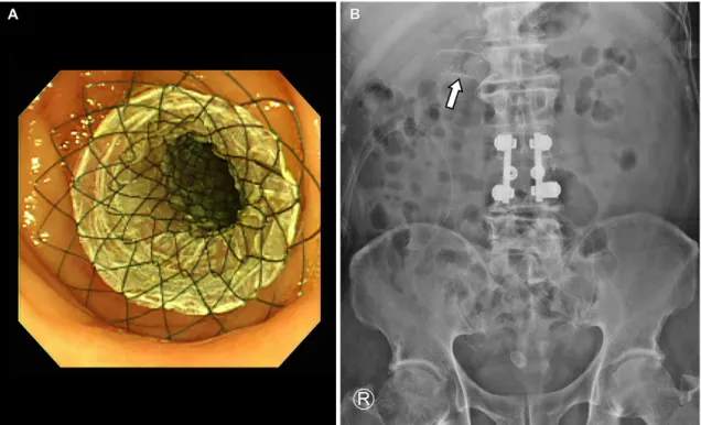
Fig. 3. Esophagogastroduodenoscopy and abdomen X-ray findings after SEMS insertion. (A) The SEMS covered with the leakage site of the duodenum. (B) The SEMS is located at the first portion of the duodenum (arrow). SEMS, self-expandable metallic stent.
sheet (Neoveil, Gunze Ltd., Kyoto, Japan) were applied in- evitably above the primary repair site in order that enhances the strength of sutures and prevents postoperative leakage.
Additionally, a negative suction drain was placed in the sub- hepatic area, primary repair site, and pelvic cavity.
At postoperative day 3, fresh bile drainage in the negative suction drain was noted, and fever spiked to 38.1°C. At post- operative day 5, diatrizoate sodium and diatrizoate meglu- mine solution (Gastrografin, Bayer Schering Pharma, Berlin, Germany) procedure was performed, and the diatrizoate so- dium and diatrizoate meglumine solution leakage at the pri- mary repair site was identified (Fig. 2). We decided on a multi-
disciplinary team to manage the problem and discussed with a gastroenterologist. After the meeting, we concluded that the endoscopic insertion of covered stent would be an alternative treatment for postoperative leakage. At postoperative day 6, a 6-cm-sized covered SEMS (Taewoong Medical, Seoul, Korea) was applied gently on the leakage site with fluoroscopic assis- tance (Fig. 3). After SEMS insertion, the patient was tolerable, and a decreased frequency of fever spiking was noted.
Additionally, the amount of drainage in the negative suction drain had decreased to below 50 mL/day after 7 days of SEMS insertion (Fig. 4). The stent was removed by esoph- agogastroduodenoscopy with endoscopic snare at post- operative day 35, and on the following day, the patient was discharged in good condition.
DISCUSSION
Surgery is widely used as the first-line treatment for duode- nal ulcer perforation.8 However, postoperative leakage is a life-threatening complication, and leakage rates remain high (2-16%).1,9,10 In addition, re-operation of postoperative leak- age is technically difficult and challenging for older patients and those with co-morbidity and poor general condition at immediate post-operation.7 However, our case demonstrated A
A BB
that it may be possible to use endoscopic-covered SEMS in- sertion as a rescue treatment for perforated duodenal ulcer with postoperative leakage. Moreover, there was no complica- tion associated with SEMS.
Endoscopic stent insertion is an attractive alternative to surgery. Re-operation is a burdensome procedure to surgeons, especially for those with poor general condition and co- morbidities. Endoscopic stent insertion may be advantageous in that patients do not require general anesthesia and it can be performed in patients with poor general condition. Therefore, endoscopic stent placement is already a standard procedure, mainly for intestinal strictures; however, it is now also an ap- proved treatment for esophageal perforations.11,12 Additionally, endoscopic stent insertion allows early oral intake, which de- creases the need for parenteral nutrition and risk of bacterial translocation from the gut, facilitating early recovery.13
However, despite many of its advantages, endoscopic SEMS insertion also has several disadvantages compared with surgery. There is a potential risk that esophagogastroduodeno- scopy may aggravate the perforation or leakage by gas insufflation.14 In addition, SEMS insertion offers the dis- advantages of stent-induced stenosis and ulcer aggravation.7,15 A major drawback of the procedure is distal migration of the stent. Previous study reported stent removal through operation due to stent migration at the small bowel.16 Therefore, SEMS may be removed early before the development of complications associated with stents. Furthermore, frequent check by abdomi- nal X-ray should be performed after SEMS insertion to confirm the stent location and prevent its migration to the small bowel.
Despite its advantages, whether covered SEMS insertion with simultaneous abdominal drainage will replace surgery as the primary treatment method of duodenal ulcer perfo- ration remains uncertain.17,18 Bergström et al.2 demonstrated favorable results of primary covered SEMS insertion with ab- dominal drainage in patients with perforated duodenal ulcer and comorbidities or technically difficult surgery. However, the procedure was only performed in a few patients, and further research may be required. Thus, surgery should still be con- sidered as the primary mode of treatment in patients with a preserved performance status and early detection of duode- nal ulcer perforation to achieve early recovery. Additionally, endoscopic SEMS insertion with simultaneous abdominal drainage may be an alternative to surgery only in patients with severe co-morbidities or delayed diagnosis.
The current report demonstrated that endoscopic covered SEMS insertion can be used as a rescue treatment for a perfo- rated duodenal ulcer with postoperative leakage after primary repair. Endoscopic SEMS insertion is a less invasive procedure comparing re-operation and increased survival chance for pa- tients with co-morbidities or technically difficult surgery. Thus, endoscopic covered SEMS insertion may be an alternative procedure for re-operation in selected patients with post- operative leakage after primary repair of duodenal ulcer perforation.
REFERENCES
1. Gupta S, Kaushik R, Sharma R, Attri A. The management of large perforations of duodenal ulcers. BMC Surg 2005;5:15.
2. Bergström M, Arroyo Vázquez JA, Park PO. Self-expandable metal stents as a new treatment option for perforated duodenal ulcer.
Endoscopy 2013;45:222-225.
3. Eubanks S, Edwards CA, Fearing NM, et al. Use of endoscopic stents to treat anastomotic complications after bariatric surgery.
J Am Coll Surg 2008;206:935-938; discussion 938-939.
4. Puli SR, Spofford IS, Thompson CC. Use of self-expandable stents in the treatment of bariatric surgery leaks: a systematic review and meta-analysis. Gastrointest Endosc 2012;75:287-293.
5. Schubert D, Scheidbach H, Kuhn R, et al. Endoscopic treatment of thoracic esophageal anastomotic leaks by using silicone-cov- ered, self-expanding polyester stents. Gastrointest Endosc 2005;61:891-896.
6. Serra C, Baltasar A, Andreo L, et al. Treatment of gastric leaks with coated self-expanding stents after sleeve gastrectomy.
Obes Surg 2007;17:866-872.
7. Swinnen J, Eisendrath P, Rigaux J, et al. Self-expandable metal stents for the treatment of benign upper GI leaks and perforations.
Gastrointest Endosc 2011;73:890-899.
8. Søreide K, Thorsen K, Harrison EM, et al. Perforated peptic ulcer.
Lancet 2015;386:1288-1298.
9. Søreide K, Thorsen K, Søreide JA. Strategies to improve the out- come of emergency surgery for perforated peptic ulcer. Br J Surg 2014;101:e51-e64.
10. Lunevicius R, Morkevicius M. Management strategies, early re- sults, benefits, and risk factors of laparoscopic repair of perfo- rated peptic ulcer. World J Surg 2005;29:1299-1310.
11. Kim KY, Tsauo J, Song HY, Kim PH, Park JH. Self-expandable met- allic stent placement for the palliation of esophageal cancer. J Korean Med Sci 2017;32:1062-1071.
12. Johnsson E, Lundell L, Liedman B. Sealing of esophageal perfo- ration or ruptures with expandable metallic stents: a prospective controlled study on treatment efficacy and limitations. Dis Esophagus 2005;18:262-266.
13. Reintam Blaser A, Starkopf J, Alhazzani W, et al. Early enteral nu- trition in critically ill patients: ESICM clinical practice guidelines.
Intensive Care Med 2017;43:380-398.
14. Chertoff J, Khullar V, Burke L. Duodenal perforation following esophagogastroduodenoscopy (EGD) with cautery and epi- nephrine injection for peptic ulcer disease: an interesting case of nonoperative management in the medical intensive care unit (MICU). Int J Surg Case Rep 2015;10:121-125.
15. Wai CT, Khor C, Lim SE, Ho KY. Post-metallic stent placement bleeding caused by stent-induced ulcers. World J Gastroenterol 2005;11:5739-5741.
16. Holm TE, Rosseland AR, Lundin KA, et al. Endoscopic stent treat-
ment of a duodenal ulcer perforation. Endoscopy 2011;43 Suppl 2 UCTN:E60.
17. Mouly C, Chati R, Scotté M, Regimbeau JM. Therapeutic manage- ment of perforated gastro-duodenal ulcer: literature review. J Visc Surg 2013;150:333-340.
18. Babor R, Talbot M, Tyndal A. Treatment of upper gastrointestinal leaks with a removable, covered, self-expanding metallic stent.
Surg Laparosc Endosc Percutan Tech 2009;19:e1-e4.