www.jpis.org
pISSN 2093-2278 eISSN 2093-2286 Copyright © 2011 Korean Academy of PeriodontologyThis is an Open Access article distributed under the terms of the Creative Commons Attribution Non-Commercial License (http://creativecommons.org/licenses/by-nc/3.0/).
Initial adhesion of bone marrow stromal cells to various bone graft substitutes
Young-Jae Jo, Kyoung-Hwa Kim, Ki-Tae Koo, Tae-Il Kim, Yang-Jo Seol, Yong-Moo Lee, Young Ku, Chong-Pyoung Chung, In-Chul Rhyu* Department of Periodontology, Seoul National University School of Dentistry, Seoul, Korea
Purpose: The aim of this study is to determine whether certain biomaterials have the potential to support cell attachment.
After seeding bone marrow stromal cells onto the biomaterials, we investigated their responses to each material in vitro.
Methods: Rat bone marrow derived stromal cells were used. The biomaterials were deproteinized bovine bone mineral (DBBM), DBBM coated with fibronectin (FN), synthetic hydroxyapatite (HA), HA coated with FN, HA coated with β-tricalcium phosphate (TCP), and pure β-TCP. With confocal laser scanning microscopy, actin filaments and vinculin were observed after 6, 12, and 24 hours of cell seeding. The morphological features of cells on each biomaterial were observed using scanning electron microscopy at day 1 and 7.
Results: The cells on HA/FN and HA spread widely and showed better defined actin cytoskeletons than those on the other biomaterials. At the initial phase, FN seemed to have a favorable effect on cell adhesion. In DBBM, very few cells adhered to the surface.
Conclusions: Within the limitations of this study, we can conclude that in contrast with DBBM not supporting cell attachment, HA provided a more favorable environment with respect to cell attachment.
Keywords: Bone substitutes, Cell adhesion, Fibronectins, Stem cells.
INTRODUCTION
Various studies have been performed to regenerate or re- pair bone defects in the craniofacial region. In particular, one strategy among the methods that have been employed in bone tissue engineering is to utilize biomaterial as a scaffold for cell attachment. The scaffold material functions to pro- vide 3-dimensional structure for cell attachment and cell de- livery. This enables the biomaterial to provide cells needed for bone regeneration directly into bone defects. To acquire a suitable scaffold for cell attachment, it is important to inves- tigate the cell-material interactions [1-4].
Calcium phosphate is one of the commonly used bone sub-
stitutes because of its high biocompatibility, osteoconductiv- ity, and non-toxicity [5]. Among the calcium phosphates, hy- droxyapatite (HA) and β-tricalcium phosphate (TCP) have been widely investigated and verified by in vitro and in vivo studies. HA has shown considerable osteogenic ability in the presence of bone marrow cells [6-8]. Also, it has been report- ed that TCP provides an excellent environment for bone re- generation despite its rapid degradation and weak mechani- cal properties [9-12]. Okumura et al. [6] demonstrated that the initial attachment of osteoblasts to HA was faster than to oth- er materials and this might result in earlier osteogenesis on HA. Kon et al. [8] also reported that autologous bone marrow stromal cells transplanted on HA-based carriers led to exten- Received: Jan. 1, 2011; Accepted: Feb. 9, 2011
*Correspondence: In-Chul Rhyu
Department of Periodontology, Seoul National University Dental Hospital, Seoul National University School of Dentistry, 275-1 Yeongeon-dong, Jongno-gu, Seoul 110-768, Korea
Email: [email protected], Tel: +82-2-2072-2640, Fax: +82-2-744-0051
play a role as a scaffold for bone formation and be useful for reconstructing bone.
High porosity and micro-architecture of deproteinized bo- vine bone mineral is like that of the human bone. It facilitates the invasion of blood vessels and cell attachment to the bo- vine bone [13,14]. It was reported that anorganic bovine bone was highly biocompatible and osteoconductive [15].
Fibronectin (FN) is one of the extracellular matrix glycopro- teins, which play various roles in bone formation. It has been known that FN plays an important role in promoting cell attchment, cell spreading, and cell differentiation. Many at- tempts have been made to improve cell attchment by means of FN [1,16,17]. It was shown that FN coated biomaterials may be able to promote initial cell attachment.
The aim of this study is to determine whether these mate- rials have potential as scaffolding material to support cell at- tachment. After seeding bone marrow stromal cells onto the biomaterials, we investigated their initial attachment in vitro.
MATERIALS AND METHODS
Cell isolation and culture
Rat bone marrow derived stromal cells were obtained ac- cording to the methods described previously by Maniato- poulos et al. [18]. Briefly, femora and tibiae were aseptically dissected from Sprague Dawley rats (130 to 150 g). The epiph- yses were cut away and the bone marrow was flushed out by α-MEM medium (Gibco, Grand Island, NY, USA), which was expelled from a 10 mL syringe. The bone marrow cells were cultured in standard culture medium at 37°C in 5% CO2. The culture medium contained α-MEM supplement with 10%
fetal bovine serum (Gibco), 100 U/mL penicillin and 100 μg/
mL streptomycin (Gibco). Hematopoietic cells and other un- attached cells were washed away after 3 days. The culture medium was replaced twice a week. Near confluency, bone marrow stromal cells had been passaged and expanded until passage 4 or 5. Subsequently, they were seeded on the bio- materials.
Biomaterials
The tested biomaterials were divided into six groups. The first group was deproteinized bovine bone mineral (Bio-Oss, Geistlich Biomaterials, Wolhusen, Switzerland) as natural bone. In addition, the deproteinized bovine bone mineral (DBBM) was also studied after being coated with 100 μM hu- man plasma fibronectin (FN; Gibco). The third material was synthetic HA, which is developed at Dentium Co. in Korea.
Additionally, HA was investigated after coating it with 100 μM FN or β-TCP. Coating DBBM or HA with FN was per-
at 4°C. In the case of HA coated with β-TCP (HA/TCP), the material (Osteon, Dentium Co., Seoul, Korea) was obtained from Dentium Co. and consisted of 70% HA and 30% β-TCP.
The sixth material was pure β-TCP particles (Cerasorb, Cu- rasan AG, Kleinostheim, Germany), which were used after crushing and passing them through sieves. These biomateri- als had particle sizes of 250 to 1,000 μm.
Confocal laser scanning microscopy
The bone marrow stromal cells were cultured on the bio- materials at a cell density of 5×10⁵ cells/well on a 48-well plate. 0.25 g of a given biomaterial was used per well. After 6, 12, and 24 hours of cell seeding, actin filaments, vinculin, and nuclei were stained with rhodamine-labelled phalloidin (Molecular Probes, Eugene, OR, USA), anti-vinculin antibody (Sigma-Aldrich Co., St. Louis, MO, USA), and DAPI (Molecular Probes), respectively, according to the manufacturer’s proto- col. In brief, cells were washed with phosphate buffered sa- line (PBS) and fixed in 10% neutral buffered formalin for 20 minutes and subsequently permeabilized with 0.1% Triton X-100 solution. The samples were incubated with 1% bovine serum albumin for 60 minutes. The vinculin was stained with mouse anti-vinculin antibody for 1 hour. Then the actin cytoskeleton was stained using rhodamine-labelled phalloi- din for 20 minutes, followed by incubation with DAPI for 5 minutes. The samples were observed in confocal laser scan- ning microscopy (Olympus-FV300, Olympus, Tokyo, Japan).
Scanning electron microscopy
The bone marrow stromal cells were seeded on each bio- material at a density of 5×10⁵ cells/well on a 48-well culture plate. 0.25 g of a biomaterial was used per well. After 1 and 7 days, the morphology of the cells was observed using a scan- ning electron microscope (S-4700, Hitach, Tokyo, Japan). The biomaterials were rinsed twice with PBS and fixed with 2.5%
glutaraldehyde at 4°C for 20 minutes. Each sample was fixed with 1% OsO4 at 4°C for 20 minutes. The samples were dehy- drated by ethanol in increasing concentrations of 70 to 100%
at 4°C, and were also dehydrated with 1,1,1,3,3,3-hexamethyl- disilazane (Sigma-Aldrich Co.) for 20 minutes. The speci- mens were sputter-coated with gold and observed in the scanning electron microscope.
RESULTS
Confocal laser scanning microscopy
At 6 hours after cell seeding, the cells, which adhered on all the materials but HA/FN, were round or oval. On HA coated with FN (HA/FN), the cells seemed to be elongated (Fig. 1).
oval shapes. On the other materials, the cells had spread more widely at 12 hours compared to the cells after 6 hours. Nota- bly, actin filaments were widely distributed in cells adhered on HA and HA/FN. Cells on HA and HA/FN overlapped one another. Although cells on HA/TCP and TCP showed pro- nounced cell extensions for anchoring to the surface, they did not seem to overlap one another (Fig. 2). On DBBM and DBBM/FN, the cells after 24 hours showed similar shapes to those after 12 hours. The cells did not seem to spread exten- sively on DBBM. In addition, it was difficult to detect the cells adhered on DBBM and DBBM/FN. The cells on the other
well-defined actin cytoskeletons after 24 hours (Fig. 3).
Scanning electron microscopy
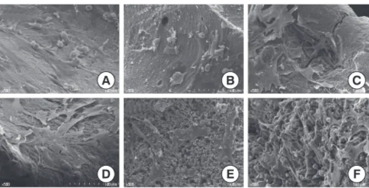
The morphological features of cells on each biomaterial were observed with scanning electron microscopy at day 1 and 7, respectively. After 1 day of incubation, even if some cells adhered flatly on the surface of DBBM and DBBM/FN, many cells were observed to have a circular morphology. The cells on HA and HA/FN appeared to be flattened out throughout the surface and were elongated. They seemed to be in close attachment to each other. In the case of HA/TCP, it was ob- served that β-TCP particles were coated on the HA surfaces and several layers of cells adhered to these surfaces. It was found cell density on HA/TCP and TCP were lower than it was on HA and HA/FN (Fig. 4). After 7 days, it was impossible to find well-spread cells on DBBM and DBBM/FN. In con- trast, cells on the other biomaterials adhered close together.
Cells adhered to HA and HA/FN developed lamellipodia and were widely spread all over the material. Some cells on HA and HA/FN formed multicellular layers. On HA/TCP and TCP, the cells showed a flat and polygonal shape. They had irregularly branched cytoplasm (Fig. 5).
DISCUSSION
In this study, we investigated rat bone marrow stromal cell responses to various bone substitutes. When biomaterials supporting bone formation are developed, attention should
50.0 μm 50.0 μm 50.0 μm
50.0 μm 50.0 μm 50.0 μm
A
D
B
E
C
F Figure 1. Immunofluorescence images showing cells stained for vinculin (green), actin (red) and the nucleus (blue) at 6 hours (×400).
On all the materials but hydroxyapatite/fibronectin (HA/FN), the cells showed round or oval shapes. (A) Deproteinized bovine bone mineral (DBBM). (B) DBBM/FN. (C) HA. (D) HA/FN. (E) HA/ tricalci- um phosphate (TCP). (F) TCP.
50.0 μm 50.0 μm 50.0 μm
50.0 μm 50.0 μm 50.0 μm
A
D
B
E
C
F Figure 3. Immunofluorescence images showing cells stained for vinculin (green), actin (red) and the nucleus (blue) at 24 hours ( ×400). On deproteinized bovine bone mineral (DBBM) and DBBM/FN, cells at 24 hours appeared to have round or oval shapes similar to the cells at 6 hours. The cells on the other biomaterials showed more extensive spreading and expressed well-defined actin cytoskeletons. (A) DBBM. (B) DBBM/fibronectin (FN). (C) Hydroxy- apatite (HA). (D) HA/FN. (E) HA/tricalcium phosphate (TCP). (F) TCP.
50.0 μm 50.0 μm 50.0 μm
50.0 μm 50.0 μm 50.0 μm
A
D
B
E
C
F Figure 2. Immunofluorescence images showing cells stained for vinculin (green), actin (red) and the nucleus (blue) at 12 hours (×400). In contrast to cells cultured on deproteinized bovine bone mineral (DBBM), cells on the other biomaterials appeared to be widely spread. (A) DBBM. (B) DBBM/fibronectin (FN). (C) Hydroxy- apatite (HA). (D) HA/FN. (E) HA/tricalcium phosphate (TCP). (F) TCP.
be paid to the cell-material interactions, such as cell attach- ment, proliferation, and differentiation. Particularly, cell at- tachment on biomaterial is the first step in cell-material in- teractions. Depending on the degree of cell attachment, the capacity to proliferate and differentiate may be determined [19].
The results of this study demonstrated that the degree of attachment was different according to bone substitutes. At day 7, the cells on DBBM showed round shapes and the iso- lation of individual cells was observed (Fig. 5). This may mean the initiation of apoptosis [20]. The cells grown on DBBM/
FN also had similar shapes to those on DBBM. These results for Bio-Oss are in agreement with other in vitro studies [5,20- 24]. Many studies have demonstrated that the viability of os- teoblasts grown on Bio-Oss decreased over time [5,22]. How- ever, some in vitro experiments [14,25,26] and clinical results [13,15,27,28] reported that DBBM produced a favorable cell re- sponse. These conflicting results about DBBM could stem from different culture conditions (e.g., seeding density, culti- vation duration) among the studies [23]. Another study, which reported on cell viability with DBBM, demonstrated that the surface properties of hydroxyapatite changed when it was in contact with blood proteins and extracellular matrix compo- nents [22]. Additionally, oxygen and nutrients in vivo would provide support for cell proliferation by developing the vas- cularization. However, the in vitro environment was usually set up under static culture conditions [20]. These factors may result in the differences among the studies’ results.
Fibronectin (FN) is an extracellular matrix protein that pro- motes cell adhesion. The binding of FN and other adhesion proteins to cell surface receptors enhances cell spreading, fo- cal contact formation, and strength of adhesion [19,29]. In this study, 6 hours after cell seeding, the cells on HA/FN showed
actin filaments, unlike those on HA (Fig. 1). At 12 hours, cells on DBBM/FN seemed to express actin filaments unlike those on DBBM. Grzesik and Robey [16] found that FN that contains the integrin-binding Arginine-Glycine-Aspartate sequence promoted bone cell attachment after 24 hours of incubation.
In our study, at the initial phase, FN seemed to have a favor- able effect on cell adhesion. Nevertheless, when the attach- ment tendencies were analyzed subsequently, the effect of FN seemed to be lost. After 12 hours, cells with or without fi- bronectin seemed to display no differences in cell morphol- ogy. The FN-coating method for biomaterials might have an effect on these results. To coat the biomaterials with FN, they were dried in FN solution overnight. Deduced from the re- sults mentioned above, it is possible that FN, which attached to the surfaces of biomaterials, would be released over time.
Therefore, the amount of FN on a surface might decrease, and this could reduce its effect on cell attachment. This is supported by previous studies [1,17]. Van den Dolder et al. [1]
also explained that the surface concentration of FN might decrease continuously because of the competitive adsorp- tion-desorption process of serum proteins. This could di- minish the beneficial effects on the seeded cells. Another possible cause, which decreased the effectiveness of FN, is that we used whole FN to treat the biomaterials with FN. Ac- cording to Grzesik et al. [17], the application of whole glyco- proteins has limitations for various reasons, such as the op- posite effect of multiple domains of the same factor, low availability of material, and loss of activity.
In the case of HA/TCP and TCP, cells seem to spread less extensively than those on pure HA in the initial phase (Fig. 2).
HA/TCP, an alloplastic material, was covered with TCP parti- cles on HA surfaces. Due to this surface characteristic of HA/
TCP, a similar phenomenon was observed on TCP and HA/
Figure 4. Scanning electron microscopy images showing cells on (A) deproteinized bovine bone mineral (DBBM), (B) DBBM/fibronectin (FN), (C) hydroxyapatite (HA), (D) HA/FN, (E) HA/tricalcium phos- phate (TCP), and (F) TCP at day 1 ( ×500). Many of the cells on DBBM and DBBM/FN were less spread out and more round shape.
In contrast, the cells on HA and HA/FN appeared to be flattened out throughout the surface.
×500 100 μm ×500 100 μm ×500 100 μm
×500 100 μm ×500 100 μm ×500 100 μm
A B C
D E F
Figure 5. Scanning electron microscopy images showing cells on (A) deproteinized bovine bone mineral (DBBM), (B) DBBM/fibronectin (FN), (C) hydroxyapatite (HA), (D) HA/FN, (E) HA/tricalcium phos- phate (TCP), and (F) TCP at day 7 (×500). Only a few, round cells ad- hered to the surface of DBBM and DBBM/FN. The cells on the oth- er biomaterials adhered close together and showed polygonal shapes.
×500 100 μm ×500 100 μm ×500 100 μm
A B C
D E F
×500 100 μm ×500 100 μm ×500 100 μm
rial is the first stage for cell proliferation and differentiation.
Therefore, these results have implications for generating the proliferation of cells on biomaterials. Suzuki et al. [30] report- ed that osteoblast-like cells on pure HA exhibited higher growth rates than those on other ceramic plates including β-TCP. HA in the culture medium tend to have high pH on the thin surface layer of the ceramic carrier and to accelerate calcinations by trapping Ca ions from the medium, which stimulates the growth of osteoblast-like cells on HA.
Within the limitations of this study, we can conclude that while DBBM does not support cell attachment, HA provides a more favorable environment for cell attachment.
CONFLICT OF INTEREST
No potential conflict of interest relevant to this article was reported.
ACKNOWLEDGEMENTS
This work was supported by a National Research Founda- tion of Korea Grant funded by the Government of the Re- public of Korea (2008-E00580).
REFERENCES
1. van den Dolder J, Bancroft GN, Sikavitsas VI, Spauwen PH, Mikos AG, Jansen JA. Effect of fibronectin- and collagen I- coated titanium fiber mesh on proliferation and differen- tiation of osteogenic cells. Tissue Eng 2003;9:505-15.
2. Yoshimoto H, Shin YM, Terai H, Vacanti JP. A biodegrad- able nanofiber scaffold by electrospinning and its potential for bone tissue engineering. Biomaterials 2003;24:2077-82.
3. Woo KM, Jun JH, Chen VJ, Seo J, Baek JH, Ryoo HM, et al.
Nano-fibrous scaffolding promotes osteoblast differenti- ation and biomineralization. Biomaterials 2007;28:335-43.
4. Langer R, Vacanti JP. Tissue engineering. Science 1993;260:
920-6.
5. Turhani D, Weissenböck M, Watzinger E, Yerit K, Cvikl B, Ewers R, et al. Invitro study of adherent mandibular os- teoblast-like cells on carrier materials. Int J Oral Maxillo- fac Surg 2005;34:543-50.
6. Okumura A, Goto M, Goto T, Yoshinari M, Masuko S, Kat- suki T, et al. Substrate affects the initial attachment and subsequent behavior of human osteoblastic cells (Saos-2).
Biomaterials 2001;22:2263-71.
7. Ohgushi H, Okumura M, Tamai S, Shors EC, Caplan AI.
Marrow cell induced osteogenesis in porous hydroxyapa- tite and tricalcium phosphate: a comparative histomor-
Mater Res 1990;24:1563-70.
8. Kon E, Muraglia A, Corsi A, Bianco P, Marcacci M, Martin I, et al. Autologous bone marrow stromal cells loaded onto porous hydroxyapatite ceramic accelerate bone repair in critical-size defects of sheep long bones. J Biomed Mater Res 2000;49:328-37.
9. Hattori H, Masuoka K, Sato M, Ishihara M, Asazuma T, Takase B, et al. Bone formation using human adipose tis- sue-derived stromal cells and a biodegradable scaffold. J Biomed Mater Res B Appl Biomater 2006;76:230-9.
10. Marino G, Rosso F, Cafiero G, Tortora C, Moraci M, Barba- risi M, et al. Beta-tricalcium phosphate 3D scaffold pro- mote alone osteogenic differentiation of human adipose stem cells: in vitro study. J Mater Sci Mater Med 2010;21:
353-63.
11. Dong J, Uemura T, Shirasaki Y, Tateishi T. Promotion of bone formation using highly pure porous beta-TCP com- bined with bone marrow-derived osteoprogenitor cells.
Biomaterials 2002;23:4493-502.
12. Boo JS, Yamada Y, Okazaki Y, Hibino Y, Okada K, Hata K, et al. Tissue-engineered bone using mesenchymal stem cells and a biodegradable scaffold. J Craniofac Surg 2002;13:
231-9.
13. Camelo M, Nevins ML, Schenk RK, Simion M, Rasperini G, Lynch SE, et al. Clinical, radiographic, and histologic evaluation of human periodontal defects treated with Bio-Oss and Bio-Gide. Int J Periodontics Restorative Dent 1998;18:321-31.
14. Stephan EB, Jiang D, Lynch S, Bush P, Dziak R. Anorganic bovine bone supports osteoblastic cell attachment and proliferation. J Periodontol 1999;70:364-9.
15. Piattelli M, Favero GA, Scarano A, Orsini G, Piattelli A. Bone reactions to anorganic bovine bone (Bio-Oss) used in si- nus augmentation procedures: a histologic long-term re- port of 20 cases in humans. Int J Oral Maxillofac Implants 1999;14:835-40.
16. Grzesik WJ, Robey PG. Bone matrix RGD glycoproteins:
immunolocalization and interaction with human primary osteoblastic bone cells in vitro. J Bone Miner Res 1994;9:
487-96.
17. Grzesik WJ, Ivanov B, Robey FA, Southerland J, Yamauchi M. Synthetic integrin-binding peptides promote adhesion and proliferation of human periodontal ligament cells in vitro. J Dent Res 1998;77:1606-12.
18. Maniatopoulos C, Sodek J, Melcher AH. Bone formation in vitro by stromal cells obtained from bone marrow of young adult rats. Cell Tissue Res 1988;254:317-30.
19. Anselme K. Osteoblast adhesion on biomaterials. Bioma- terials 2000;21:667-81.
blast interactions with calcium phosphate ceramics modi- fied by coating with type I collagen. J Biomed Mater Res A 2005;73:409-21.
21. Petrovic L, Schlegel AK, Schultze-Mosgau S, Wiltfang J.
Different substitute biomaterials as potential scaffolds in tissue engineering. Int J Oral Maxillofac Implants 2006;21:
225-31.
22. Herten M, Rothamel D, Schwarz F, Friesen K, Koegler G, Becker J. Surface- and nonsurface-dependent in vitro ef- fects of bone substitutes on cell viability. Clin Oral Inves- tig 2009;13:149-55.
23. Wiedmann-Al-Ahmad M, Gutwald R, Gellrich NC, Hüb- ner U, Schmelzeisen R. Search for ideal biomaterials to cultivate human osteoblast-like cells for reconstructive surgery. J Mater Sci Mater Med 2005;16:57-66.
24. Kübler A, Neugebauer J, Oh JH, Scheer M, Zöller JE. Growth and proliferation of human osteoblasts on different bone graft substitutes: an in vitro study. Implant Dent 2004;13:
171-9.
25. Açil Y, Terheyden H, Dunsche A, Fleiner B, Jepsen S. Three- dimensional cultivation of human osteoblast-like cells on
2000;51:703-10.
26. Ignjatovic@ N, Ninkov P, Kojic@ V, Bokurov M, Srdic@ V, Krnojelac D, et al. Cytotoxicity and fibroblast properties during in vitro test of biphasic calcium phosphate/poly- dl-lactide-co-glycolide biocomposites and different phos- phate materials. Microsc Res Tech 2006;69:976-82.
27. Carmagnola D, Adriaens P, Berglundh T. Healing of hu- man extraction sockets filled with Bio-Oss. Clin Oral Im- plants Res 2003;14:137-43.
28. Zitzmann NU, Schärer P, Marinello CP, Schüpbach P, Ber- glundh T. Alveolar ridge augmentation with Bio-Oss: a histologic study in humans. Int J Periodontics Restorative Dent 2001;21:288-95.
29. Burmeister JS, Vrany JD, Reichert WM, Truskey GA. Effect of fibronectin amount and conformation on the strength of endothelial cell adhesion to HEMA/EMA copolymers. J Biomed Mater Res 1996;30:13-22.
30. Suzuki T, Hukkanen M, Ohashi R, Yokogawa Y, Nishizawa K, Nagata F, et al. Growth and adhesion of osteoblast-like cells derived from neonatal rat calvaria on calcium phos- phate ceramics. J Biosci Bioeng 2000;89:18-26.