Research Article Open Access http://dx.doi.org/10.5423/RPD.2014.20.2.112
Research in Plant Disease
The Korean Society of Plant Pathology pISSN 1598-2262, eISSN 2233-9191
키틴분해세균, 키틴 및 그들의 산물이 함유된 미생물제에 의한 오이의 뿌리혹선충(Meloidogyne spp.) 방제
Control of the Root-Knot Nematode (Meloidogyne spp.) on
Cucumber by a Liquid Bio-Formulation Containing Chitinolytic Bacteria, Chitin and Their Products
하우종1 · 김영철1 · 정현채2 · 박서기3*
1전남대학교 식물생명공학부, 2순천대학교 산림자원학과, 3순천대학교 식물의학과
Woo Jong Ha1, Young Cheol Kim1, Hyunchae Jung2 and Seur Kee Park3*
1Department of Plant Biotechnology and Biotechnology Research Institute, Chonnam National University, Gwangju 500-757, Korea
2Department of Forest Resources, Sunchon National University, Sunchon 540-742, Korea
3Department of Agricultural Biology, Sunchon National University, Sunchon 540-742, Korea
A liquid bio-formulation containing chitinolytic bacteria, chitin and their products was assessed for its po- tential biological control against root-knot nematodes on cucumber. The bio-formulation was prepared by cultures of three chitinolytic bacteria, Chromobacterium sp. strain C-61, Lysobacter engymogenes and Serratia plymuthica in minimal medium supplemented with chitin. Under pot conditions, the bio-formulation showed better growth of cucumber plants, and less root galls and population density of Meloidogyne spp. than control media without the bio-formulation. In a greenhouse, 75-fold diluted bio-formulations were treated instead of water around cucumber plants through hoses for drip irrigation six times at 5-day intervals from the transplanting date. After 30 and 60 days, the treatment provided about 7% and 10% enhancement in the plant height and about 78% and 69% reduction in population density of Meloidogyne spp. in the rhizosphere, respectively. In addition, the experiments showed that the control effects occurred only in the soils contacted with the bio-formulation. Undiluted bio-formulations were drenched three times at 10-day intervals around cucumber plants severely infested with Meloidogyne spp. The treatment showed about 37% plant enhance- ment without dead plants compared with 37% death in the untreated control, and about 82% nematode reduction. These results suggest that the bio-formulation can be practically used to control the root-knot nematode on cucumber.
Keywords : Bio-formulation, Chitinolytic bacteria, Cucumber, Root-knot nematode
서 론
뿌리혹선충(Meloidogyne spp.)은 전 세계적으로 96종이 기술 되었는데, Meloidogyne incognita, M. javanica, M. arenaria 및 M.
hapla의 4종이 약 95%를 차지하며, 3000종 이상의 식물을 침입
한다(Rich 등, 2008). 국내에는 6종의 뿌리혹선충이 서식하는데 (Cho 등, 2000) M. incognita, M. arenaria 및 M. hapla의 3종이 문 제시 되고, 기주식물로서 오이, 참외, 토마토, 고추, 배추, 인삼 등 24종이 보고되었다(Choi와 Choo, 1978). 이러한 뿌리혹선충 은 국내 시설재배 포장의 약 54%에 감염되어 있고, 밀도도 상당 히 높은데(Cho 등, 2000; Kim, 2001), 오이의 경우 정식 전 뿌리 혹선충이 100 cm3 당 10마리 이상이면 수량이 약 39% 감소되 는 것으로 알려져 있다(Kim과 Lee, 2008). 특히, 전남 순천 인근
*Corresponding author Tel : +82-61-750-3864 Fax: +82-61-750-3268 E-mail: [email protected]
Received March 31, 2014 Revised May 23, 2014 Accepted May 27, 2014
의 시설재배지역에서는 연중 내내 오이, 호박, 토마토 등을 재 배하고 있기 때문에 뿌리혹선충에 의한 피해가 크다.
이러한 뿌리혹선충을 방제하기 위하여 유기합성농약이 많이 사용되고 있다. 그러나 유기합성농약은 인간의 건강뿐만 아니 라 자연 환경을 파괴하기 때문에 사용이 금지되거나 제한되어 가는 추세이다(Abawi와 Windmer, 2000). 이를 대체하기 위한 수단으로 태양열처리, 객토, 윤작 등을 실시할 수 있으나(Kim 등, 2001) 비용, 노력 및 시간 등의 제약이 따른다. 또한 생물적 방제에 대한 연구가 많이 이루어졌는데(Kim 등, 2011; Lee 등, 2013; Radwan 등, 2012), 유기합성농약에 비하여 방제효과가 낮고 안정된 방제효과를 보여주지 못하는 실정이다(Lamovsek 등, 2013). 따라서 유기합성농약의 사용을 줄이기 위해서는 방 제효과가 더 높으면서도 더 적은 비용으로 생산될 수 있는 생물 적 방제원의 개발이 필요하다.
선충에 길항하는 미생물들은 키틴분해미생물에서 많이 보 고되었고, 이들은 주로 선충의 큐티클과 난각의 침입을 용이 하게 수 있는 chitinase, protease 및 collagenase를 분비하거나 (Khan 등, 2004; Liang 등, 2010), 선충을 죽이는 항생물질을 분 비한다(Park 등, 2004). 특히, 키틴은 선충 난각의 주요 구성성분 으로서 chitinase는 난각을 분해하여 부화를 억제하고(Zhang 등, 2009), 그것의 생산은 키틴이나 키토산에 의해서 유도된다 (Palma-Guerrero 등, 2010). 또한 토양에 키틴을 첨가하면 키 틴분해미생물들이 증가되어 선충이 억제되거나(Hallmann 등, 1998) 키틴이 분해되면서 방출된 암모니아에 의해서 억제된다 (Godoy 등, 1983). 이러한 효과 이외에 키틴은 식물의 영양원으 로 작용하고 식물의 생장을 촉진하며 식물의 방어반응을 유도 하는 것으로 보고되었다(Sharp, 2013). 따라서 키틴분해미생물 과 키틴을 함께 처리하면 단독으로 처리했을 때보다 선충에 대 한 방제효과가 더 좋고(Mittal 등, 1995; Tian 등, 2000), 그러한 효과는 선충에 대한 억제와 식물에 유도된 저항성 때문이라는 보고도 있다(Kalaiarasan 등, 2006). 따라서 선충 방제용 길항미 생물들은 항생물질(독소), protease와 chitinase 등의 분비에 의 한 직접적인 억제와 선충 인식부위의 방해, 영양분에 대한 경 쟁, 식물에 대한 저항성 유도 등과 같은 간접적인 효과를 지닌 다(Lamovsek 등, 2013).
이러한 기작들에 근거하면 키틴분해 미생물들을 포함한 식 물병 방제용 미생물제가 뿌리혹선충의 방제에도 효과가 있을 것으로 예상된다. 즉, 이 미생물제는 키틴함유배지에서 배양한 액이기 때문에 키틴분해세균, 키틴 및 키틴올리고당, chitinase 를 비롯한 다양한 분해효소, 항균물질들이 들어 있다(Kim 등, 2008; 2010; 2014). 또한 미생물제에 들어 있는 Chromobacterium sp. 균주는 감자씨스트선충의 부화를 억제하고(Chronin 등, 1997), Lysobacter enzymogenes는 뿌리혹선충을 비롯한 다양 한 선충(Chen 등, 2006), Serratia plymuthica는 뿌리혹선충을 억 제하는 것으로 보고되었고(Alballay 등, 2013), 이들로부터 다양 한 항생물질, protease 및 chitinase 등도 보고되었다(Dunne 등,
1997; Frankowski 등, 2001; Yuen 등, 2006).
이 연구에서는 식물병 방제용으로 개발된 미생물제가 뿌리 혹선충의 방제에도 활용될 수 있는지 알아보기 위하여 포트 상 태에서의 방제효과를 확인하고, 포장에서 오이 정식일 부터 물 대신에 희석된 미생물제를 살포하였을 경우와 뿌리혹선충에 심하게 감염된 오이에 미생물제 원액을 살포하였을 경우의 방 제효과를 조사하였다.
재료 및 방법
미생물제의 조제와 뿌리혹선충의 증식. 미생물제는 3종의 키틴분해세균을 500 l용 미생물배양기(흙살림)를 이용하여 키 틴이 포함된 최소영양배지에 배양하여 다음과 같이 조제하였 다. 배양기는 물을 채우고 80oC의 온도로 높여서 소독한 다음 물 1 l당 키틴 2 g과 (NH4)2SO4 0.6 g, KH2PO4 0.8 g, K2HPO4 0.6 g, MgSO4 · 7H2O 0.04 g을 첨가한 후 Nutrient broth에 서 1일간 배양(28oC, 180 rpm)한 Chromobacterium sp. C-61, L.
enzymogenes 및 S. plymuthica의 배양액 100 ml씩을 접종하고 28oC에서 10일 동안 배양하여 시험에 사용하였다. 공시균주는 식물병 방제에 사용되었던 균주들로서 기존의 방법들(Kim 등, 2008; 2010)에 근거하여 배양되었다.
뿌리혹선충은 전남 순천지역 시설재배 내 오이 뿌리의 혹에 서 분리, 증식하여 사용하였다. 즉, 혹이 형성된 뿌리를 물로 씻 어 절단한 다음 0.5% sodium hypochloride에서 3분간 흔들어 200 mesh와 500 mesh를 이용하여 알을 세척하고 모아서 오이 뿌리에서 증식하였다.
뿌리혹선충에 대한 미생물제의 방제효과 검정(포트 시험).
뿌리혹선충에 대한 미생물제의 방제효과를 검정하기 위하여 순천대학교 구내 온실에서 포트 시험을 수행하였다. 시험은 뿌 리혹선충을 접종한 토양에 미생물제 처리구와 대조구로서 배 지 처리구로 나누고, 뿌리혹선충을 접종하지 않은 토양에 배지 를 처리하여 상호 비교하였다. 오이에서 순수 증식된 뿌리혹선 충 함유 토양은 새로운 상토와 1 : 1(v : v)의 비율로 섞어서 플라 스틱 포트(길이 58 cm × 폭 9 cm × 높이 15 cm)에 넣었다. 여 기에 발아 후 30일된 오이 유묘(‘장형낙합’)를 포트 당 4주씩 심고, 각 주에 미생물제 원액 또는 배지를 10일 간격 3회 처리 하였다.
일반적으로 뿌리혹선충 1세대는 적정 온도에서 45일 정도 소요되지만, 이번 시험의 경우 뿌리혹선충이 증식되고 있는 토 양에 오이를 이식하였기 때문에 뿌리혹이 더 빨리 관찰되었다.
따라서 마지막 처리 10일 후 오이 줄기, 잎, 뿌리의 생육과 뿌리 에 형성된 혹의 수, 토양에 있는 뿌리혹선충의 밀도를 비교, 분 석하였다. 뿌리혹선충 밀도는 각 처리구로부터 토양시료를 채 취하여 2 mm 표준체로 걸렀다. 체를 통과한 토양은 Sieve and baermann funnel technique을 이용하여 선충을 분리하였다
(Southey, 1986). 분리된 선충은 24-well culture plate에 옮겨 도 립현미경(Leica. DMIL)하에서 뿌리혹선충 밀도를 조사하였다.
뿌리의 혹은 뿌리 5 cm에 붙어 있는 수를 세어서 평균하였다.
모든 실험은 각 처리별로 3반복 수행하였다.
뿌리혹선충에 대한 미생물제의 방제효과 검정(포장 시험).
전남 순천시 대대동 비닐하우스 재배단지에서 오이, 호박을 친 환경적으로 재배하면서 뿌리혹선충에 피해가 심했던 600평의 포장을 선정하여 2012년 3월 23일에 오이 유묘(‘장형낙합’)를 정식하였다. 포장의 1/2(300평)에 정식 후 미생물제(75배 희석) 를 1개월 동안 5일 간격으로 점적 관수하고, 나머지 300평은 동 일 조건으로 물을 점적 관수 후, 정식 일부터 한 달 간격으로 2개월간 오이의 초장과 근권의 선충밀도를 조사하였다.
정식 3개월 후쯤 오이 주변에 분포하는 주름잎(Mazus pumilus) 의 뿌리에서 많은 혹이 관찰되었다. 이 주름잎은 점적관수에 의 하여 물(미생물제제)이 살포되었을 부분과 살포되지 않았을 부 분의 토양에 고루 분포하고 있었다. 즉, 바로 인접해 있지만 미 생물제를 함유한 물이 도달되었을 부분과 도달되지 않았을 부 분 모두에 분포하고 있었다. 따라서 미생물제의 접촉여부에 따 른 뿌리혹선충 감염여부를 평가해 보기 위하여 미생물처리구 와 무처리구에서 물이 도달되었을 부분과 도달되지 않았을 부 분의 주름잎 뿌리에 대한 혹 형성 정도를 조사하였다.
정식 45일 후에는 무처리구에서 뿌리혹선충에 의해서 생육 이 현저히 낮은 오이들이 관찰되었다. 따라서 이들 오이의 반은 미생물제 원액을 10일 간격 3회 관주하고, 나머지 반은 동량의 물을 관주하여 10일 간격으로 오이의 초장, 생존율 및 근권의 선충 밀도를 조사하였다. 근권의 선충은 Sieve and baermann funnel technique을 이용하여 분리 후, 80oC에서 중탕하여 사멸
시켜 24-well plate에 옮겨 1 ml씩 분주하고 도립현미경하에서 총 선충 밀도, 식물기생선충 밀도, 뿌리혹선충 밀도를 조사하고 식물기생선충도감(Mai, 1996; Mekete 등, 2012)과 비교하여 분 류하였다.
결 과
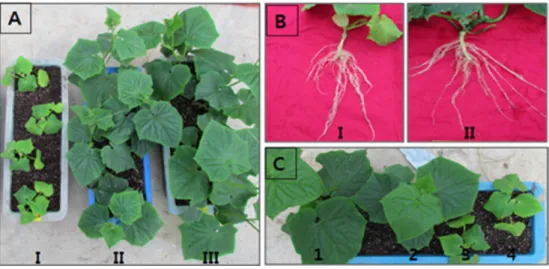
포트 조건에서 뿌리혹선충 감염 오이에 대한 미생물제의 방 제효과. 뿌리혹선충을 접종한 토양의 오이에 미생물제를 처 리한 결과, 배지를 처리한 대조구에 비하여 잎, 줄기의 생육이 월등히 우수하였다. 그러나 뿌리혹선충을 접종하지 않은 토양 보다는 생육이 적었다. 따라서 뿌리혹선충은 오이의 생육을 크 게 억제하고, 미생물제는 뿌리혹선충에 의한 생육 억제를 막아 준다는 것을 알 수 있었다(Fig. 1A). 또한 본 미생물제는 뿌리의 생육을 더 좋게 하고, 뿌리의 선충 혹도 더 적게 한다는 것을 알 수 있었다(Fig. 1B).
이러한 미생물제가 어느 정도의 범위에 영향을 미치는가를 알아보기 위하여 하나의 포트 내에 미생물제와 배지를 각각 처 리한 후 비교하였다. 그 결과, 미생물 처리주는 매우 우수한 생 육을 보인 반면 배지를 처리한 나머지 주는 미생물 처리주와의 거리에 따라 중간 정도의 생육 또는 아주 저조한 생육을 보였 다. 이것은 미생물제가 주변의 식물까지는 어느 정도 스며들어 영향을 미치나 그렇지 않은 식물에는 전혀 영향을 미치지 못한 것으로 판단되었다. 즉, 본 미생물제는 직접 접촉되어야만 효과 가 발휘될 수 있다는 것을 알 수 있었다(Fig. 1C).
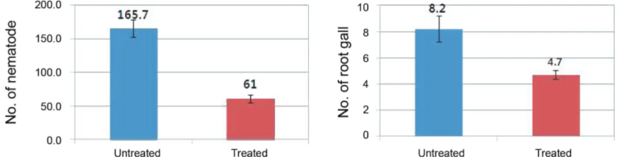
미생물제가 토양의 뿌리혹선충에 미치는 영향을 알아보기 위하여 토양의 선충 밀도와 뿌리에 감염된 혹의 수를 조사하였 다. 그 결과, 뿌리혹선충은 토양 100 cm3 당 미생물 처리구에서
Fig. 1. Effects of the bio-formulation on growth (A, C) and root gall (B) of cucumber plant in the soils infested with Meloidogyne spp. The bio-formulation was prepared by culture of three chitinolytic bacteria in minimal synthetic medium containing chitin. The bio-formulation and medium as a control were treated three times at 10-day-interval from transplanting date, and evaluated at 10 days after last treatment. In A and B, I and II were treated by medium and bio-formulation, respectively, on the soils infested with Meloidogyne spp., and III was not infested with Meloidogyne spp.
In C, first plant (1) was treated with the bio-formulation, and 2,3,4th plants were treated with medium on soil infested with Meloidogyne spp.
61마리, 배지 처리구에서 165.7마리로서 미생물제 처리에 의해 서 약 63%가 감소되었다. 뿌리의 혹은 미생물 처리구에서 5 cm 당 4.7개, 배지 처리구에서 8.2개가 관찰되어 미생물제를 처리 함으로서 뿌리혹의 약 43%가 감소된 것을 알 수 있었다(Fig. 2).
비닐하우스 재배 오이에서 뿌리혹선충에 대한 미생물제의 방제효과. 비닐하우스재배 오이에서 정식한 날부터 물 대신 에 75배 희석된 미생물제를 한 달간 점적 관수하고, 오이의 생 육과 토양 속의 선충 밀도를 조사하였다. 그 결과, 미생물제를 처리하면 물만 처리한 대조구에 비하여 오이의 초장은 정식 30일에 6.5%, 60일 후에는 10.1% 증가하고, 근권의 뿌리혹선 충밀도는 정식 30일에 77.6%, 60일 후에 69.3% 감소되었다(Table 1).
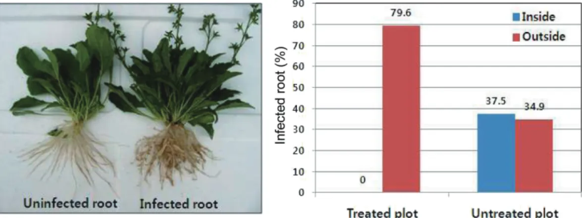
정식 3개월 후 미생물제의 접촉여부에 따른 방제 효과를 평 가해 보기 위하여 다양한 위치에서 선충 감염 여부를 조사하였 다. 그 결과, 미생물 처리구의 경우 물(미생물제)이 충분히 살포 되었을 점적호스 안쪽에서는 조사한 모든 뿌리(47개)가 전혀
감염되어 있지 않았으나 물이 도달하지 않았을 점적호스 바깥 쪽에서는 80% 정도가 감염되어 있었다. 반면, 물만 처리한 대조 구에서는 점적호스 안쪽에서 37.5%, 바깥쪽에서 34.7% 가 감염 되어 있었다(Fig. 3).
정식 45일 후경 무처리구에서 뿌리혹선충에 심하게 감염되 어 오이의 초장이 현저히 작은 곳이 군데군데 관찰되었다. 이러 한 오이에 미생물제 원액을 10일 간격 3회 관주하고 오이의 초 장 및 생존과 토양의 선충 밀도를 조사하였다. 오이의 초장은 첫 번째 처리 후 약 22%, 두 번째 처리 후 약 36%, 세 번째 처리 후 약 37% 증가하였다. 또한 무처구에서는 죽은 식물이 나오기 시작하여 30일 후에는 37%가 고사되었는데, 미생물 처리구에 서는 한 주도 죽지 않았다. 근권에 분포하는 뿌리혹선충의 밀도 도 미생물제를 처리함으로서 감소하였는데, 첫 번째, 2번째 및 3번째 처리 후 각각 37.9%, 68.3%, 81.6% 감소되었다(Table 2).
그러나 미생물제 처리에 의해서 회복된 오이의 생육은 뿌리 혹선충에 감염되지 않은 다른 오이들에 비하여 왕성하지 못 하였다.
Fig. 2. Effects of the bio-formulation on population density of Meloidogyne spp. and root gall of cucumber plant in the soils infested with Meloidogyne spp. under the pot conditions. The bio-formulation (Treated) and medium as a control (Untreated) were applied three times at 10-day-interval from transplanting date, and evaluated 10 days after last treatment. No. of nematode is the number of Meloidogyne spp. per 100 cm3 soil and no.
of root gall is the number of galls per 5 cm root. Vertical bars represent standard deviation.
Table 1. Effects of the bio-formulation on the growth of cucumber plant and population density of Meloidogyne spp. in the rhizosphere soil under greenhouse conditionsa
Days after Transplanting
Plant height (cm) Increaseb
(%)
No. of J2/100 cm3 soil Reductionc
Treated Untreated Treated Untreated (%)
0 19.2 ± 0.35 19.3 ± 0.53 -0.5 26.3 ± 4.04 24.0 ± 7.940 -
30 55.5 ± 3.56 52.1 ± 3.76 6.5 19.7 ± 6.11 80.3 ± 10.02 77.6
60 127.8 ± 6.40 116.1 ± 9.58 10.1 59.3 ± 9.71 177.0 ± 19.23 69.3
aA greenhouse field was divided into two plots, which are treated by the bio-formulation (Treated) and water as a control (Untreated). The bio- formulation diluted at ratio of 1:75 and water were treated through hoses for drip irrigation six times at 5-day-interval from transplanting date (March 23) of cucumber plant.
bIncrease (%) was calculated by the following formula: [(Plant height of the treated plot - Plant height of the untreated plot)/ Plant height of the untreated plot] × 100
cReduction (%) was calculated according to Henderson and Tilton formula (1955) as follows:
Reduction (%) = 1 - Ta Ub × 100 Tb Ua
Where : Tb and Ta are no. of J2 before (0 day )and after (30, 60 days) transplanting, respectively. in the treated plot. Ub and Ua are no. of J2 before and after transplanting, respectively, in the untreated plot.
고 찰
이번 실험에 공시된 미생물제는 뿌리혹선충에 의해서 억제 된 오이의 생육을 회복시키고 뿌리에 형성된 혹과 토양속의 선 충도 감소시킴을 알 수 있었다. 본 미생물제는 키틴분해세균을 키틴 함유배지에서 배양하여 얻어진 혼합물로 이미 보고되었 다(Kim 등, 2008). 특히, 여기에 들어 있는 Chromobacterium sp., L. enzymogenes 및 S. plymuthica는 뿌리혹선충을 비롯한 여러 선충을 억제하는 것으로 보고되었다(Alballay 등, 2013; Chen 등, 2006; Chronin 등, 1997). 또한 이들 균주는 chitinase 및 pro- tease를 비롯한 여러 분해효소와 항생물질를 분비하는 것으로 보고되었다(Dunne 등, 1997; Frankowski 등, 001; Kim 등, 2014;
Yuen 등, 2006). 따라서 본 미생물제는 선충을 억제하는 3종의 키틴분해세균뿐만 아니라 선충 방제에 주요한 역할을 할 수 있
는 키틴과 키틴올리고머, chitinase와 protease를 비롯한 여러 분 해효소, 그리고 다양한 항생물질들이 들어 있을 수 있다. 본 실 험에서 이들 각각의 요소들에 대한 방제 효과는 평가되지 않았 다. 그러나 보고된 문헌들에 의하면 이들 각 요소들이 상호 복 합적으로 작용하여 우수한 방제효과를 보여 주었을 것으로 판 단된다.
본 미생물제의 방제효과는 직접 접촉된 곳에서만 일어난다 는 것을 알 수 있었다. 즉, 아무리 가까이 있더라도 미생물제가 직접 접촉되지 않은 오이의 생육은 전혀 촉진되지 않았다. 또 한 물만 점적관수한 지역에서는 물이 도달되었을 부위나 도달 되지 않았을 부위의 잡초(주름잎)들에 뿌리혹선충이 고루 감염 되어 있었으나 미생물제제를 점적관수한 지역에서는 물(미생 물제)이 도달되었을 부위에는 전혀 감염되지 않은 반면 도달하 지 않았을 바로 인접 부위에서는 훨씬 더 높은 비율로 감염되어 Fig. 3. The percentage of root infected with Meloidogyne spp. according to position of weed (Mazus pumilus) in the treated plot with the bio-formulation and the untreated control plot. The percentage of infected root (root with galls) was calculated as number of infected root/number of investigated root
× 100. Inside and outside indicate inner and outer area, respectively, of two hoses for drip irrigation.
Table 2. Effects of the bio-formulation on the growth and survival of cucumber plant, and population density of Meloidogyne spp. in the rhizosphere soil under greenhouse conditionsa
Days after 1st treat
Plant height (cm) Increaseb (%)
Dead plant (%) No of J2/100 cm3 soil Reductionc
Treated Untreated Treated Untreated Treated Untreated (%)
0 51.4 ± 5.95 50.7 ± 6.77 1.4 0 0 171.9 ± 12.31 162.9 ± 13.16 -
10 67.4 ± 5.03 55.1 ± 7.27 22.3 0 5.0 ± 5.00 124.6 ± 22.46 190.0 ± 25.01 37.9 20 87.2 ± 6.69 64.0 ± 8.53 36.3 0 13.3 ± 2.89 87.7 ± 15.00 262.5 ± 24.75 68.3 30 91.3 ± 7.48 66.8 ± 6.78 36.7 0 36.7 ± 7.64 72.8 ± 13.95 376.0 ± 80.76 81.6
aThe bio-formulation (undilution) was drenched three times at 10-day-interval from May 8 in soil of cucumber plant severely infested with Meloidogyne spp., and evaluated at 10-day-interval after first treatment.
bIncrease (%) was calculated by the following formula: [(Plant height of the treated plot - Plant height of the untreated plot)/ Plant height of the untreated plot] × 100
cReduction (%) was calculated according to Henderson and Tilton formula (1955) as follows:
Reduction (%) = 1 - Ta Ub × 100 Tb Ua
Where : Tb and Ta are no. of J2 before (0 day) and after (10, 20, 30 days) 1st treatment, respectively. in the treated plot. Ub and Ua are no. of J2 before and after 1st treatment, respectively, in the untreated plot.
있었다. 즉, 미생물제가 살포되면 뿌리혹선충들이 인접지역으 로 이동했을 수도 있다. 본 연구에서 미생물제가 뿌리혹선충을 직접 죽이는지, 다른 곳으로 이동시키는지에 대해서는 조사하 지 않았다. 그러나 본 미생물제의 방제효과는 직접 접촉된 곳에 서만 일어났기 때문에 이 미생물제를 포장 전 지역의 선충 제거 목적으로 사용하기는 어려울 것으로 판단되었다. 현재 태양열 소독이나 벼와의 윤작 등은 포장의 뿌리혹선충을 90% 이상 감 소시키는 것으로 알려져 있다(Kim 등, 2001). 따라서 포장 전체 의 선충 제거 목적이라면 그러한 방법들을 이용하고, 본 미생물 제는 재배중인 작물에 처리하여 선충이 감염되지 못하게 하거 나 감염된 선충의 피해를 감소시킬 목적으로 사용하는 것이 바 람직할 것으로 판단되었다.
비닐하우스에서 오이를 정식하고 본 미생물제를 75배 희석 하여 물 대신에 한 달간 5일 간격으로 점적 관수하면, 무처리에 비하여 오이의 생육이 증진되고 뿌리혹선충 밀도도 감소되었 다. 여기에서 75배 희석액이란 600평 비닐하우스의 오이에 살 포될 약 5톤의 물에 70 l의 미생물제제를 첨가한 것이다. 따라서 배양기에서 500 l 배양하면 5일 간격으로 한 달간 살포할 수 있 는 양이 되는데, 이 제제는 상온에서 한 달 정도 두더라도 세균 밀도나 chitinase 활성이 크게 변화되지 않았다(Kim 등, 2008).
따라서 배양된 미생물제는 상온에 보존하면서 필요에 따라 사 용하더라도 방제 효과에 큰 영향을 미치지 않을 것으로 판단된 다. 한편, 뿌리혹선충에 감염되어 초장이 현저히 작은 오이에 미생물제 원액을 관주해 주면 생육이 크게 회복되고 오이도 죽 지 않았다. 그러나 뿌리혹선충에 감염되지 않았던 오이들에 비 해서는 생육이 좋지 않았다. 따라서 뿌리혹선충의 피해가 예상 되는 포장에서는 미생물제를 물에 희석하여 정식일 부터 물 대 신에 살포해 주는 것이 효과적이라 판단되었다.
본 미생물제는 조제하는데 많은 노력과 비용이 소요되지 않 고 여기에 소요되는 재료값도 극히 저렴하다. 또한 물에 희석 하여 점적 관수하므로 살포에 대한 별도의 노동도 소요되지 않 는다. 특히, 본 미생물제는 토양전염성병을 비롯한 다양한 식물 의 병 방제에도 효과가 인정되었다 (Seo 등, 2007; Kim 등, 2008;
2010). 따라서 농민들이 배양하는데 어려움이 없도록 배지와 균주를 제품화하면 오이의 뿌리혹선충을 방제할 수 있는 친환 경적인 방제법이 될 수 있을 것이다.
요 약
본 연구는 키틴분해세균, 키틴 및 그들의 산물이 함유된 미생 물제가 오이의 뿌리혹선충을 어느 정도 방제할 수 있는가를 알 아보기 위하여 실시되었다. 미생물제는 3종의 키틴분해세균 (Chromobacterium sp. C-61, Lysobacter engymogenes 및 Serratia plymuthica)을 키틴 + 최소기본배지에서 배양함으로서 조제되 었다. 포트 조건에서 미생물제는 대조구에 비하여 오이의 생육 을 증진시키고, 뿌리혹과 뿌리혹선충의 밀도를 감소시켰다. 비
닐하우스에서 정식한 날부터 물 대신에 75배 희석된 미생물제 를 5일 간격으로 6회 오이에 점적 관수하면, 30일 후 오이의 초 장이 7% 증가하고 근권의 뿌리혹선충 밀도는 78% 감소되며, 60일 후에는 10%의 초장 증가와 69%의 밀도 감소를 나타냈다.
아울러, 이러한 효과는 미생물제가 직접 접촉된 부분에서만 일 어났다. 한편, 뿌리혹선충에 심하게 감염되어 생육이 저조한 오 이에 미생물제 원액을 10일 간격 3회 토양 관주하였을 경우에 는 무처리구에서 37%의 오이가 죽었지만 처리구에서는 한 주 도 죽지 않고, 37%의 생육 증가와 82%의 선충 감소를 보여 주 었다. 따라서 본 미생물제는 오이 뿌리혹선충의 방제에 활용 가 능하리라 판단되었다.
Acknowledgements
This research was supported by Basic Science Research Pro- gram through the National Research Foundation of Korea (NRF) funded by the Ministry of Education, Science and Technology (2009-0072331) and by a grant from the Korea Institute of Plan- ning and Evaluation for Technology in Food, Agriculture, For- estry, and Fisheries (311019-03), Ministry for Food, Agriculture, Forest, and Fisheries.
References
Aballay, E., Ordenes, P., Mårtensson, A. and Persson, P. 2013. Effects of rhizobacteria on parasitism by Meloidogyne ethiopica on grapevines. Eur. J. Plant Pathol. 135: 137-145.
Abawi, G. S. and Widmer, T. L. 2000. Impact of soil health manage- ment practices on soilborne pathogens, nematodes and root diseases of vegetable crops. Appl. Soil Ecol. 17: 37-47.
Chen, J., Moore, W. H., Yuen, G. Y., Kobayashi, D. and Caswell-Chen, E. P. 2006. Influence of Lysobacter enzymogenes strain C3 on nematodes. J. Nematol. 38: 233-239.
Cho, M. R., Lee, B. C., Kim, D. S., Jeon, H. Y., Yiem, M. S. and Lee, J. O.
2000. Distribution of plant-parasitic nematodes in fruit veg- etable production areas in Korea and identification of root-knot nematodes by enzyme phenotypes. Korean J. Appl. Entomol. 39:
123-129. (In Korean)
Choi, Y. E. and Choo, H. Y. 1978. A study on the root-knot nematodes (Meloidogyne spp.) affecting economic crops in Korea. Korean J.
Plant Prot. 17: 89-98. (In Korean)
Cronin, D., Moënne-Loccoz, Y., Dunne, C. and O’Gara, F. 1997. Inhibi- tion of egg hatch of the potato cyst nematode Globodera rostochiensis by chitinase-producing bacteria. Eur. J. Plant Pathol. 103: 433-440.
Dunne, C., Crowley, J. J., Monne-Loccoz, Y., Dowling, D. N., de Bruijn, F. J. and O’Gara, F. 1997. Biological control of Pythium ultimum by Stenotrophomonas maltophilia W81 is mediated by an extra- cellular proteolytic activity. Microbiology 143: 3921–3931.
Frankowski, J., Lorito, M., Scala, F., Schmid, R., Berg, G. and Bahl, H.
2001. Purification and properties of two chitinolytic enzymes of Serratia plymuthica HRO-C48. Arch. Microbiol. 176: 421–426.
Godoy, G., Rodriguez-Kabana, R., Shelby, R. and Morgan-Jones, G.
1983. Chitin amendments for control of Meloidogyne arenaria in infested soil. II. Effects on microbial population biological control, root-knot nematode, Florida. Nematropica 13: 63-74.
Hallmann, J., Rodríguez-Kábana, R. and Kloepper, J. W. 1998. Chitin- mediated changes in bacterial communities of the soil, rhi- zosphere and within roots of cotton in relation to nematode control. Soil Biol. Biochem. 31: 551-560.
Henderson, C. F. and Tilton, E. W. 1955. Tests with acaricides against the brown wheat mite. J. Econ. Entomol. 48: 157-160.
Kalaiarasan, P., Lakshmanan, P. I., Rajendran, G. and Samiyappan, R.
2006. Chitin and chitinolytic biocontrol agents for the manage- ment of root knot nematode, Meloidogyne arenaria in groundnut (Arachis hypogaea L.) cv. Co3. Indian J. Nematol. 36: 181-186.
Khan, A., Willianms, K. and Nevalainen, H. 2004. Effects of Paecilomy- ces lilacinus protease and chitinase on the egg shell structures and hatching of Meloidogyne javanica juveniles. Biol. Control 31:
346-352.
Kim, D. G. 2001. Occurrence of root-knot nematodes on fruit veg- etables under greenhouse conditions in Korea. Res. Plant Dis. 7:
69-79. (In Korean)
Kim, D. G., Choi, D. R. and Lee, S. B. 2001. Effects of control methods on yields of oriental melon in fields infested with Meloidogyne arenaria. Res. Plant Dis. 7: 42-48. (In Korean)
Kim, D. G. and Lee, J. H. 2008. Economic threshold of Meloidogyne incognita for greenhouse grown cucumber in Korea. Res. Plant Dis. 14: 117-121. (In Korean)
Kim, H. J., Choi, H. S., Yang, S. Y., Kim, I. S., Yamaguchi, T., Sohng, J.
K., Park, S. K., Kim, J. C., Lee, C. H., Gardener, B. M. and Kim, Y. C.
2014. Both extracellular chitinase and a new cyclic lipopeptide, chromobactomycin, contribute to the biocontrol activity of Chromobacterium sp. C61. Mol. Plant Pathol. 15: 122-132.
Kim, S. S., Kang, S. I., Kim, J. S., Lee, Y. S., Hong, S. H., Naing, K. W. and Kim, K. Y. 2011. Biological control of root-knot nematode by Streptomyces sampsonii KK1024. Korean J. Soil Sci. Fert. 44: 1150- 1157. (In Korean)
Kim, Y. C., Jung, H. C., Kim, K. Y. and Park, S. K. 2008. An effective bio- control bioformulation against Phytophthora blight of pepper using growth mixtures of combined chitinolytic bacteria under different field conditions. Eur. J. Plant Pathol. 120: 373-382.
Kim, Y. C., Lee, J. H., Bae, Y.-S., Sohn, B.-K. and Park, S. K. 2010. Devel- opment of effective environmentally-friendly approaches to control Alternaria blight and anthracnose diseases of Korean ginseng. Eur. J. Plant Pathol. 127: 443-450.
Lamovsek, J., Urek, G. and Trdan, S. 2013. Biological control of root- knot nematode (Meloidogyne spp.) : microbes against the pests.
Acta Agri. Slov. 101: 263-275.
Lee, Y. S., Park, Y. S., Kim, S. B. and Kim, K. Y. 2013. Biological control of root-knot nematode by Lysobacter capsici YS1215. Korean J.
Soil Sci. Fert. 46: 105-111. (In Korean)
Liang, L., Meng, Z., Ye, F., Yang, J., Liu, S., Sun, Y., Guo,Y., Mi, Q., Huang, X., Zou, C., Rao, Z., Lou, Z. and Zhang, K. Q. 2010. The crystal structures of two cuticle-degrading proteases from nema- tophagous fungi and their contribution to infection against nematodes. FASEB J. 24: 1391-1400.
Mai, W. F. 1996. Plant-Parasitic Nematodes: A Pictorial Key to Genera, 5th ed., Cornell University Press, Ithaca, USA. 288 pp.
Mekete, T., Dababat, A., Sekora, N., Akyazi, F. and Abebe, E. 2012.
Identification Key for Agriculturally Important Plant-Parasitic Nematodes Prepared for the International Namatode Diagnosis and Identification Course 2012-A Manual for Nematology, In- ternational Maize and Wheat Improvement Center, 23 pp.
Mittal, N., Saxena, G. and Mukerji, K. G. 1995. Integrated control of root-knot disease in three crop plants using chitin and Paecilomyces lilacinus. Crop Prot. 14: 647–651.
Palma-Guerrero, J., Gómez-Vidal, S., Tikhonov, V. E., Salinas, J., Jansson, H.-B. and Lopez-Liorca, L. V. 2010. Comparative analysis of extracellular proteins form Pochonia chlamydosporia grown with chitosan or chitin as main caron and nitrogen sources. Enzyme Microb. Tech. 46: 568-574.
Park, J., Hargreaves, J., McConville, E., Stirling, G., Ghisalberti, E. and Sivasithamparam, K. 2004. Production of leucinostatins and ne- maticidal activity of Australian isolates of Paecilomyces lilacinus (Thom) Samson. Lett. Appl. Microbiol. 38: 271-276.
Radwan, M. A., Farrag, S. A. A., Abu-Elamayem, M. M. and Ahmed, N.
S. 2012. Biological control of the root-knot nematode, Meloidogyne incognita on tomato using bioproducts of microbial origin.
Appl. Soil Ecol. 56: 58-62.
Rich, J. R., Brito, J. A., Kaur, R. and Ferrell, J. A. 2008. Weed species as hosts of Meloidogyne: a review. Nematropica 39: 157-185.
Seo, C. C., Jung, H. C. and Park, S. K. 2007. Control of powdery mildew of pepper using culture solutions of chitinolytic bacteria, Chro- mobacterium sp. and Lysobacter enzymogenes. Res. Plant Dis. 13:
40-44. (In Korean)
Sharp, R. G. 2013. A review of the applications of chitin and its de- rivatives in agriculture to modify plant-microbial interactions and improve crop yields. Agronomy 3: 757-793.
Southey, J. F. 1986. Laboratory Methods for Work with Plant and Soil Nematodes, 6th ed, Her Majesty’s Stationery Office, London.
202 pp.
Tian, H., Riggs, R. D. and Crippen, D. L. 2000. Control of soybean cyst nematode by chitinolytic bacteria with chitin substrate. J.
Nematol. 32: 370-376.
Yuen, G. Y., Broderick, K., Moore, W. H. and Caswell-Chen, E. P. 2006.
Effects of Lysobacter enzymogenes C3 and its antibiotic dihydro- maltophilin on nematodes. Phytopathology 96: S128.
Zhang, C.-M., Wu, X. and Cai, X.-H. 2009. Effect of chitinases produced by Pochonia chlamydosporia on egg-hatching of Meloidogyne incognita. Scientia Agricultura Sinica 42: 3509- 3515.