Implication of High Mobility Group Box 1 (HMGB1) in Multicellular Tumor Spheroid (MTS) Culture-induced Epithelial-mesenchymal Transition
Su Yeon Lee1, Min Kyung Ju1, Hyun Min Jeon1, Cho Hee Kim1,2, Hye Gyeong Park3 and Ho Sung Kang1*
1Department of Molecular Biology, College of Natural Sciences, Pusan National University, Busan 609-735, Korea
2DNA Identification Center, National Forensic Service, Seoul 158-707, Korea 3Nanobiotechnology Center, Pusan National University, Busan 609-735, Korea
Received August 31, 2018 /Revised September 29, 2018 /Accepted November 16, 2018
As tumors develop, they encounter microenvironmental stress, such as hypoxia and glucose depletion, due to poor vascular function, thereby leading to necrosis, which is observed in solid tumors. Necrotic cells are known to release cellular cytoplasmic contents, such as high mobility group box 1 (HMGB1), into the extracellular space. The release of HMGB1, a proinflammatory and tumor-promoting cytokine, plays an important role in promoting inflammation and metabolism during tumor development.
Recently, HMGB1 was shown to induce the epithelial-mesenchymal transition (EMT) and metastasis.
However, the underlying mechanism of the HMGB1-induced EMT, invasion, and metastasis is unclear. In this study, we showed that noninvasive breast cancer cells MCF-7 formed tightly packed, rounded spheroids and that the cells in the inner regions of a multicellular tumor spheroid (MTS), an in vitro model of a solid tumor, led to necrosis due to an insufficient supply of O2 and glucose.
In addition, after 7 d of MTS culture, the EMT was induced via the transcription factor Snail. We also showed that HMGB1 receptors, including RAGE, TLR2, and TLR4, were induced by MTS culture.
RAGE, TLR2, and TLR4 shRNA inhibited MTS growth, supporting the idea that RAGE/TLR2/TLR4 play critical roles in MTS growth. They also prevented MTS culture-induced Snail expression, pointing to RAGE/TLR2/TLR4-dependent Snail expression. RAGE, TLR2, and TLR4 shRNA suppressed the MTS-induced EMT. In human cancer tissues, high levels of RAGE, TLR2, and TLR4 were detected.
These findings demonstrated that the HMGB-RAGE/TLR2/TLR4-Snail axis played a crucial role in the growth of the MTS and MTS culture-induced EMT.
Key words : Epithelial-mesenchymal transition (EMT), high mobility group box 1 (HMGB1), multicellular tumor spheroid (MTS), RAGE/TLR2/4, snail
*Corresponding author
*Tel : +82-51-510-2275, Fax : +82-51-513-9258
*E-mail : [email protected]
This is an Open-Access article distributed under the terms of the Creative Commons Attribution Non-Commercial License (http://creativecommons.org/licenses/by-nc/3.0) which permits unrestricted non-commercial use, distribution, and reproduction in any medium, provided the original work is properly cited.
Journal of Life Science 2019 Vol. 29. No. 1. 9~17 DOI : https://doi.org/10.5352/JLS.2019.29.1.9
서 론
암조직의 내부에서 hypoxia와 glucose depletion 등의 met- abolic stress를 받게 되면 암세포는 necrosis로 죽으며, 실제로 암조직 내부에서 necrotic core의 형성이 관찰된다. Necrosis는 oncogenic mutation이 일어날 확률을 증가시킴으로써 상피세 포나 기질세포에서 tumor suppressor gene이나 proapoptotic gene의 유전자 발현을 억제하며, 만성염증과 함께 암의 발생 을 촉진하는 환경을 조성한다. Necrosis는 세포의 급격한 팽창 과 세포막의 파열, HMGB1을 포함한 세포 내용물의 세포외부 로의 방출 등을 수반하는 세포죽음이다[7, 9, 13, 27, 34, 42, 44, 48, 54].
HMGB1 단백질은 핵단백질이지만 necrosis에 의해 밖으로 방출되면, 인접한 세포의 RAGE와 상호작용함으로써 염증을 유발할 뿐만 아니라 암성장, angiogenesis, invasion을 촉진하 는 것으로 알려져 있다. 대다수의 암세포에서 HMGB1와 RAGE는 과발현 되어있다. HMGB1은 RAGE에 작용하여 1) p38, p42/44 MAPK kinase, stress-activated protein kin- ase/c-Jun N-terminal kinase 등과 같은 MAPKs (mitogen-ac- tivated protein kinases) 신호전달경로를 통하여 NF-kB를 활 성화함으로써 cytokine (TNF, IL-6, IFN-γ 등) 생성을 유도하 고, 2) CDC42/Rac GTPase 활성화를 통하여 cell motility를 조절한다[10, 23, 43, 47, 51].
또한 HMGB1은 TLR2와 TLR4에 결합하여 tumor meta- stasis에서 중요하게 작용하는 것으로 알려져 있다[1, 3, 50, 51].
HMGB1은 TLR2에 작용하여 NF-κB와 STAT3, Smad3의 활성 을 유도하고 IL-6 및 TGF-β를 통해 breast cancer stem cell self-renewal, tumorigenesis, metastasis에 기여한다[3]. 또한 HMGB1은 TLR2/TLR4와 상호작용하여 MyD88 (myeloid differentiation primary response protein 88) 의존적으로 NF-kB를 활성화한다[14, 19-22, 26, 33, 40].
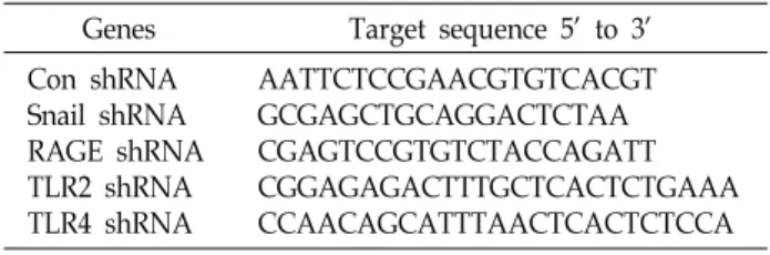
Table 1. shRNA target sequences used in this paper Genes Target sequence 5’ to 3’
Con shRNA Snail shRNA RAGE shRNA TLR2 shRNA TLR4 shRNA
AATTCTCCGAACGTGTCACGT GCGAGCTGCAGGACTCTAA CGAGTCCGTGTCTACCAGATT CGGAGAGACTTTGCTCACTCTGAAA CCAACAGCATTTAACTCACTCTCCA HMGB1 단백질은 posttranslational modification에 따라
암의 성장을 촉진하거나 억제할 수 있다. Oxidized HMGB1은 면역체계를 억제하여 암의 성장과 진행을 촉진함으로써 항암 효과에 대한 저항성을 증가시키는 반면, reduced HMGB1은 암에 대항하여 면역반응을 활성화시키는 것으로 알려져 있다.
HMGB1은 inflammatory cytokine으로 작용하여 암성장, 신 생혈관생성, EMT 및 전이를 촉진하는 것으로 알려져 있으며 HMGB1과 암 발생 기작에 대한 관심이 높아지고 있다[2, 20, 22, 33, 35, 38, 40, 53].
암세포는 invasion 및 metastasis 능력을 획득하기 위해 epi- thelial-mesenchymal transition (EMT)를 나타낸다. EMT는 암 의 전이(metastasis) 과정에서 침입과 혈관 내 침투에 필요한 새로운 특성을 암세포에게 제공함으로써 악성종양 발달에 기 여한다. 종양 세포는 EMT를 통해 epithelial morphology를 잃어버리고, motile, invasive fibroblast-mesenchymal pheno- type을 나타내며 cell adhesion molecule인 E-cadherin 발현 감소를 수반한다. E-cadherin의 전사억제자로는 Snail/Slug family, ZEB1/δEF1, ZEB2/SIP1, Twist1과 Twist2, E12/E47 factor 등이 알려져 있다[5, 29, 45, 46].
Snail은 EMT의 대표적인 early marker로 알려져 있으며, epithelial markers인 E-cadherin 뿐만 아니라 desmoplakin, epithelial mucin Muc-1, cytokeratin-18, occludins, claudins, ZO-1 유전자의 발현을 억제하고, mesenchymal markers인 N-cadherin, vimentin, fibronectin, vitronectin, alpha-smooth muscle actin (ɑ-SMA), fibroblast specific protein 1 (FSP1) 유 전자의 발현을 활성화하여 EMT를 유도한다[5, 29, 45, 46]. 또 한 Snail은 glycolytic switch를 유도한다[30]. Glycolytic switch 는 악성종양세포(malignant cell)에서 나타나는 특징으로 암세 포가 산소가 충분한 상태에서도 미토콘드리아에 의존하지 않 고 해당과정(glycolysis)을 통해 대부분의 ATP 에너지를 생성 하는 현상으로 알려져 있다[4, 6, 8, 11, 12, 18, 25, 37, 39, 49, 52]. Snail은 미토콘드리아 산호호흡 및 전자전달계 complex IV 활성을 억제하며 fructose-1,6-bisphosphatase (FBP1) 발현 또한 감소시킨다[4, 30].
본 연구진은 necrosis에 의해서 방출되는 HMGB1이 EMT 에 미치는 영향을 분석하기 위해, multicellular tumor sphe- roid (MTS)를 이용하였다. MTS는 in vitro의 monolayer cul- ture와 in vivo의 tumors 간의 중간단계의 특성을 나타낸다.
MTS는 바깥 테두리 주변의 cell population은 proliferation이 왕성한 반면, 중간 부위는 quiescent cell을 형성하며, 충분한 산소와 영양분 공급이 이루어 지지 못하는 core부분은 ne- crosis를 일으키는 등 solid tumor의 성질을 공유하고 있어 tu- mor 연구에 적합한 모델로 알려져 있다[17, 20].
본 연구에서는 Snail, RAGE/TLR2/TLR4의 발현이 MTS culture에 의해 증가됨을 관찰하였다. MTS에 의해 유도되는 Snail 발현은 RAGE/TLR2/TLR4 의존적으로 일어남을 발견
하였다. 또한 Snail 및 RAGE/TLR2/TLR4 shRNA가 MTS 성 장 및 MTS-induced EMT를 억제함을 관찰하였다. 실제, 인간 암조직에서도 RAGE/TLR2/TLR4발현이 증가됨을 확인하였 다. 이러한 결과를 통해 HMGB1이 RAGE/TLR2/TLR4/Snail axis를 통해 MTS 성장 및 MTS culture-induced EMT에 중요 하게 작용할 것으로 여겨진다.
재료 및 방법
세포배양
MCF-7은 American Type Culture Collection (ATCC, USA) 으로부터 구입하여 사용하였으며, 실험실에서 확립한 방법에 의해 배양하였다[24]. 세포는 10% fetal bovine serum (FBS) 및 1% penicillin/streptomycin이 포함된 Eagle's Minimum Essential Medium (EMEM)을 사용하여 37℃, 5% CO2 조건 하에서 배양하였다.
Multicellular tumor spheroid (MTS) 배양 및 염색 MTS는 실험실에서 확립한 방법으로 배양하였다[24, 31].
MTS 형성을 위해 MCF-7 cells를 1.2% agarose-coated 96-well plates에 400 cells/200 ul media/well로 배양하였다. 배양 후 3일째부터 2일 간격으로 media change를 진행하였다. MTS growth를 측정하기 위해 spheroid의 diameters을 2일 간격으 로 측정하였다. MTS를 회수하여 formalin으로 fixing한 후 paraffin section하여 H & E 염색과 HO-PI 이중 염색은 실험실 에서 확립한 방법으로 수행하였다[24, 31]. MTS 배양이 MCF- 7에 미치는 영향을 조사하기 위해, MCF-7 MTS를 회수하여 0.5X trypsin으로 dissociation 한 후 MTS/MCF-7 cells을 re- plating하여 2D culture 하였다.
Transfection 및 short hairpin RNA (shRNA) interfer- ence
MCF-7에 jetPEI (Polyplus transfection, SA, USA)를 이용하 여 Control 및 Snail, RAGE, TLR2, TLR4에 특이적으로 반응하 는 shRNA/pSUPER vectors를 실험실에서 확립한 방법에 의 해 제작하여 transfection 하였다[24]. shRNA target sequences 는 Table 1에 정리되어 있다.
Western blotting 및 real time qRT-PCR
Western blotting 및 real time qRT-PCR은 실험실에서 확립
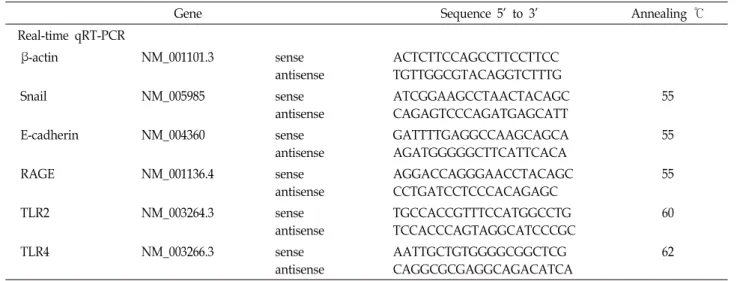
Table 2. Primer sequences used in this study
Gene Sequence 5’ to 3’ Annealing ℃
Real-time qRT-PCR
β-actin NM_001101.3 sense
antisense
ACTCTTCCAGCCTTCCTTCC TGTTGGCGTACAGGTCTTTG
Snail NM_005985 sense
antisense
ATCGGAAGCCTAACTACAGC CAGAGTCCCAGATGAGCATT
55
E-cadherin NM_004360 sense
antisense
GATTTTGAGGCCAAGCAGCA AGATGGGGGCTTCATTCACA
55
RAGE NM_001136.4 sense
antisense
AGGACCAGGGAACCTACAGC CCTGATCCTCCCACAGAGC
55
TLR2 NM_003264.3 sense
antisense
TGCCACCGTTTCCATGGCCTG TCCACCCAGTAGGCATCCCGC
60
TLR4 NM_003266.3 sense
antisense
AATTGCTGTGGGGCGGCTCG CAGGCGCGAGGCAGACATCA
62
한 방법으로 수행하였다[24]. Western blotting에 사용한 항체 (antibodies)는 RAGE (Santa Cruz, CA, USA); α-tubulin (Bio- genex, CA, USA)이다. TRIzol (Invitrogen, Carlsbad, CA, USA)을 이용하여 total RNA를 세포에서 분리하였다. Tran- scription levels은 real time qRT-PCR로 측정하였다. 사용한 primer sequences는 Table 2에 정리되어 있다. 값은 β-actin으 로 normalization하였다.
인간 암조직 연구
모든 인간 조직은 기관생명윤리위원회(IRB)의 심의(IRB no. 2011-20)를 받은 후 한국인체자원은행 네트워크에 소속된 부산대학교 병원 인체자원은행(National Biobank of Korea, PNUH)에서 제공받았다. 유방암(breast cancer)은 환자 #70331,
#70648 (infiltrating ductal carcinoma)과 #69965 (invasive ductal carcinoma), #69941 (metaplastic carcinoma), #70168 (pleomorphic lobular carcinoma)이고, 대장암(colon cancer) 은 환자 #71335, #71593, #70852, #71304 (adenocarcinoma)과
#70825 (mucinous adenocarcinoma)이다. 그리고 난소암 (ovarian cancer)은 환자 #1963, #1844 (clear cell carcinoma)과
#1903 (mucinous cystadenocarcinoma), #2281, #2297 (serous adenocarcinoma)이다. 각각의 암조직은 매치된 정상 조직과 대조하여 분석하였다. 조직 50-100 mg 당 TRIzol (Invitrogen, Carlsbad, CA, USA) 1 ml을 사용하여, tissuelyser (QIAGEN) 로 30 Hz에서 2-3분 homogenization한 후, total RNA를 분리 하였다. Real time qRT-PCR을 실시하여 인간 암 조직에서의 유전자 발현을 조사하였다.
통계학적 분석
Real time qRT-PCR은 최소 3번 수행하였으며, 모든 실험은 두 번 이상 반복하였다. 분석된 실험 데이터의 통계적 유의성 은 Student’s t test를 통하여 검증하였다. 모든 결과는 mean
± SE로 나타내었으며, p<0.05는 통계학적으로 유의하다고 간 주하였다.
결과 및 고찰
MTS 배양을 통해 MCF-7에 미치는 영향 분석
본 연구진은 non-invasive breast cancer cell lines MCF-7이 MTS 배양 동안 tumor progression를 위해 요구되는 bio- logical properties을 획득할 가능성을 조사하였다. MCF-7 세 포를 1% agarose에서 배양한 결과, cell-to-matrix interaction 은 소실되고 cell-to-cell interaction만을 통하여 완전한 구형의 MTS를 형성함을 관찰하였다(Fig. 1A, Fig. 1B).
MTS는 성장함에 따라 inner region에 있는 세포는 hypoxia 와 low glucose 상태에 놓이게 되는데, 이는 전형적으로 solid tumor에서 관찰되는 현상이다[16, 36, 41]. MTS의 inner re- gion에 있는 세포는 산소와 포도당(glucose)의 불충분한 공급 으로 인해 microenvironmental stress를 받게 되고 necrotic cell death를 야기한다[24]. Necrotic cells은 HMGB1를 ex- tracellular space로 방출하는 것으로 알려져 있다. 방출된 HMGB1은 tumor-promoting cytokine으로 작용함으로써 tu- mor development 시 inflammation, metabolism 및 meta- stasis에 기여한다[14, 23, 32]. 그러므로, MTS에서 necrosis에 의해 방출되는 HMGB이 tumor progression에 필요한 특성을 암세포에 부여할 것으로 여겨진다.
MTS 배양이 necrosis에 미치는 영향을 조사하기 위해 MTS 를 paraffin section하여 H & E 염색과 HO-PI 이중 염색을 하였다. 그 결과 PI-positive cells이 MTS 8 days에 관찰되었다 (Fig. 1C). 이러한 결과는 MTS에서 necrotic core는 8 days에 형성됨을 알 수 있었다.
더 나아가 MTS 배양이 MCF-7 세포에 미치는 영향을 조사 하기 위해, MCF-7 MTS를 회수하여 0.5X trypsin으로 dis-
A B
C
D replating 48 hr E
replating 48 hr replating 48 hr Fig. 1. MCF-7 cells acquire the potential to exhibit EMT during MTS culture. (A) MCF-7 cells were seeded onto 1.2% agarose-coated
96-well plates at a density of 400 cells per well and cultured up to 9 days. Formation, growth, and morphology of MTSs from MCF-7 cells were measured. (B) To calculate MTS size, diameters of five spheroids were measured at intervals of two days. Results are expressed as mean ± SE from three independent experiments. **P<0.01 versus MTS 3 days. (C) MCF-7 cells were grown as MTSs and the spheroids were sectioned and stained with H & E staining and HO/PI staining after 7 days and 8 days of culture. (D) MTSs were developed using MCF-7 cells that were cultured up to 9 days. MTSs were dissociated and transferred back to 2-D culture plates for 48 hr. The dissociated cells were analyzed by phase-contrast for cell morphology. (E) MTSs were developed using MCF-7 cells that were cultured up to 9 days and then MTSs were dissociated and transferred back to 2-D culture plates for 48 h. The cells were analyzed by real-time qRT-PCR using the indicated primers.
**P<0.01 versus 2-D culture. All error bars represent the SE. All scale bars represent 100 μm.
sociation 한 후 MTS/MCF-7 세포를 replating하여 2D culture 하였다. 그 결과, MTS/MCF-7 7 days과 9 days에서 EMT가 유도됨을 mesenchymal cells로의 세포 모양 변화를 통하여 관찰하였고(Fig. 1D), Snail 발현이 증가하고 E-cadherin 발현 이 감소됨을 발견하였다(Fig. 1E). 즉 이러한 결과는 solid tu- mor의 성질을 공유하고 있는 in vitro tumor model인 MTS가 EMT 연구를 위한 model system으로 사용될 수 있음을 보여 준다. 또한 MTS/MCF-7 세포의 EMT는 necrosis가 일어나기 전인 7 days에 일어나는 것으로 보아 MTS 성장에 따른 산소와 영양분 고갈에 의해 발생하는 necrosis가 EMT와 연관되어 있 을 것으로 보인다.
HMGB1-RAGE/TLR2/4/Snail이 MTS 배양에 의한 EMT 에 미치는 영향 분석
HMGB1과 HMGB1 수용체(receptor)인 RAGE, TLR2,
TLR4는 inflammation 및 metastasis, tumorigenesis에 중요하 게 작용하는 것으로 알려져 있다. 또한 HMGB1과 RAGE 발현 은 다양한 tumor에서 증가되어 있으며 tumor invasiveness와 밀접하게 연관되어 있는 것으로 알려져 있다[10, 23, 43, 47, 51]. 또한 이전 연구에서 종양내부에서 발견되는 hypoxia 및 glucose depletion에 의해 necrosis 유도 및 HMGB1 방출을 확인하였다[24, 31].
먼저 EMT를 유도하는 대표적인 전사인자인 Snail이 MTS 성장에 미치는 영향을 분석한 결과 Snail shRNA가 MTS 성장 을 억제함을 관찰하였다(Fig. 2A, Fig. 2B). 또한 Snail 발현이 necrotic core 형성되기 전인 MTS 7 days에서 발현이 증가됨 을 real-time qRT-PCR을 통해 확인하였다(Fig. 2C).
HMGB1이 MTS 성장에 미치는 영향을 조사하기 위해 RAGE, TLR2, TLR4 shRNA의 영향을 조사한 결과 RAGE, TLR2, TLR4 shRNA가 MTS 성장을 억제함을 관찰하였다(Fig. 2A,
A B
C D
E F
replating 48 hr replating 48 hr
Fig. 2. RAGE/TLR2/4 signaling is involved in MTS culture-induced EMT via Snail activation. (A) The formation, growth, and morphology of MTSs from MCF-7 cells transfected with shRNAs for Snail, RAGE, and TLR2/4 were measured. (B) To calculate MTS size, diameters of five spheroids were measured 7 days and 9 days. Results are expressed as mean ± SE from three independent experiments. **P<0.01 versus control shRNA; ##P<0.01 MTS 7 days. The bars in the graph represent mean ± SE. (C) MTSs were produced using MCF-7 cells transfected with shRNAs for Snail, RAGE, and TLR2/4 and cultured for 7 days and 9 days. Then, the cells were dissociated and analyzed by real-time qRT-PCR using the indicated primers.
*P<0.05; **P<0.01 versus 2-D culture, #P<0.05; ##P<0.01 versus control shRNA. (D) MCF-7 spheroids cultured for the indicated times were analyzed using immunoblotting with antibodies against RAGE and α-tubulin. MTSs were produced using MCF-7 cells transfected with shRNAs for Snail, RAGE, and TLR2/4. MTSs were dissociated and transferred back to 2-D culture plates for 48 h. The cells were analyzed by phase-contrast microscopy to assess cell morphology (E) and real-time qRT-PCR using the indicated primers (F). **P<0.01 versus 2-D culture, ##P<0.01 versus control shRNA. All error bars represent the SE. All scale bars represent 100 μm.
Fig. 2B). 또한 RAGE, TLR2, TLR4의 발현이 MTS 9 days에 증가됨을 관찰하였다(Fig. 2C, Fig. 2D). MTS에 의한 Snail 발 현에 미치는 RAGE/TLR2/4의 영향을 확인한 결과, RAGE/
TLR2/4 shRNA가 MTS 7 days와 9 days에서의 Snail 발현을 억제함을 발견하였다(Fig. 2C). 이러한 결과를 바탕으로 MTS 에서 Snail 발현이 RAGE/TLR2/4 의존적으로 유도됨을 알 수 있었다. 이를 통해 necrosis가 일어나기 전인 MTS 7 days에 서 Snail 발현이 RAGE/TLR2/TLR4에 의해 조절됨으로써 RAGE/TLR2/TLR4-Snail이 MTS 성장 및 EMT에 영향을 미 칠 가능성을 제시한다.
RAGE/TLR2/4-Snail가 MTS 배양에 의해 유도되는 EMT 에 미치는 영향을 조사한 결과, Snail, RAGE, TLR2, TLR4 shRNA가 MTS 배양에 의해 유도되는 EMT를 억제함을 mes- enchymal cells로의 세포 모양 변화를 통하여 관찰하였으며 (Fig. 2E), Snail 발현 감소 및 E-cadherin 발현 증가를 발견하였
다(Fig. 2F). 이를 통해 RAGE/TLR2/4-Snail axis가 MTS cul- ture-induced EMT와 밀접하게 연관되어 있음을 알 수 있다.
이러한 결과들은 MTS에서 necrosis가 일어나기 전에 HMGB1 이 EMT에 중요하게 작용할 가능성을 제시한다.
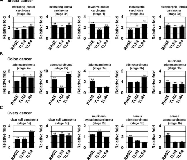
인간 암조직에서 RAGE/TLR2/4 분자의 발현 분석 Cancer와의 연관성을 조사하기 위해 실제 인간 암조직에서 RAGE/TLR2/TLR4의 발현 변화를 조사하였다. 그 결과 breast 및 colon, ovarian cancer에서 RAGE, TLR2, TLR4 발현이 증가 됨을 관찰하였다(Fig. 3).
실제 gastric cancer과 colorectal cancer에서 RAGE levels은 invasive 및 metastatic phenotype와 연관되어 있는 것으로 알 려져 있다[10, 23, 43, 47, 51]. HMGB1은 hepatocellular carci- noma에서 RAGE signaling pathway 및 NF-кB를 통해 cel- lular proliferation과 invasion, metastasis에 기여하는 것으로
A
B
C
Fig. 3. The expression of RAGE, and TLR2/4 in human tumors. real-time qRT-PCR data showing the expression of RAGE and TLR2/4 mRNA from the indicated tumor types and histological stages (TNM classification) of breast (A), colon (B), and ovarian cancers (C). Relative levels of mRNAs were normalized to those in the corresponding normal tissues. *P<0.05; **P<0.01 versus matched normal (N) tissues. All error bars represent the SE.
보고되어 있다[2, 15, 35, 38, 53]. TLR2와 TLR4 발현 또한 gas- tric cancer과 colorectal cancer, breast cancer에서 높게 관찰되 는 것으로 밝혀져 있다[10, 23, 43, 47, 51]. 이러한 결과들은 암성장 및 MTS-induced EMT에서 HMGB1-RAGE/TLR2/
TLR4-Snail이 중요하게 작용함을 제시한다.
이러한 결과는 solid tumor의 성질을 공유하고 있는 in vitro tumor model인 MTS가 cells에서 aggressive phenotype 뿐만 아니라 EMT, invasion, cancer stem cells, metastasis등의 연구 를 위한 model system으로 사용될 수 있음을 보여준다[28].
본 연구에서 HMGB1이 MTS 성장에 관여할 뿐만 아니라 MTS 배양에 의해 유도되는 EMT에도 관여함을 알 수 있었다. 이러 한 결과를 바탕으로 HMGB1-RAGE/TLR2/TLR4-Snail axis 에 의한 전이 및 tumor progression 조절 기전이 암 발생 기전 을 분석하고 항암 치료법 개발의 중요한 target이 될 수 있음을 시사한다.
감사의 글
이 논문은 부산대학교 기본연구지원사업(2년)에 의하여 연 구되었음.
References
1. Bald, T., Quast, T., Landsberg, J., Rogava, M., Glodde, N., Lopez-Ramos, D., Kohlmeyer, J., Riesenberg, S., van den Boorn-Konijnenberg, D., Homig-Holzel, C., Reuten, R., Schadow, B., Weighardt, H., Wenzel, D., Helfrich, I., Scha- dendorf, D., Bloch, W., Bianchi, M. E., Lugassy, C., Barnhill, R. L., Koch, M., Fleischmann, B. K., Forster, I., Kastenmuller, W., Kolanus, W., Holzel, M., Gaffal, E. and Tuting, T. 2014.
Ultraviolet-radiation-induced inflammation promotes an- giotropism and metastasis in melanoma. Nature 507, 109- 113.
2. Chen, R. C., Yi, P. P., Zhou, R. R., Xiao, M. F., Huang, Z.
B., Tang, D. L., Huang, Y. and Fan, X. G. 2014. The role of HMGB1-RAGE axis in migration and invasion of hep- atocellular carcinoma cell lines. Mol. Cell. Biochem. 390, 271- 280.
3. Conti, L., Lanzardo, S., Arigoni, M., Antonazzo, R., Radaelli, E., Cantarella, D., Calogero, R. A. and Cavallo, F. 2013. The noninflammatory role of high mobility group box 1/Toll- like receptor 2 axis in the self-renewal of mammary cancer stem cells. FASEB J. 27, 4731-4744.
4. Dang, C. V., Kim, J. W., Gao, P. and Yustein, J. 2008. The interplay between MYC and HIF in cancer. Nat. Rev. Cancer 8, 51-56.
5. De Craene, B. and Berx, G. 2013. Regulatory networks defin- ing EMT during cancer initiation and progression. Nat. Rev.
Cancer 13, 97-110.
6. DeBerardinis, R. J., Lum, J. J., Hatzivassiliou, G. and Thompson, C. B. 2008. The biology of cancer: metabolic re- programming fuels cell growth and proliferation. Cell Metab.
7, 11-20.
7. Degenhardt, K., Mathew, R., Beaudoin, B., Bray, K., Ander- son, D., Chen, G., Mukherjee, C., Shi, Y., Gelinas, C., Fan, Y., Nelson, D. A., Jin, S. and White, E. 2006. Autophagy promotes tumor cell survival and restricts necrosis, in- flammation, and tumorigenesis. Cancer Cell 10, 51-64.
8. Denko, N. C. 2008. Hypoxia, HIF1 and glucose metabolism in the solid tumour. Nat. Rev. Cancer 8, 705-713.
9. Edinger, A. L. and Thompson, C. B. 2004. Death by design:
apoptosis, necrosis and autophagy. Curr. Opin. Cell Biol. 16, 663-669.
10. Fukata, M., Chen, A., Vamadevan, A. S., Cohen, J., Breglio, K., Krishnareddy, S., Hsu, D., Xu, R., Harpaz, N., Dannen- berg, A. J., Subbaramaiah, K., Cooper, H. S., Itzkowitz, S.
H. and Abreu, M. T. 2007. Toll-like receptor-4 promotes the development of colitis-associated colorectal tumors. Gas- troenterology 133, 1869-1881.
11. Fukuda, R., Zhang, H., Kim, J. W., Shimoda, L., Dang, C.
V. and Semenza, G. L. 2007. HIF-1 regulates cytochrome oxi- dase subunits to optimize efficiency of respiration in hy- poxic cells. Cell 129, 111-122.
12. Gatenby, R. A. and Gillies, R. J. 2004. Why do cancers have high aerobic glycolysis? Nat. Rev. Cancer 4, 891-899.
13. Golstein, P. and Kroemer, G. 2007. Cell death by necrosis:
towards a molecular definition. Trends Biochem. Sci. 32, 37- 43.
14. Guo, Z. S., Liu, Z., Bartlett, D. L., Tang, D. and Lotze, M.
T. 2013. Life after death: targeting high mobility group box 1 in emergent cancer therapies. Am. J. Cancer Res. 3, 1-20.
15. He, M., Kubo, H., Ishizawa, K., Hegab, A. E., Yamamoto, Y., Yamamoto, H. and Yamaya, M. 2007. The role of the receptor for advanced glycation end-products in lung fibrosis. Am. J. Physiol. Lung Cell Mol. Physiol. 293, L1427- 1436.
16. Hielscher, A. and Gerecht, S. 2015. Hypoxia and free radi- cals: role in tumor progression and the use of engineer- ing-based platforms to address these relationships. Free
Radic. Biol. Med. 79, 281-291.
17. Horning, J. L., Sahoo, S. K., Vijayaraghavalu, S., Dimitrijevic, S., Vasir, J. K., Jain, T. K., Panda, A. K. and Labhasetwar, V. 2008. 3-D tumor model for in vitro evaluation of anti- cancer drugs. Mol. Pharm. 5, 849-862.
18. Hsu, P. P. and Sabatini, D. M. 2008. Cancer cell metabolism:
Warburg and beyond. Cell 134, 703-707.
19. Hua, D., Liu, M. Y., Cheng, Z. D., Qin, X. J., Zhang, H. M., Chen, Y., Qin, G. J., Liang, G., Li, J. N., Han, X. F. and Liu, D. X. 2009. Small interfering RNA-directed targeting of Toll-like receptor 4 inhibits human prostate cancer cell in- vasion, survival, and tumorigenicity. Mol. Immunol. 46, 2876- 2884.
20. Ivascu, A. and Kubbies, M. 2007. Diversity of cell-mediated adhesions in breast cancer spheroids. Int. J. Oncol. 31, 1403- 1413.
21. Kang, R., Tang, D., Schapiro, N. E., Livesey, K. M., Farkas, A., Loughran, P., Bierhaus, A., Lotze, M. T. and Zeh, H.
J. 2010. The receptor for advanced glycation end products (RAGE) sustains autophagy and limits apoptosis, promoting pancreatic tumor cell survival. Cell Death Differ. 17, 666-676.
22. Kang, R., Tang, D., Schapiro, N. E., Loux, T., Livesey, K.
M., Billiar, T. R., Wang, H., Van Houten, B., Lotze, M. T.
and Zeh, H. J. 2014. The HMGB1/RAGE inflammatory path- way promotes pancreatic tumor growth by regulating mi- tochondrial bioenergetics. Oncogene 33, 567-577.
23. Kang, R., Zhang, Q., Zeh, H. J. 3rd., Lotze, M. T. and Tang, D. 2013. HMGB1 in cancer: good, bad, or both? Clin. Cancer Res. 19, 4046-4057.
24. Kim, C. H., Jeon, H. M., Lee, S. Y., Ju, M. K., Moon, J. Y., Park, H. G., Yoo, M. A., Choi, B. T., Yook, J. I., Lim, S.
C., Han, S. I. and Kang, H. S. 2011. Implication of snail in metabolic stress-induced necrosis. PLoS One 6, e18000.
25. Kim, J. W., Tchernyshyov, I., Semenza, G. L. and Dang, C.
V. 2006. HIF-1-mediated expression of pyruvate dehydroge- nase kinase: a metabolic switch required for cellular adapta- tion to hypoxia. Cell Metab. 3, 177-185.
26. Kim, S., Takahashi, H., Lin, W. W., Descargues, P., Grivenni- kov, S., Kim, Y., Luo, J. L. and Karin, M. 2009. Carcinoma- produced factors activate myeloid cells through TLR2 to stimulate metastasis. Nature 457, 102-106.
27. Kondo, Y., Kanzawa, T., Sawaya, R. and Kondo, S. 2005.
The role of autophagy in cancer development and response to therapy. Nat. Rev. Cancer 5, 726-734.
28. Kunjithapatham, R., Karthikeyan, S., Geschwind, J. F., Kieserman, E., Lin, M., Fu, D. X. and Ganapathy-Kanniap- pan, S. 2014. Reversal of anchorage-independent multi- cellular spheroid into a monolayer mimics a metastatic model. Sci. Rep. 4, 6816.
29. Lamouille, S., Xu, J. and Derynck, R. 2014. Molecular mecha- nisms of epithelial-mesenchymal transition. Nat. Rev. Mol.
Cell Biol. 15, 178-196.
30. Lee, S. Y., Jeon, H. M., Ju, M. K., Kim, C. H., Yoon, G., Han, S. I., Park, H. G. and Kang, H. S. 2012. Wnt/Snail signaling regulates cytochrome C oxidase and glucose metabolism.
Cancer Res. 72, 3607-3617.
31. Lee, S. Y., Jeon, H. M., Kim, C. H., Ju, M. K., Bae, H. S., Park, H. G., Lim, S. C., Han, S. I. and Kang, H. S. 2011.
Homeobox gene Dlx-2 is implicated in metabolic stress-in- duced necrosis. Mol. Cancer 10, 113.
32. Lee, S. Y., Ju, M. K., Jeon, H. M., Jeong, E. K., Lee, Y. J., Kim, C. H., Park, H. G., Han, S. I. and Kang, H. S. 2018.
Regulation of Tumor Progression by Programmed Necrosis.
Oxid. Med. Cell. Longev. 2018, 3537471.
33. Liu, A., Fang, H., Dirsch, O., Jin, H. and Dahmen, U. 2012.
Oxidation of HMGB1 causes attenuation of its pro-in- flammatory activity and occurs during liver ischemia and reperfusion. PLoS One 7, e35379.
34. Lotze, M. T. and Tracey, K. J. 2005. High-mobility group box 1 protein (HMGB1): nuclear weapon in the immune arsenal. Nat. Rev. Immunol. 5, 331-342.
35. Lynch, J., Nolan, S., Slattery, C., Feighery, R., Ryan, M. P.
and McMorrow, T. 2010. High-mobility group box protein 1: a novel mediator of inflammatory-induced renal epi- thelial-mesenchymal transition. Am. J. Nephrol. 32, 590-602.
36. Marin-Hernandez, A., Gallardo-Perez, J. C., Hernandez- Resendiz, I., Del Mazo-Monsalvo, I., Robledo-Cadena, D. X., Moreno-Sanchez, R. and Rodriguez-Enriquez, S. 2016.
Hypoglycemia enhances epithelial-mesenchymal transition and invasiveness, and restrains the warburg phenotype, in hypoxic HeLa cell cultures and microspheroids. J. Cell.
Physiol. 232, 1346-1359.
37. Matoba, S., Kang, J. G., Patino, W. D., Wragg, A., Boehm, M., Gavrilova, O., Hurley, P. J., Bunz, F. and Hwang, P.
M. 2006. p53 regulates mitochondrial respiration. Science 312, 1650-1653.
38. Palumbo, R., Sampaolesi, M., De Marchis, F., Tonlorenzi, R., Colombetti, S., Mondino, A., Cossu, G. and Bianchi, M.
E. 2004. Extracellular HMGB1, a signal of tissue damage, induces mesoangioblast migration and proliferation. J. Cell Biol. 164, 441-449.
39. Papandreou, I., Cairns, R. A., Fontana, L., Lim, A. L. and Denko, N. C. 2006. HIF-1 mediates adaptation to hypoxia by actively downregulating mitochondrial oxygen consumption.
Cell Metab. 3, 187-197.
40. Rouhiainen, A., Kuja-Panula, J., Tumova, S. and Rauvala, H. 2013. RAGE-mediated cell signaling. Methods Mol. Biol.
963, 239-263.
41. Sabharwal, S. S. and Schumacker, P. T. 2014. Mitochondrial ROS in cancer: initiators, amplifiers or an Achilles' heel?
Nat. Rev. Cancer 14, 709-721.
42. Scaffidi, P., Misteli, T. and Bianchi, M. E. 2002. Release of chromatin protein HMGB1 by necrotic cells triggers in-
flammation. Nature 418, 191-195.
43. Sims, G. P., Rowe, D. C., Rietdijk, S. T., Herbst, R. and Coyle, A. J. 2010. HMGB1 and RAGE in inflammation and cancer. Annu. Rev. Immunol. 28, 367-388.
44. Taguchi, A., Blood, D. C., del Toro, G., Canet, A., Lee, D.
C., Qu, W., Tanji, N., Lu, Y., Lalla, E., Fu, C., Hofmann, M.
A., Kislinger, T., Ingram, M., Lu, A., Tanaka, H., Hori, O., Ogawa, S., Stern, D. M. and Schmidt, A. M. 2000. Blockade of RAGE-amphoterin signalling suppresses tumour growth and metastases. Nature 405, 354-360.
45. Thiery, J. P. and Sleeman, J. P. 2006. Complex networks or- chestrate epithelial-mesenchymal transitions. Nat. Rev. Mol.
Cell Biol. 7, 131-142.
46. Tsai, J. H. and Yang, J. 2013. Epithelial-mesenchymal plasti- city in carcinoma metastasis. Genes Dev. 27, 2192-2206.
47. Tye, H., Kennedy, C. L., Najdovska, M., McLeod, L., Mc Cormack, W., Hughes, N., Dev, A., Sievert, W., Ooi, C. H., Ishikawa, T. O., Oshima, H., Bhathal, P. S., Parker, A. E., Oshima, M., Tan, P. and Jenkins, B. J. 2012. STAT3-driven upregulation of TLR2 promotes gastric tumorigenesis in- dependent of tumor inflammation. Cancer Cell 22, 466-478.
48. Vakkila, J. and Lotze, M. T. 2004. Inflammation and necrosis promote tumour growth. Nat. Rev. Immunol. 4, 641-648.
49. Vander Heiden, M. G., Cantley, L. C. and Thompson, C.
B. 2009. Understanding the Warburg effect: the metabolic requirements of cell proliferation. Science 324, 1029-1033.
50. Yan, W., Chang, Y., Liang, X., Cardinal, J. S., Huang, H., Thorne, S. H., Monga, S. P., Geller, D. A., Lotze, M. T. and Tsung, A. 2012. High-mobility group box 1 activates cas- pase-1 and promotes hepatocellular carcinoma invasiveness and metastases. Hepatology 55, 1863-1875.
51. Yu, L. X., Yan, L., Yang, W., Wu, F. Q., Ling, Y., Chen, S.
Z., Tang, L., Tan, Y. X., Cao, D., Wu, M. C., Yan, H. X.
and Wang, H. Y. 2014. Platelets promote tumour metastasis via interaction between TLR4 and tumour cell-released high-mobility group box1 protein. Nat. Commun. 5, 5256.
52. Zhang, H., Gao, P., Fukuda, R., Kumar, G., Krishnamachary, B., Zeller, K. I., Dang, C. V. and Semenza, G. L. 2007. HIF-1 inhibits mitochondrial biogenesis and cellular respiration in VHL-deficient renal cell carcinoma by repression of C-MYC activity. Cancer Cell 11, 407-420.
53. Zhu, L., Li, X., Chen, Y., Fang, J. and Ge, Z. 2015. High-mo- bility group box 1: a novel inducer of the epithelial-mesen- chymal transition in colorectal carcinoma. Cancer Lett. 357, 527-534.
54. Zong, W. X. and Thompson, C. B. 2006. Necrotic death as a cell fate. Genes Dev. 20, 1-15.
초록:Multicellular tumor spheroid (MTS) 배양에 의한 EMT에서 HMGB1의 역할
이수연1․주민경1․전현민1․김초희1․박혜경2․강호성1*
(1부산대학교 자연과학대학 분자생물학과, 2한국나노바이오테크놀러지센터)
암조직의 내부에서 hypoxia와 glucose depletion 등의 microenvironmental stress를 받게 되면 necrosis가 유도 되고, 실제로 암 조직 내부에서 necrotic core 형성이 관찰된다. Necrotic cells은 high mobility group box 1 (HMGB1)를 extracellular space로 방출하는 것으로 알려져 있다. 방출된 HMGB1은 tumor-promoting cytokine으 로 작용함으로써 tumor development 시 inflammation, metabolism 및 metastasis에 기여한다. 본 연구에서 non-invasive breast cancer cells MCF-7이 solid tumor의 in vitro model인 multicellular tumor spheroid (MTS) 배양을 통해 완전한 구형의 MTS를 형성하며 MTS가 성장함에 따라 inner region에 necrosis가 유도됨을 밝혔다.
또한 MCF-7 세포의 MTS 배양은 Snail 의존적으로 epithelial-mesenchymal transition (EMT)를 유도함을 관찰하 였다. HMGB1의 cell surface receptors인 RAGE, TLR2, TLR4 발현이 MTS 배양에 의해 증가됨을 발견하였다.
RAGE, TLR2, TLR4 를 knockdown한 결과 MTS 성장을 억제할 뿐만 아니라 MTS에 의해 증가되는 Snail 발현을 억제함을 밝혔다. 이는 MTS-induced Snail 발현이 RAGE/TLR2/TLR4의존적으로 조절되며 RAGE/TLR2/TLR4- Snail이 MTS 성장에 관여하는 것으로 보인다. 또한 Snail, RAGE, TLR2, TLR4 shRNA는 MTS 배양에 의해 유도되 는 EMT를 억제함을 밝혔다. 실제 인간 암조직에서 정상조직에 비해 RAGE, TLR2, TLR4 유전자의 발현이 높음을 관찰하였다. 따라서 HMGB1이 RAGE/TLR2/4-Snail axis를 통해 MTS 배양에 따른 성장 및 EMT에 중요하게 작 용할 것으로 예상된다.