—125—
INTRODUCTION
The brown croaker, Miichthys miiuy Basilewsky has adapted well to the western coastal waters of Korea, which are characterized by high turbidity resulting from strong tidal currents and low water temperatures in win- ter (Park et al., 2007). Given the commercial importance of brown croaker, especially to the aquaculture industry (Seo, 2004), information on aspects of its biology and early morphological development are of great interest (Park et al., 2007).
Detailed morphological information is important for the early detection of both morphological and physiologi- cal abnormalities in reared fish (Mana and Kawamura, 2002). Fish reared in hatcheries for release as juveniles into the wild experience conditions during the maturation process that differ markedly from those encountered by
fish raised in the wild (Hard et al., 2000). As hatchery- reared (hereafter, reared) fish are not well equipped to survive in the natural environment, most die during early stages of development (Hughes et al., 1992; Mana and Kawamura, 2002; Seo, 2004), or often harbor a variety of morphological abnormalities if they survive (Kanazawa, 1993; Dedi et al., 1997).
Although more information about the developmental stages of this fish is required because of the high levels of hatchery mortality that commonly occur during the early life stages of reared larvae, there have been no detailed reports of the anatomy of this species other than those on egg development and morphological changes in brown croaker larvae (Han et al., 2002). The early life stages in ichthyoplankton have been characterized in surveys of developmental series of specimens (Russell, 1976; Dunn, 1984). To construct a morphological database of early life stages, we studied fin development, head dimensions, and squamation in a series of laboratory- reared specimens.
Early Morphological Development of the Brown Croaker, Miichthys miiuy (Basilewsky): Fin Differentiation,
Head Dimensions, and Squamation
By In-Seok Park*, Young Ja Kim
1, In Bon Goo and Dong Soo Kim
2Division of Marine Environment and Bioscience, College of Ocean Science and Technology, Korea Maritime University, 727 Taejong-ro, Yeong do-gu, Busan 606-791, Korea
1Korea Environmental Industry & Technology Institute (KEITI), 215 Jinheung-ro, Eunpyeong-gu, Seoul 122-706, Korea
2Institute of Marine Living Modified Organisms (iMLMO), Pukyoung National University, 45 Yongso-ro, Nam-gu, Busan 608-737, Korea
ABSTRACT We describe early morphological development in laboratory-reared specimens of the brown croaker, Miichthys miiuy, in relation to fin differentiation, head dimensions, and squamation.
From the yolk sac stage to the flexion larval stage (a period of 12 days following hatching, at which time the larvae were ⁄⁄4.2 mm in total length; TL) we observed the presence of a fin-fold around the body, while the caudal fin appeared rounded and lacked scales. Rays developed in the dorsal, anal, and pectoral fins in a process that was almost complete in larvae 12 days, while ray segmentation occurred between 26 and 29 days of age. Elongation of the middle rays of the caudal fin was initiated at 32 days, and the rays were remarkably elongated by 37 days. By 68 days the caudal fin was lanceo- lated (50.7 mm TL). Scales began to develop from the midlateral lines of the caudal peduncle at 9.1 mm TL (28 days), eventually encompassing the entire operculum (22.1 mm TL; 44 days). The head dimensions were largely stabilized at ¤¤12 mm TL (30 day).
Key words : Fin differentiation, head dimensions, Miichthys miiuy, squamation
*Corresponding author: In-Seok Park Tel: 82-51-410-4321, Fax: 82-51-405-4322, E-mail: [email protected]
ISSN: 1225-8598
Accepted: April 3, 2012
http://www.fishkorea.or.kr
MATERIALS AND METHODS
The brown croaker, Miichthys miiuy were produced from naturally fertilized eggs of wild adults. The eggs were stocked in 20-tonne concrete tanks and reared accord- ing to established commercial procedures. Briefly, hatched larvae were fed enriched rotifers from 4 days, and then fed Artemia nauplii with artificial diets from 15 days.
The water temperature during the fish production period was not controlled and ranged from 25.1 to 27.3�C. All observations and measurements were made on pre-larvae (n==72) of 3.0 mm total length (TL), and juveniles (n== 48; TL==60.0 mm). The two size groups were preserved in 5% and 10% formalin solution, respectively.
As shown in Fig. 1, for specimens of ¤40.0 mm TL, four head dimensions were measured to the nearest 0.1 mm using a digital vernier caliper (CD-20CP, Mitutoyo, Japan). These included: head length (HL; the most ante- rior extension of the head to the most posterior point of the operculum); postorbital length (PL; the most poster- ior point of the eye to the most posterior point of the operculum); snout length (SNL; the most anterior exten- sion of the head to the most anterior point of the eye);
and eye diameter (ED). Larvae and juveniles of ⁄40.0 mm TL were observed using a microscope (Axioskop 40 FL, Zeiss, Germany) with a mounted video camera (AxioCam MRm, Zeiss, Germany) connected to a com- puter; images were interpreted using image analysis soft- ware (Axiovision4, Zeiss, Germany). Specimens were periodically sampled for assessment of fin differentiation and squamation, following staining with alizarin red S
(Sigma, USA). The developmental stages were identified according to the criteria described by Russell (1976) and Han et al. (2002).
RESULTS AND DISCUSSION
The dorsal fin anlage in brown croaker, Miichthys miiuy appeared when the larvae were at 3.7 mm TL (8 days);
ray formation occurred at 4.2 mm TL, 12 days after hatch- ing; ray segmentation was initiated at 11.9 mm TL (29 days); and the formation of ray branching was initiated at 38.5 mm TL (¤51 days). Anal fin anlage appeared at 3.7 mm TL (8 days); ray formation was initiated at 4.2 mm TL (12 days); and ray segmentation was initiated at 7.4 mm TL (26 days). The pectoral fin bud appeared at 3.5 mm TL (2 days) and was initially fanlike. Ray forma- tion was initiated at 4.2 mm TL (12 days), and elongated with elongation of the middle ray of the caudal fin ray at 13.2 mm TL (35 days). The caudal fin became rounded immediately following hatching, and at 5.9 mm TL, dur- ing the postlarval stage (16 days), it was particularly long and rounded at the end margin. The middle fin ray was prominently elongated at ¤32 days, but then became lanceolated at 50.7 mm TL in juveniles 68 days. The full complement of ray counts was initially observed in the caudal ray, followed by the anal, dorsal, and pectoral rays.
The use of reared larvae in studying fish ontogeny has been previously proposed (Hunter, 1984; Myung et al., 2004). However, some critical considerations must be taken account of because the rearing method has signi- ficant effects on development (Hunter, 1984; Koumoun- douros et al., 2001). In this study we found slightly dif- ferent values for several parameters relative to those reported by Han et al. (2002). These include length of fish, day of hatching, and initiation and completion of ray formation, segmentation, and branching. The differ- ences may be a consequence of differences in the sampl- ing and rearing methods used. In the present study all fin rays were observed to have been completely developed between 30 and 37 days of age, and the size correspond- ing to these ages largely encompassed the juvenile stage, as reported by Han et al. (2002). Specifically, the length of the middle rays of the caudal fin began to increase at 32 days, and then became prominently elongated after 37 days.
Fin elongation and spination have roles in maintaining buoyancy, but may also be central to the species’ predator avoidance strategy (Moser, 1981). Fin development is an important process in the early life stages of fish, and has been intimately correlated with both swimming speed, and feeding techniques and preferences (Fukuhara, 1992).
Consequently, specific fin ray elongation is assumed to be key to distinguishing development stages among
Fig. 1. Head dimensions used in this study. HL, head length (from the most anterior extension of the head to the most posterior point of the operculum); PL, postorbital length (from the most posterior point of the eye to the most posterior point of the operculum); SNL, snout length (most anterior extension of the head to the most anterior point of the eye); ED, eye diameter.
HL
PL ED
SNL
related species.
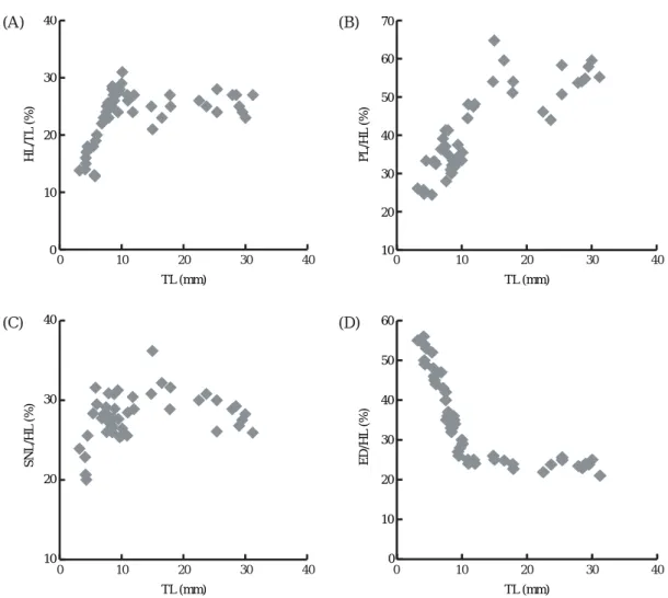
The initial head length to total length (HL/TL) propor- tion was 13.0~17.4% in specimens of 3.2~4.9 mm TL, and this increased gradually with increasing size, event- ually reaching 23.8~37.4% at 5.0~6.3 mm TL (Fig. 2A).
In specimens of 6.6~7.8 mm TL, the relative degree to which growth in head length had occurred was quite variable (23.8~32.0%), but it stabilized gradually with further growth to an average of 25.0%.
The postorbital length to head length (PL/HL) propor- tion was found to increase with size, reaching 33.1% in specimens of ⁄8.0 mm TL, but then stabilized with fur- ther growth, eventually ranging from 50.0~60.3% (Fig.
2B). The snout length to head length (SNL/HL) propor- tion increased markedly in specimens of ¤6.2 mm TL (from 20.0% to 32.1%), decreased to 25.3% at 8.0 mm TL, and stabilizing at 25.9~31.9% with further growth (Fig. 2C). The eye diameter to head length (ED/HL) pro- portion was approximately 50.0~55.6% in specimens of 8.0 mm HL (Fig. 2D). The growth of all head dimen-
sions stabilized at approximately 30 days of age and a HL¤12 mm, coinciding with extension of the middle ray of the caudal fin.
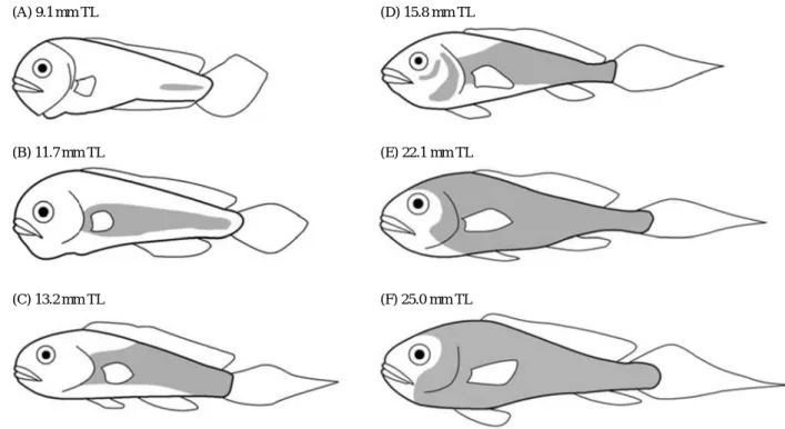
The origin of scales and squamation in the brown croaker is illustrated in Fig. 2. The initial site of scale formation was parallel to the longitudinal principal axis of the caudal peduncle, and occurred at 9.1 mm TL (28 days); no scales were observed on any fish younger than this age. Scale development progressed in an anterior direction along the midlateral line, and reached the mid- lateral line area posterior to the head at 11.7 mm TL (29 days).
New scales then developed on the dorsal and ventral regions, with scale formation around the circumference of the caudal peduncle being completed at 13.2 mm TL (35 days). Scales subsequently emerged around the gill cover and ventral region of the eye at 15.8 mm TL (37 days), and the entire operculum was covered with scales at 22.1 mm TL (44 days). Squamation in juveniles was complete at 25.0 mm TL (57 days). The single patch place
0 10 20 30 40
HL/TL(%)
TL (mm)
10 20 30 40 50 60 70
0 10 20 30 40
TL (mm)
0 10 20 30 40
10 20 30 40
SNL/HL(%) PL/HL(%)
0 10 20 30 40 50 60
ED/HL(%)
TL (mm)
0 10 20 30 40
TL (mm)
0 10 20 30 40
(A) (B)
(C) (D)
Fig. 2. Scatter diagrams showing the proportion of various dimensions to total length (TL) in fingerlings of reared brown croaker, Miichthys miiuy. (A) head length (HL/TL), (B) postorbital length (PL/HL), (C) snout length (SNL/HL), and (D) eye diameter (ED/HL).
on the caudle peduncle is a common pattern (White, 1977).
However, scale development varies among fish, espe- cially with regard to the number and location of patches, as does the correlation of scale formation with the length and age of the fish (Fukuhara and Fushimi, 1988; Park and Lee, 1988; Fukuhara, 1992). In this study, the speci- mens tended to develop patches in three places on the body. Squamation typically progressed along the lateral line, and then moved dorsally and ventrally. Scale devel- opment initially occurred laterally on the central portion of the caudal peduncle, progressed along the midlateral line, then extended dorsally and ventrally. A similar pat- tern was observed in Rivulus marmoratus (Cyprinodon- tidae), but without the caudal peduncle being involved initially (Park and Lee, 1988).
Larger and longer fish develop more squamation than smaller fish, including the zebra fish Brachydanio rerio (Armstrong, 1973) and R. marmoratus (Park and Lee, 1988). We noted some differences among fish with respect to the scale patch region. Yellow sail red bass, Callanthias japonicus (Kim and Okiyama, 1989), red sea bream, Pagrus major, and black sea bream, Acanthopagrus schlegeli (Fukuhara, 1992) initially develop scales on the middle of the body. However, in the Japanese amber- jack, Seriola quinqueradialta, scale patches first emerge around the caudal peduncle and the anal fin (Fukuhara, 1992). Table 1 was shown sequence of fin differentiation, and head and scale development in relation to TL in the brown croaker. The identification of early life stages in
ichthyoplankton was previously surveyed by examination of developmental stages of specimens (Russell, 1976).
Table 1. Sequence of fin differentiation, and head and scale develop- ment in relation to total length in the brown croaker, Miichthys miiuy
Stage Range (mm) Age
Stage A (Yolksac larva to flexion)
Finfold exist 2.3~3.8 ⁄10-d
No scales 2.3~8.8 ⁄26-d
Stage B (Post flexion larva) Fin ray formation begins
Dosal fin 4.2~4.8 12-d
Anal fin 4.0~4.5 12-d
Pectoral fin 4.0~5.7 12-d
Caudal fin is getting longer 5.9~6.5 16-d Stage C (Post flexion larva)
Fin segmentation initiated
Dorsal fin 11.9~12.0 29-d
Anal fin 7.4~8.0 26-d
Elongation of middle ray of caudal fin
Initiated 12.3~13.9 30-d
Remarkably elongated 15.9~16.8 ⁄32-d
Scale developed along midlateral lines 9.1~13.2 28-d to 35-d Gill cover and ventral region of eye 15.8~22.1 37-d to 44-d Entirely covering the operculum 25.0~27.0 57-d Growth of head dimension stabilized 12.3~30.8 ¤30-d Stage D (Juvenile)
Caudal fin initially lanceolated 56.1~70.0 68-d
Squamation completed 25.0 57-d
(A) 9.1 mm TL (D) 15.8 mm TL
(B) 11.7 mm TL (E) 22.1 mm TL
(C) 13.2 mm TL (F) 25.0 mm TL
Fig. 3. Development of squamation in the brown croaker, Miichthys miiuy. The area of the body covered by scales is shaded in the diagrams. TL, total length.
These developmental data are of critical importance to the early detection and elimination of morphological deformities in reared fish (Koumoundouros et al., 2001).
These result may prove to be useful indicators in the successful rearing of brown croaker fingerlings.
ACKNOWLEDGMENTS
This study was supported by a research fund (Project No. #20088033-1) from the Ministry of Land, Transport and Maritime Affairs, Korea. Comments from anonymous reviewers greatly improved the quality of this manuscript.
We declare that all experiments in this study comply with the current laws of Korea (Ordinance of Agriculture, Food and Fisheries, No. 1-the law regarding experimen- tal animals, No. 9932).
REFERENCES
Armstrong, J.G. 1973. Squamation chronology of the zebra- fish (Cyprinidae), Brachydnio rerio. Copeia, 4: 823- 824.
Dedi, J., T. Takeuchi, T. Seikai, T. Watanabe and K. Hosoya.
1997. Hypervitaminosis A during vertebral morpho- genesis in larval Japanese flounder. Fish. Sci., 63:
466-473.
Dunn, J.R. 1984. Developmental osteology. In: Moser, H.G., W.J. Richards, D.M. Cohen, M.P. Fahay, A.W. Ken- dal and S.L. Richardson (eds.), Ontogeny and syste- matics of fishes. American Society of Ichthyologists and Herpetologists, Special publication. 1, Allen, Lawrence, KS, pp. 48-50.
Fukuhara, O. 1992. Study on the development of functional morphology and behaviour of the larvae and juvenile Limanda yokohamae (Pisces: Pleuronectidae) reared in the laboratory. Mar. Biol., 99: 271-281.
Fukuhara, O. and T. Fushimi. 1988. Fin differentiation and squamation of artificially reared grouper, Epinephelus akaara. Aquaculture, 69: 379-386.
Han, K.H., S.H. Oh, D.S. Hwang, Y.H. Cho and D.C. Seo.
2002. Egg development and morphological change of larvae of the brown croaker, Miichthys miiuy.
Korean J. Ichthyol., 14: 93-99.
Hard, J.J., B.A. Berejikian, E.P. Tezak, S.L. Schroder, C.M.
Knudsen and L.T. Parker. 2000. Evidence for morpho- metric differentiation of wild and captively reared adult coho salmon: a genometric analysis. Environ.
Biol. Fishes., 58: 61-73.
Hughes, R.N., M.J. Kaiser, P.A. Mackney and K. Warburton.
1992. Optimizing foraging behavior through learning.
J. Fish Biol., 41: 77-91.
Hunter, J.R. 1984. Synopsis of culture methods for marine fish larvae. In: Moser, H.G., W.J. Richards, D.M.
Cohen, M.P. Fahay, A.W. Kendal and S.L. Richardson (eds.), Ontogeny and systematics of fishes, American Society of Ichthyologists and Herpetologists, Special pub. 1, Allen, Lawrence, KS, pp. 24-27.
Kanazawa, A. 1993. Nutritional mechanism involved in the occurrence of abnormal pigmentation in hatchery- reared flatfish. J. World Aquacul. Soc., 24: 162-166.
Kim, J.-M. and M. Okiyama. 1989. Larval morphology and distribution of Callanthias japonicus (Franz) (Serrani- dae). Ocean Res., 11: 1-7.
Koumoundouros, G., P. Divanach and M. Kentouri. 2001.
Osteological development of Dentex dentex (Ostei- chthyes: Sparidae): dorsal, anal, paired fins and squa- mation. Mar. Biol., 138: 399-406.
Mana, R.R. and G. Kawamura. 2002. A comparative study on morphological differences in the olfactory system of red sea bream (Pagrus major) and black sea bream (Acanthopagrus schlegeli) from wild and cultured stocks. Aquaculture, 209: 285-306.
Moser, H.G. 1981. Morphological and functional aspects of marine fish larvae. In: Lasker, R. (ed.), Marine Fish Larvae, Washington Sea Grant Program, Washington University Press, Seattle, USA, pp. 99-131.
Myung, J.G., Y.U. Kim, Y.J. Park, P.K. Kim, J.M. Kim and H.T. Huh. 2004. Embryonic development, larvae and juveniles of the small yellow croaker (Larimichthys polyactis) reared in aquarium. J. Korean Fish. Soc., 37: 478-484.
Park, E.H. and S.H. Lee. 1988. Scale growth and squama- tion chronology for the laboratory-reared hermaphro- ditic fish Rivulus marmoratus (Cyprinodontidae).
Japanese J. Ichthyol. 34: 476-482.
Park, I.-S., Y.J. Kim, H.J. Choi, S.Y. Oh, C.H. Noh and S.H.
Lee. 2007. Total length estimation from head dimen- sions of artificially propagated brown croaker Miichthys miiuy. Korean J. Ichthyol., 19: 128-131.
Russell, F.S. 1976. The eggs and planktonic stages of Bri- tish marine fishes. Academic Press, London, 524pp.
Seo, D.C. 2004. Developmental ecology and early life growth of brown croaker Miichthys miiuy. Doctoral disser- tation, Yosu National University, Yosu, Korea, pp.
1-49.
White, D.S. 1977. Early development and pattern of scale formation in the spotted sucker, Minytrema melanops (Catostomidae). Copeia, 19: 400-403.
민어, Miichthys miiuy의 초기 형태 발달: 지느러미 분화, 두부 계측 및 비늘 도포
박인석∙김영자1∙구인본∙김동수2
한국해양대학교 해양환경∙생명과학부, 1한국환경산업기술원, 2부경대학교 해양수산형질전환생물연구소
요 약 :민어, Miichthys miiuy (Basilewsky)에서의 지느러미 분화, 두부 계측 및 비늘 도포 양상의 초기 형태학 적 발달을 조사하였다. 부화 후 12일(전장 4.2 mm 미만)에, 어체 주위로 fin-fold의 존재가 관찰되었다. 등지느러 미, 뒷지느러미 및 가슴지느러미의 기조 형성은 부화 후 12일에 거의 완전히 이루어진 반면, 지느러미 분절은 부화 후 26일과 29일 사이에 이루어졌다. 꼬리지느러미 중간 기조의 신장은 부화 후 32일에 시작되었으며 부화 후 37일에 현저하였다. 부화 후 68일에 꼬리지느러미가 뾰족해지기 시작하였다(전장 70.7 mm). 비늘의 발달은 전장이 9.1 mm (부화 후 28일)일 때 미병의 측선부위로부터 시작되어 결국 아가미덮개 전체를 도포하였다(전장 22.1 mm, 부화 후 44일). 두부 계측치들은 전장이 12 mm 이상(부화 후 30일)에서 거의 안정화되었다.
찾아보기 낱말 :민어, 두부 계측, 비늘 도포, 지느러미 분화