CASE REPORT
Cerebral Toxoplasmosis in a Patient with AIDS on F-18 FDG PET/CT
Hae Won Kim&Kyoung Sook Won&
Byung Wook Choi&Seok Kil Zeon
Received: 12 August 2009 / Revised: 27 October 2009 / Accepted: 23 November 2009 / Published online: 26 February 2010
# Korean Society of Nuclear Medicine 2010
Abstract The distinction between primary central nervous system (CNS) lymphoma and nonmalignant lesions due to opportunistic infections, in particular cerebral toxoplasmo- sis, is important because of the different treatments involved. A 32-year-old patient with AIDS was hospital- ized for intermittent headaches. Brain magnetic resonance imaging (MRI) showed a small well-enhanced nodular lesion in the right frontal lobe. A fluorine-18 fluorodeox- yglucose (F-18 FDG) positron emission tomography (PET)/
computed tomography (CT) scan showed moderate FDG uptake in the nodular lesion of the right frontal lobe. We present a case of cerebral toxoplasmosis in a patient with acquired immunodeficiency syndrome (AIDS) and the usefulness of F-18 FDG PET/CT in the differential diagnosis of the cerebral toxoplasmosis will be discussed.
Keywords FDG . PET . CT . AIDS . Cerebral toxoplasmosis
Introduction
Acquired immunodeficiency syndrome (AIDS) is a set of symptoms and infections resulting from damage to the human immune system caused by the human immunodefi- ciency lentivirus-1 (HIV-1). In 2007, an estimated 33.2 million people lived with the disease worldwide, over 2.7 million individuals became newly infected with HIV-1, and approximately 2 million AIDS-related deaths occurred [1].
HIV is neurotrophilic and is involved in the pathogenesis of
several of the neurologic syndromes seen with HIV infection, including HIV encephalopathy and progressive dementia. The central nervous system (CNS) may also be involved with opportunistic infections or malignancies associated with progressive immunosuppression [2, 3].
Toxoplasma gondii causes an opportunistic infection, which most commonly involves the CNS in patients with AIDS [4,5]. To our knowledge, no positron emission tomography (PET)/computed tomography (CT) finding of the cerebral toxoplasmosis has been reported yet in Korea.
We present a case of cerebral toxoplasmosis in a patient with AIDS and the usefulness of fluorine-18 fluorodeox- yglucose (F-18 FDG) PET/CT in the differential diagnosis of the cerebral toxoplasmosis will be discussed.
Case Report
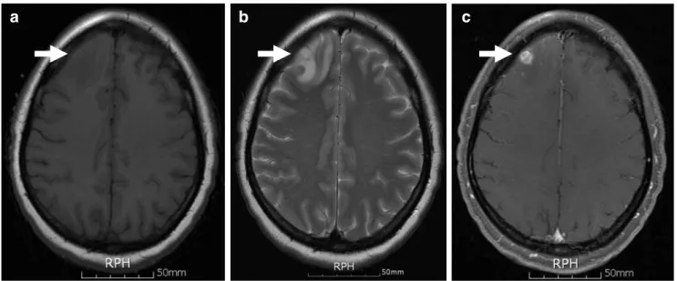
A 32-year-old woman was hospitalized for intermittent headaches for the past 3 months. She had no previous illness history and no abnormal results from neurologic examinations. The HIV and toxoplasma antibody tests were positive. Gadolinium-enhanced brain magnetic resonance imaging (MRI) showed a small well-enhanced nodular lesion with edema in the right frontal lobe (Fig. 1). It was difficult to differentiate cerebral toxoplasmosis from prima- ry CNS lymphoma. A F-18 FDG PET/CT scan of the brain was performed for a differential diagnosis. Images were obtained 30 min after an intravenous injection of 380 MBq of F-18 FDG with a PET/CT scanner (Discovery STE, General Electric Medical Systems, Milwaukee, USA).
Moderate FDG uptake was noted in the nodular lesion of the right frontal lobe (Fig. 2). The maximum standardized uptake value (SUVmax) of the lesion was 7.5 and the SUVmax of the contralateral homologous brain region was H. W. Kim:K. S. Won (*):B. W. Choi:S. K. Zeon
Department of Nuclear Medicine, Keimyung University, School of Medicine, 194 Dongsan-Dong, Jung-Gu, Daegu, Korea
e-mail: [email protected]
Nucl Med Mol Imaging (2010) 44:75–77 DOI 10.1007/s13139-009-0014-3
10.2. A count ratio of the lesion-to-contralateral homologus brain was 0.74.
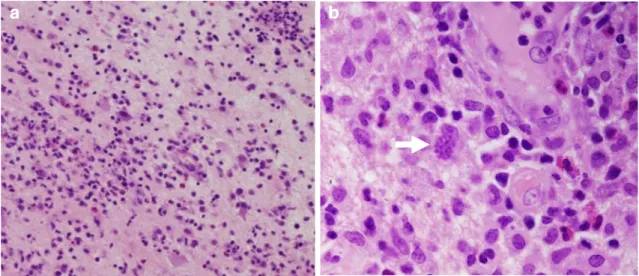
Because it was still difficult to differentiate cerebral toxoplasmosis from primary CNS lymphoma, the lesion of the right frontal lobe was removed surgically. The lesion was 1.2×1.2×1.0 cm in size and the cut surface was edematous and focally hemorrhagic. The pathologic exam- ination showed bradyzoites of Toxoplasma gondii with inflammatory cells and confirmed diagnosis of the cerebral toxoplasmosis (Fig.3).
Discussion
HIV primarily infects and kills CD4+T cells, macrophages, and dendritic cells [6]. When CD4+T cell numbers decline below a critical level, cell-mediated immunity is lost, and the body becomes progressively more susceptible to opportunistic infections including Toxoplasma gondii, cytomegalovirus, Cryptococcus neoformans and tuberculo- sis, and to malignancies [7]. The distinction between primary CNS lymphoma and nonmalignant lesions due to
a b c
Fig. 1 a T1-weighted axial brain MR image showing a hypointense lesion (arrow) in the right frontal lobe and (b) T2-weighted axial brain MR image showing a hyperintense lesion (arrow) with edema in the
right frontal lobe. c A gadolinium-enhanced axial brain MR image showing a small well-enhanced nodular lesion (arrow) in the right frontal lobe
a b c
Fig. 2 Axial F-18 FDG (a) PET and (b) PET/CT images of the brain show moderate FDG uptake (arrow) in the right frontal lobe. c An axial F-18 FDG PET/MRI coregistration image shows moderate FDG
uptake in the enhanced nodular lesion (arrow) of the right frontal lobe.
The SUVmax of the lesion was 7.5 and the SUVmax of contralateral homologous brain region (arrowhead) was 10.2
76 Nucl Med Mol Imaging (2010) 44:75–77
opportunistic infections, in particular cerebral toxoplasmo- sis, is important because treatment is different. Cerebral toxoplasmosis can be effectively treated with medication, whereas primary CNS lymphomas are treated with radiation therapy and corticosteroids. Neither CT nor MRI scans can reliably distinguish CNS infections, such as toxoplasmosis, from lymphoma in HIV-1-positive patients [8].
Several authors reported the usefulness of FDG PET to differentiate cerebral toxoplasmosis and other infectious diseases from primary CNS lymphoma. Villringer et al. [9]
examined 11 AIDS patients, six with toxoplasmosis, one with a tuberculoma, and four with primary CNS lymphoma.
The FDG uptake within the lesion was compared with the uptake in a contralateral brain area. In all subjects with cerebral infections, the FDG uptake ratio was significantly lower than the FDG ratio in patients with lymphoma with no overlap of the uptake values. Hoffman et al. [10] also studied 11 individuals with AIDS and CNS lesions using FDG PET. Significant difference was noted between FDG uptake in lymphoma and cerebral toxoplasmosis. The count ratio in lymphoma was 1.8±0.6 and the count ratio in toxoplasmosis was 0.65±0.3.
In our case, it was difficult to differentiate cerebral toxoplasmosis from primary CNS lymphoma by visual analysis of PET/CT. The SUVmax of the cerebral toxoplas- mosis was 7.5 and a count ratio was 0.74. This result is consistent with the results of earlier studies which FDG PET is useful to accurately differentiate between primary CNS lymphoma and cerebral toxoplasmosis. However, primary CNS lymphoma can have variable FDG uptakes according to their pathologic nature [11–13]. Thus, increased FDG uptake of the cerebral lesion in patients with AIDS should be interpreted with caution in differentiating cerebral toxoplas- mosis from primary CNS lymphoma.
References
1. Wainberg MA, Jeang KT (2008) 25 years of HIV-1 research—
progress and perspectives. BMC Med 6:31
2. Hult B, Chana G, Masliah E, Everall I (2008) Neurobiology of HIV. Int Rev Psychiatry 20:3–13
3. Wright E, Brew B, Arayawichanont A, Robertson K, Saminthar- apanya K, Kongsaengdao S et al (2008) Neurologic disorders are prevalent in HIV-positive outpatients in the asia-pacific region.
Neurology 71:50–56
4. Peregudova AB, Shakhgil’dian VI, Goncharov DB, Ermak TN, Tishkevich IM, Shipulina O et al (2007) Cerebral toxoplasmosis in HIV-infected patients. Ter Arkh 79:36–39
5. Ong EL (2008) Common aids-associated opportunistic infections.
Clin Med 8:539–543
6. Kelleher AD, Zaunders JJ (2006) Decimated or missing in action:
Cd4+ T cells as targets and effectors in the pathogenesis of primary HIV infection. Curr HIV/AIDS Rep 3:5–12
7. Samuel R, Bettiker RL, Suh B (2002) AIDS related opportunistic infections, going but not gone. Arch Pharm Res 25:215–228 8. Sathekge M, Goethals I, Maes A, van de Wiele C (2009) Positron
emission tomography in patients suffering from HIV-1 infection.
Eur J Nucl Med Mol Imaging 36:1176–1184
9. Villringer K, Jager H, Dichgans M, Ziegler S, Poppinger J, Herz M et al (1995) Differential diagnosis of CNS lesions in aids patients by FDG-PET. J Comput Assist Tomogr 19:532–536 10. Hoffman JM, Waskin HA, Schifter T, Hanson MW, Gray L,
Rosenfeld S et al (1993) FDG-PET in differentiating lymphoma from nonmalignant central nervous system lesions in patients with AIDS. J Nucl Med 34:567–575
11. Mohile NA, Deangelis LM, Abrey LE (2008) The utility of body FDG PET in staging primary central nervous system lymphoma.
Neuro Oncol 10:223–228
12. Nishiyama Y, Yamamoto Y, Monden T, Sasakawa Y, Kawai N, Satoh K et al (2007) Diagnostic value of kinetic analysis using dynamic FDG PET in immunocompetent patients with primary CNS lymphoma. Eur J Nucl Med Mol Imaging 34:78–86
13. Kang YH, Lim ST, Kim DW, Jeong HW, Sohn MH, Yim CY (2008) Usefulness of F-18 FDG PET/CT in staging of peripheral T cell lymphoma. Nucl Med Mol Imaging 42:369–374
a b
Fig. 3 a Pathologic examination shows inflammatory tissues infiltrated with lymphocytes, macrophages and plasma cells, areas with microglial nodules and lymphocytic perivascular infiltration and hyalinization, and
thickening of arteriolar wall (H & E stain, ×100). (b) Bradyzoite of Toxoplasma gondii (arrow) is noted (H & E stain, ×200). These findings are consistent with cerebral toxoplasmosis
Nucl Med Mol Imaging (2010) 44:75–77 77