대한두경부종양학회지, 제37권 제1호, 2021. pp.57-61 Korean Journal of Head & Neck Oncology, Vol.37, No.1
https://doi.org/10.21593/kjhno/2021.37.1.57 ISSN 1229-5183(Print) / ISSN 2586-2553(Online)
전두-안와 구역에 발생한 섬유성 이형성증의 근치적 절제술 및 자가두개골 이식을 이용한 재건을 통한 치료: 증례보고
최지안⋅곽정하⋅윤청민
동아대학교 의과대학 성형외과학교실
Treatment of Fibrous Dysplasia of the Fronto-Orbital Area with Radical Resection and Autogenous Reconstruction Using Split Calvarial Bone Graft: A Case Report
Ji-An Choi, MD, Jung-Ha Kwak, MD, Chung-Min Yoon, MD
Department of Plastic & Reconstructive Surgery, Dong-A University School of Medicine, Busan, Korea
= Abstract =
Fibrous dysplasia is a bone condition characterized by the replacement of normal bone tissue and the medullary cavity by abnormal fibrous tissues. Craniofacial fibrous dysplasia causes facial asymmetry compromising the aes- thetics as well as vision and hearing. A 21-year-old male visited the clinic due to vertical orbital dystopia and exophthalmos that had developed over the previous 2 months. The patient was diagnosed with a fibrous dysplasia of the frontal, ethmoid bones and superior orbital wall. By a bicoronal incision on the scalp, the radical resection of the lesions was done. After harvesting the remaining frontal bone, we did the autogenous reconstruction using split calvarial bone graft. Postoperatively, the vertical orbital dystopia and exophthalmos significantly improved.
The patient is satisfied with the surgical outcomes and has not reported any recurrence.
Key Words : Bone transplantation⋅Craniofacial fibrous dysplasia⋅Reconstructive surgical procedures
Received R e v i s e d Accepted
: February 23, 2021 : March 23, 2021 : March 30, 2021
+Corresponding author: Chung-Min Yoon, MD
Department of Plastic and Reconstructive Surgery, Dong-A University school of Medicine. 26, Daesingongwon-Ro, Seo-Gu, Busan, 49201, Korea
Tel: +82-51-240-2744, Fax: +82-51-243-5416 E-mail: [email protected]
Introduction
Fibrous dysplasia is a benign developmental bone con- dition in which abnormal fibrous tissues gradually proliferate and replace spongy bone, filling the medullary cavity.1,2) Fibrous dysplasia of the orbit and skull base can invade the periorbital bone and induce orbital displacements and eye protrusion, consequently causing craniofacial deformities.3) Therefore, appropriate resection followed by craniofacial re-
construction is necessary to treat fibrous dysplasia of the orbit and skull base. This study reports a case of facial fi- brous dysplasia that developed in the frontal and ethmoid bones and superior orbital wall in a non-native patient that was successfully treated by radical resection followed by reconstruction using an autogenous split calvarial bone graft.
Case
A 21-year-old Uzbek male visited the present institution with a feeling that his left eye had started bulging 2 months ago. A clinical examination revealed downward and lateral displacements of the left eye by 5 mm and 2 mm, respectively, and eye protrusion by 3 mm (Fig. 1). No visual disturbance, malocclusion, or neurological symptoms were observed in the patient. Computed tomography (CT) images showed
Fig. 1. Preoperative photograph. Vertical orbital dystopia is observed.
A
B
Fig. 2. Preoperative computed tomography. Focal bony scle- rosis and thickening of the frontal bone and left ethmoidal sinus (A) axial view. (B) coronal view.
Fig. 3. Intraoperative photograph. Frontal bone was harvested following the radical resection of the fibrous dysplasia lesion in the fronto-orbital region.
evidence of cortical thickening and osteosclerosis of the frontal bone and left ethmoid sinus (Fig. 2A, 2B). The pa- tient was diagnosed with fibrous dysplasia based on these findings, and radical resection was planned after considering the patient’s age and location of the lesion.
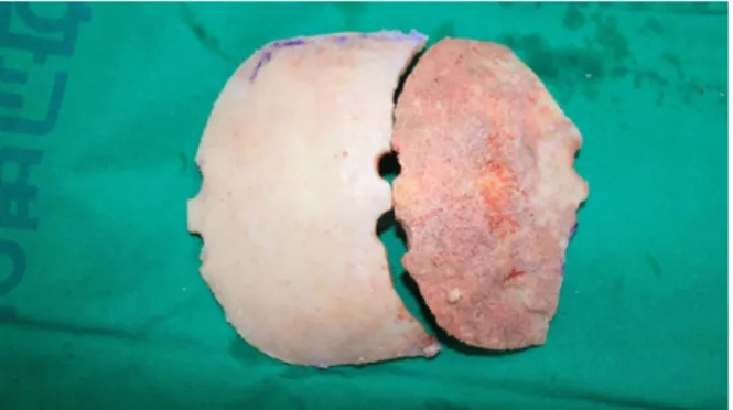
A bicoronal incision was made following induction with general anesthesia. Dissection was performed up to the gla- bellar area to secure a sufficient operative field. The lesion measured approximately 6.5 cm × 4 cm × 3.5 cm and in- volved the frontal and ethmoid bones and the s uperior orbi- tal wall. An oscillating saw and rongeur were used to care- fully perform radical resection of the lesion. Further, an air craniotome was used to harvest the frontal bone, in col- laboration with the department of neurosurgery (Fig. 3). The harvested frontal bone was divided into inner and outer cor- tices using an oscillating saw, and the outer cortex was re- served for donor site coverage. A part of the inner cortex flap was excised into a convex triangle shape of approx- imately 1.5 cm × 2 cm and approximately 2 cm × 3.5 cm and fixed with a wire to the right ethmoid defect, left eth- moid bone and the bone defect area on the superior orbital wall. A new contour was formed by moving and fixing the inner cortex flap to the inner upper side so that the position of the newly formed upper orbital wall on the left side was similar to that of the right upper orbital wall. The remaining
Fig. 4. Intraoperative photograph. The harvested frontal bone was split and reassembled for reconstruction at the defect site.
A B C
Fig. 5. Postoperative computed tomography image 8 days postoperatively. The frontal bone, left superior orbital wall and rim, and ethmoid bone were well-reconstructed (A) axial view. (B-C) 3D C-T images.
Fig. 6. Postoperative photograph 1 year and 5 months postoperatively. No recurrence and good aesthetic results.
inner cortex flaps were cut in the shape of the frontal bone defect by maintaining the convex shape and then fixed with a wire to the newly formed superior orbital margin and the lower margin of the outer cortex flap used for donor site coverage (Fig. 4). A drain was inserted into the bicoronal incision area and wound closure was performed accordingly.
On the axial view of the CT taken 8 days after the operation, the inner cortex flap fixed on the defect of ethmoid bones on both sides was well maintained while the 3D image showed that the frontal area and both superior orbital rims
were found to maintain their symmetric contours (Fig. 5A, 5B, 5C). No specific complications such as vision problems, facial nerve disorders, and infections were observed in the patient. He has now been postoperatively followed up for 1 year and 5 months and has not experienced any recurrence.
A clinical examination revealed a reduction in the down- ward displacement of the left eye from 5 mm to 1 mm and a reduction in lateral displacement from 2 mm to 0.5 mm. Exophthalmos also improved from 3 mm to 0.5 mm.
The patient was satisfied with the functional and aesthetic
outcomes of the surgery (Fig. 6). Written consent was re- ceived from the patient to publish this case report.
Discussion
Fibrous dysplasia is a benign bone condition characterized by the replacement of normal bone by the connective tissues of fibrous bone. An activated mutation on the α-subunit of the G-signaling protein within osteoblastic cells is known to induce fibrous dysplasia by disrupting the functions of cells in the osteogenic lineage.1,2) Fibrous dysplasia usually develops in childhood and accounts for 2.5% of all bone tumors and 7.5% of benign bone tumors .3-5) It is clinically classified as monostotic or polyostotic. Seventy percent of patients with fibrous dysplasia have monostotic lesions, and craniofacial involvement is observed in 10% of monostotic lesions and 50-100% of polyostotic lesions.3)
Swelling and facial asymmetry are the most common symptoms of craniofacial fibrous dysplasia.6,7) Different signs and symptoms are observed depending on the anatomical location involved.3) Involvement of the temporal bone or otic capsule can lead to hearing loss, and that of the frontal, sphenoid, ethmoid, and maxillary bone can lead to sinusitis and nasal congestion.6,8) Cranial bone or orbital involvement can lead to headaches, orbital dystopia, vision loss, paresthesia, eye protrusion, epiphora, and blindness.8,9)
No clear guidelines have been established for the treat- ment of craniofacial fibrous dysplasia.3) Surgery is the only option for cranial fibrous dysplasia, excluding for those le- sions that develop at the central cranial base with involve- ment of major ves s els and cranial nerves .3,7) In cases where surgery is not possible, a radiologic examination is per- formed and symptoms are monitored accordingly. There have been attempts to pharmacologically treat craniofacial fibrous dysplasia using potent osteoclast inhibitors such as pamironate and bisphosphonate; however, their therapeutic effects are yet to be verified.3)
Whether surgical treatment is more appropriate than con- servative or radical treatment remains controversial.7) When choosing a treatment method, it is necessary to aim for not only achievement of functional and aesthetic improvements but also to minimize the risk of recurrence and reoperation.
The patient’s age, lesion location, scope of lesion, orbital displacement, patient preferences, and the level of experi-
ence of the surgical team must also be considered before the initiation of treatment.7,10)
Radical resection has been reported to achieve lower re- currence and reoperation rates in craniofacial fibrous dys- plasia than conservative treatment.11) Complete resection of a craniofacial fibrous dysplasia lesion is necessary since ma- lignant changes of fibrous dysplasia have been reported, al- beit rarely.12) In cases of lesions in the fronto-orbital region accompanied by orbital dystopia such as the one in this case report, radical resection is absolutely needed since the lesions can progress toward the inside of the eye or the cranial base to apply pressure on the optic canals or the neurovascular structures of the superior orbital fissure, con- sequently causing vision loss and serious complications.3) A recent study has also reported radical resection to be the most appropriate method of treatment for adult patients with zone I, II, and IV lesions under the Chen and Noordhoff grading system.7)
However, it has been reported that conservative proce- dures such as reduction burring resolved functional and aes- thetic problems and minimized facial changes that occurred over the course of development in patients aged less than 7 years without vision problems more effectively than radi- cal resection.7) It has also been reported that early surgery before skeletal maturity reduces the rate of recurrence afterwards.13) However, if a les ion has already invaded the fronto-orbital region and acute visual deficits have occurred within one month as was the case with the patient in this report, radical resection is still necessary, regardless of age.7) It is important to appropriately determine the globe posi- tion and maintain the orbital volume when reconstructing the calvarial bone in the fronto-orbital region.3) Aesthetic abnormalities such as dystopia can be corrected once an orbital rim has been appropriately formed, and a sufficient orbital volume must be secured to prevent functional globe problems such as diplopia and exophthalmos.14-15)
In this case report, the surgical site was widely exposed through a bicoronal incision, and a sufficient size of autoge- nous frontal bone was harvested for reconstruction. The har- vested frontal bone was reassembled to match the shape of the defect s ite us ing a s plit calvarial bone graft and was transplanted onto the sites of defect on the frontal bone and superior orbital wall and rim to achieve convexity sim- ilar to that on the side opposite to the lesion. Using this
approach, an appropriate orbital contour and volume could be obtained. In cases of lesions in the frontal sinuses or ethmoid sinuses where there is a potential risk of con- tamination or in lesions accompanied by nearby defects in the calvarial bone, using an autogenous bone graft as was done in this case may more effectively lower the risk of infections as opposed to using alloplastic materials.
Many factors must be considered during craniofacial bone reconstruction. Several surgical methods have been pro- posed to predict the appropriate graft shape and reduce the operation time. In a recent study, Ahn et al. introduced a reconstruction approach in which a rapid prototyped model created by a three-dimensional printer was used to plan the shape of an autologous calvarial bone graft following the radical resection of fibrous dysplasia in the zygomaticomax- illary region.10)
With exceptions, radical resection and reconstruction is necessary for craniofacial fibrous dysplasia and especially so in lesions in the fronto-orbital region to prevent aesthetic and functional complications, recurrences, and malignant changes. This paper reports a case of fibrous dysplasia that occurred in the fronto-orbital region, which was success- fully treated by radical resection followed by reconstruction using an autogenous bone graft. The procedure achieved satisfactory functional and aesthetic outcomes.
Acknowledgments
This study was supported by the research funds of Dong-A University.
References
1) Riminucci M, Liu B, Corsi A, Shenker A, Spiegel AM, Robey PG, et al. The histopathology of fibrous dysplasia of bone in pa- tients with activating mutations of the Gs alpha gene: site-specif- ic patterns and recurrent histological hallmarks. J Pathol.
1999;187:249-258.
2) Hunt JA, Hobar PC. Common craniofacial anomalies: conditions
of craniofacial atrophy/hypoplasia and neoplasia. Plast Reconstr Surg. 2003;111:1497-1508.
3) Ricalde P, Horswell BB. Craniofacial fibrous dysplasia of the fronto-orbital region: a case series and literature review. J Oral Maxillofac Surg. 2001;59:157-167.
4) Hoffman S, Jacoway JR, Krolls SO. Fibrous dysplasia: benign nonodontogenic tumors of the jaws. Intraosseous and Periosteal Tumors of the Jaws. 2nd ed. Bethesda: Armed Forces Institute of Pathology. 1987;211-216.
5) Edgerton MT, Persing JA, Jane JA. The surgical treatment of fi- brous dysplasia with emphasis on recent contributions from cra- nio-maxillo-facial surgery. Ann Surg. 1985;202:459-479.
6) Moor RT, Buncic JR, Munro IR. Fibrous dysplasia of the orbit in childhood: clinical features and management. Ophthalmology.
1985;92:12-20.
7) Denadai R, Raposo-Amaral CA, Marques FF, Ghizoni E, Buzzo CL, Raposo-Amaral CE. Strategies for the optimal individualized surgical management of craniofacial fibrous dysplasia. Ann Plast Surg. 2016;77:195-200.
8) Katz BJ, Nerad JA. Ophthalmic manifestations of fibrous dys- plasia: a disease of children and adults. Ophthalmology. 1998;
105:2207-2215.
9) Morrissey DD, Talbot JM, Schleuning AJ. Fibrous dysplasia of the temporal bone: reversal of sensorineural hearing loss after decompression of the internal auditory canal. 2nd. Laryngoscope.
1997;107:1336-1340.
10) Ahn SJ, Hong JW, Kim YO, Lew DH, Lee WJ. Treatment of fi- brous dysplasia of the zygomaticomaxillary complex with radical resection and three-dimensional reconstruction with autologous calvarial bone graft. Arch Craniofac Surg. 2018;19:200-204.
11) Gabbay JS, Yuan JT, Andrews BT, Kawamoto HK, Bradley JP.
Fibrous dysplasia of the zygomaticomaxillary region: outcomes of surgical intervention. Plast Reconstr Surg. 2013;131:1329-1338.
12) Sadeghi SM, Hosseini SN. Spontaneous conversion of fibrous dysplasia into osteosarcoma. J Craniofac Surg. 2011;22:959-961.
13) Fattah A, Khechoyan D, Phillips JH, Forrest CR. Paediatric cra- niofacial fibrous dysplasia: the hospital for sick children experi- ence and treatment philosophy. J Plast Reconst Aesthet Surg.
2013;66:1345-1355.
14) Brusati R, Biglioli F, Mortini P, Raffaini M, Goisis M.
Reconstruction of the orbital walls in surgery of the skull base for benign neoplasm. Int J Oral Maxillofac Surg. 2000;29:325-330.
15) Papay FA, Zins JE, Hahn JF. Split calvarial bone graft in cra- nio-orbital sphenoid wing reconstruction. J Craniofac Surg.
1996;7:133-139.