원유 내 내냉성 미생물의 계절별, 지역별 분포 및 동정
신용국
1ㆍ이현아
1ㆍ오남수
1ㆍ남명수
2*
1서울우유협동조합중앙연구소, 2충남대학교 농업생명과학대학 동물바이오시스템과학과
Seasonal, regional distribution and identification of psychrotrophic bacteria in milk
Yong Kook Shin
1, Hyun Ah Lee
1, Nam Su Oh
1, Myoung Soo Nam
2*
1
R&D Center, Seoul Dairy Cooperative, Ansan Kyunggi-Do 425-839, Korea
2
Lab. of Milk Food Biochemistry and Biotechnology, College of Agriculture and Life Sciences, Chungnam National University, Daejeon 305-764, Korea
Received on 19 February 2013, revised on 17 March 2013, accepted on 19 March 2013
Abstract : To investigate the distribution of psychrotrophic bacteria, raw milk was collected from farms in nine different regions located around Kyunggi province in South Korea at four different seasons. Psychrotrophic counts were higher in winter than in other seasons as 3.0×10
4CFU/mL (p<0.05). Among nine regions, the population in raw milk sampled from B region was in significantly greater numbers and those from C and D province were in significantly lower numbers than any other regions, 2.4×10
5CFU/mL and 8.7×10
3CFU/mL, respectively (p<0.05). In addition, among 706 bacterial isolates, the predominant class was Gamma-proteobacteria (81.02%) and genus was Pseudomonas (32.34%), especially Pseudomonas fluorescens (39.46%). Compared to the regional predominance, Acinetobacter johnsonii in A region, Pseudomonas fluorescens in B region, Enterobacter amnigenus in C region, Psychrobacter maritimus in D region, Acinetobacter johnsonii in E region, Acinetobacter haemolyticus in F region, Pseudomonas fluorescens in G region, Acinetobacter jounsonii in H region, and Pseudomonas mucidolens in I region were found.
Key words : Psychrotrophic bacteria, Raw milk, Gamma-proteobacteria, Pseudomonas fluorescens
*Corresponding author: Tel: +82-42-821-5782 E-mail address: [email protected]
I. 서 론
우유의 미생물학적 품질은 원유의 품질, 살균조건, 2차 오염 및 저장과 유통온도에 의해 영향을 받는다(IDF, 1993). 유제품에서 나타나는 미생물은 열처리를 함에도 불 구하고 사멸되지 않고 살아있는 내열성 미생물과 우유의 포장용기와 공기로부터의 2차 오염 등으로 구분될 수 있고, 유제품의 품질은 주로 시유의 내냉성 미생물과 내열성 미 생물수에 의하여 평가할 수 있다(Kwon et al. , 1998). 원유 에 존재하는 내냉성 미생물의 수는 계절별과 지역별 차이 를 보이고 있다. 미생물이 잘 성장할 수 있는 온도인 여름철 이 겨울철보다 내냉성 미생물의 오염이 더 높다는 보고 (Cousin, 1981; Sutherland and Murdoch, 1994)와 오히
려 겨울철에 위생관리가 소홀해지기 때문에 여름철보다 원
유의 내냉성 미생물 오염 발생건수가 더 높다는 상반된 연
구 결과(Kikuchi and Matsui, 1974b; Elionora and Malka,
2007; Maria et al., 2010)가 발표되고 있다. 최근 우리나라
목장에도 냉각기가 설치되고 탱크로리에 의한 콜드체인시
스템(cold chain system)이 도입되면서 원유의 미생물학
적 품질이 크게 향상되었으나, 여전히 내냉성 미생물의 오
염으로 인한 원유의 품질에는 악영향을 미치고 있다. 원유
에서 가장 많이 검출되는 내냉성 미생물로는 그람 음성균
인 Pseudomonas 속이며(Martins et al., 2006), 그 외
Achromobacter (Credit et al., 1972), Acinetobacter (Milliere
and Veillet-Poncet, 1979), Enterobacter (Andrey and
Frazier, 1959), Escherichia (Jayashankar et al., 1966),
Flavobacterium (Andrey and Frazier, 1959), Serratia
(Ingraham, 1962) 등이 있다. 본 연구의 목적은 원유에 존
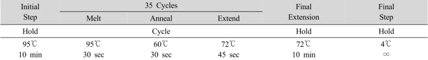
Table 1. Thermal cycling conditions for PCR.
Initial Step
35 Cycles Final
Extension
Final
Melt Anneal Extend Step
Hold Cycle Hold Hold
95℃
10 min
95℃
30 sec
60℃
30 sec
72℃
45 sec
72℃
10 min
4℃
∞
Table 2. Thermal cycling conditions for sequencing.
25 Cycles Final
Melt Anneal Extend Step
Cycle Hold
96℃
10 sec
50℃
5 sec
60℃
4 min
4℃
∞ 재하는 계절별 및 지역별 내냉성 미생물의 분포 및 종류를
조사하기 위해서 수행하였다.
II. 재료 및 방법
1. 계절별, 지역별 원유 시료와 총 내냉성 미생물의 측정 및 분리 배양
연구에 필요한 원유 시료는 계절별로 여름철 원유는 2010년 7월, 가을철 원유는 2010년 10월, 겨울철 원유는 2011년 1월, 봄철 원유는 2011년 4월에 공급받았다. 계절별 로 원유는 경기 동부, 경기 동남부, 경기 동북부, 경기 서 부, 경기남부, 경기북부, 경기중부, 안산, 경인지역 목장으 로부터 공급받았으며, 시료는 냉장보관하였고 실험을 수행 하기까지 2일이 넘지 않도록 하였다. 지역별 결과는 무작위 로 A, B, C, D, E, F, G, H, I 지역으로 표기하였으며, 확보한 시료는 멸균생리식염수에 각 농도별로 희석하여 Standard methods(Wehr and Frank, 2004)에 따라 Plate count agar(Becton, Dickson and Company, Sparks, MD, USA)를 이용하여 7℃에서 10일 동안 배양하 였다. 배양이 끝난 후, 각 원유시료의 총 내냉성미생물의 균수를 측정하였으며, 그 중 무작위로 colony를 선택하여 최소 3번 이상 plate에 계대배양하여 단일 colony를 취하 였다. 지역별로 20개의 colony를 취하여, 총 9개 지역에서 한 계절에 총 180개의 균을 분리배양하였으며, 사계절, 9개 지역으로부터 총 720개의 균을 분리하여 동정에 사용하였 다. 결과 분석을 위해 모든 통계처리는 Statistical Analysis System(SAS Institute Inc., Cary, NC, USA)을 이용하여
Duncan's Multiple Range Test를 사용하였다.
2. 분자생물학적 방법에 의한 내냉성 미생물의 동정
분리배양한 colony들은 동정을 수행하기 위해 Applied
Biosystems(Foster City, CA, USA)의 방법에 따라 PrepMan
Ultra Sample Preparation Reagent(Applied Biosystems,
Foster City, CA, USA)를 이용하여 genomic DNA를 추출
하였다. 분리배양한 균에서 1 µL loop를 이용하여 colony를
취한 후, 100 µL reagent에 30초 동안 강하게 혼합하여 9
5℃에서 10분 동안 가열하였다. 그 후 13,000 rpm/min에
서 2분 동안 원심분리하여 상징액을 DNA template로 사용
하였다. PCR은 MicroSeq 500 16S rDNA Bacterial Iden-
tification PCR Kit(Applied Biosystems) 를 사용하였으며
(Table 1), 15 µL PCR master mix와 15 λ의 DNA
templete를 넣고 반응시켰다. PCR product를 2% agarose
gel에 4 µL를 loading하여 100 V에서 2분 동안 전기영동하
여 band를 확인하였다. 확인한 PCR product는 남은 dNTP
와 primer를 제거하기 위해 ExoSAP-IT (USB, Cleveland,
Ohio, USA)으로 세척한 뒤, Microseq 16S rDNA bacterial
500 Sequencing Kit(Applied Biosystems)를 사용하여
sequencing 하였다 (Table 2). 모든 과정이 끝난 반응물
10 µL와 10 µL의 Hi-Di Formamide(Applied Biosystems)
를 혼합하여 POP 6 polymer와 4×50 cm capillary를 장착
한 3130 genetic analyzer(Applied Biosystems)를 사용하
여 분석하였다. 모든 동정 결과는 MicroSeq library version
2.1(Applied Biosystems)을 통하여 처리하였다.
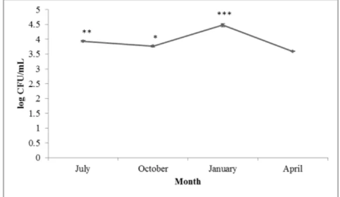
Fig. 1. Seasonal variation in the numbers of psychrotrophic bacteria.
July; summer, October; fall, January; winter, April; spring
*p<0.05 vs April, **p<0.05 vs October, ***p<0.05 vs July.
Fig. 2. Counts of psychrotrophic bacteria isolated from raw milk sampled from different provinces. A ~ I; different regions.
Significant differences expressed as different letters were analyzed using Duncan's multiple range test (p < 0.05)
III. 결과 및 고찰
1. 계절별 및 지역별 원유의 총 내냉성 미생물 균수 변화
내냉성 미생물의 총 균수를 측정하여 계절별 분포를 비 교해 본 결과, 겨울철 원유에서 3.0×10
4CFU/mL로 가장 높은 균수가 검출되었고, 여름철 원유에서 8.7×10
3CFU/mL, 가을철 원유에서 5.9×10
3CFU/mL가 검출되었 으며, 봄철 원유에서 3.9×10
3CFU/mL로 가장 낮게 균이 검출되었다( p <0.05) (Fig. 1). 이 결과는 Elionora와 Malka (2007)의 연구와도 유사한 결과로 나타났는데, 이 연구진 들은 이스라엘에서 1월, 4월, 7월 및 9월에 집유한 원유로 부터 내냉성 미생물과 중온성 미생물을 분리하여 균수를 측정하여 비교하였다. 실험 결과는 본 연구와 유사하게 계 절적 변이를 보였으며, 겨울철 원유에서 가장 높은 수준의 균수가 검출되었고, 봄철 원유에서 가장 낮은 수준의 균수 가 검출되었다. 또한 Maria 등(2010)이 보고한 연구 결과 와 유사하게 여름철에 비해 겨울철 원유에서 더 높은 내냉 성미생물 균수가 측정되었으며, 전체적인 균수 분포는 2.0×10
5CFU/mL에서 1.6×10
7CFU/mL 사이였다. 이는 본 연구 결과에서 균수분포가 3.9×10
3CFU/mL에서 3.0×10
4CFU/mL인 것과 비교하였을 때 본 연구 결과보다 더 높은 수준의 균수로 나타났다. 또한 Lee 등(1990a)이 보고한 바에 따르면 1년 동안 경상남도 북부지역의 한 집유 소에서 집유한 원유의 내냉성 미생물 분포를 측정한 결과, 본 연구 결과와 동일하게 겨울철에 가장 높은 균수가 측정 되었다. 이 연구에서는 총 내냉성 미생물수의 범위가
3.5×10
4~ 9.4×10
6CFU/mL로 나타났으며, 평균 1.5×10
6CFU/mL로 나타나 본 실험 결과보다 더 높은 수준임을 알
수 있었다. 또한 Lee 등(1990b)은 경기 지방의 농장으로부
터 수집한 원유 시료에 내냉성 미생물이 1.2×10
6CFU/mL
의 수준으로 존재한다고 발표하였다. 본 연구 결과와 비교
했을 때, 더 높은 수준임을 알 수 있었다. 일본의 Kikuchi와
Matsui(1974b)의 연구에서는 봄, 여름 및 겨울철의 원유
내 내냉성 미생물수의 변화를 측정한 결과, 여름철 원유에
서 가장 낮은 수준의 균이 검출되었고 겨울철 원유에서 가
장 높은 수준의 균이 검출되었다. 이는 여름철에 특히 착유
기구와 냉각기 등의 세척과 살균이 철저하기 때문이라고
보고하였다. 그러나 Cousin(1981)의 연구에 따르면, 여름
철에 미생물이 최적의 생장을 할 수 있는 온도이며 위생적
관리의 소홀로 인하여 일반적으로 겨울철보다 더 많은 균
수가 검출된다는 결과가 발표되었다. Sutherland와 Murdoch
(1994)는 겨울철 원유에서보다 여름철 원유에서 내냉성 미
생물이 더 많이 검출되고, 이는 중온성 미생물과 내냉성
미생물간의 길항작용 때문으로, 여름철에 중온성 미생물의
증식이 감소하고, 이에 상대적으로 내냉성 미생물이 여름
철에 더 활발하게 증식할 수 있기 때문인 것으로 분석하였
다. 이처럼 원유 에 존재하는 내냉성 미생물의 계절별 분포
는 나라별로 다른 추이를 나타내고 있으며, 원유 집유 조건
에 따라서도 계절별 분포에 차이를 나타내고 있음을 알 수
있었다. 각 지역별 균수를 비교한 결과는 B 지역 원유에서
2.4×10
5CFU/mL로 유의적으로 가장 높은 균수가 검출되
었고, C 지역과 D 지역 원유에서 8.7×10
3CFU/mL로 9개
지역 중 유의적으로 가장 낮은 균수가 검출되었다( p <0.05)
(Fig. 2). 나머지 지역은 I 지역에서 6.2×10
4CFU/mL, A
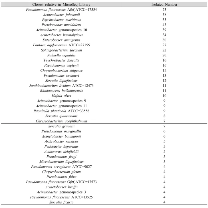
Table 3. Summary of psychrotrophic bacteria from raw milk over four seasons.
Closest relative in MicroSeq Library Isolated Number
Pseudomonas fluorescens A(bt)ATCC=17554 73
Acinetobacter johnsonii 58
Psychrobacter maritimus 53
Pseudomonas mucidolens 43
Acinetobacter genomospecies 10 39
Acinetobacter haemolyticus 34
Enterobacter amnigenus 30
Pantoea agglomerans ATCC=27155 27
Sphingobacterium faecium 22
Rahnella aquatilis 20
Psychrobacter faecalis 16
Pseudomonas asplenii 16
Chryseobacterium shigense 15
Pseudomonas brenneri 13
Serratia liquefaciens 12
Janthinobacterium lividum ATCC=12473 11
Rhodococcus baikonurensis 11
Hafnia alvei 10
Acinetobacter genomospecies 9 9
Acinetobacter genomospecies 11 9
Raoultella planticola ATCC=33558 9
Serratia quinivorans 8
Chryseobacterium scophthalmum 7
Serratia grimesii 7
Pseudomonas marginallis 6
Acinetobacter baumannii 6
Arthrobacter russicus 5
Pedobacter heparinus 5
Acidovorax delafieldii 5
Pseudomonas fragi 5
Microbacterium liquefaciens 5
Pseudomonas aeruginosa ATCC=9027 4
Chryseobacterium gleum 4
Pseudomonas fulva 4
Pseudomonas fluorescens G(bt)ATCC=17573 4
Acinetobacter lwoffii 4
Acinetobacter genomospecies 3 4
Pseudomonas fluorescens ATCC=13525 4
Serratia ficaria 4
지역과 F 지역에서 3.2×10
4CFU/mL, E 지역에서 2.9×10
4CFU/mL, G 지역에서 1.6×10
4CFU/mL, H 지역 에서 1.1×10
4CFU/mL, C 지역에서 8.9×10
3CFU/mL의 균이 원유에서 검출되었다. 또한 전체적인 균수 분포는 8.7×10
3CFU/mL에서 2.4×10
5CFU/mL 사이로 나타났다.
2. 내냉성 미생물의 계절별 및 지역별 분포
720개의 분리배양한 내냉성 미생물 중 총 706개의 균을 동정하였고 총 87가지의 서로 다른 종이 분석되었으며, 동
정 결과는 Table 3과 같다. Pseudomonas fluorescens 가
81개로 가장 높은 검출 빈도수를 나타내었으며, Acinetobacter
genomospecies가 61개, Acinetobacter johnsonii 가 58
개, Psychrobacter maritimus 가 53개 순으로 높은 검출
빈도수를 나타내었다. 동정된 psychrotrophic bacteria의
지역별 검출 빈도수가 높은 균주는 A 지역은 12개의
Acinetobacter johnsonii 가, B 지역은 22개의 Pseudomonas
fluorescens , C 지역은 13개의 Enterobacter amnigenus ,
D 지역은 19개의 Psychrobacter maritimus , E 지역은 19
개의 Acinetobacter johnsonii , F 지역은 8개의 Acinetobacter
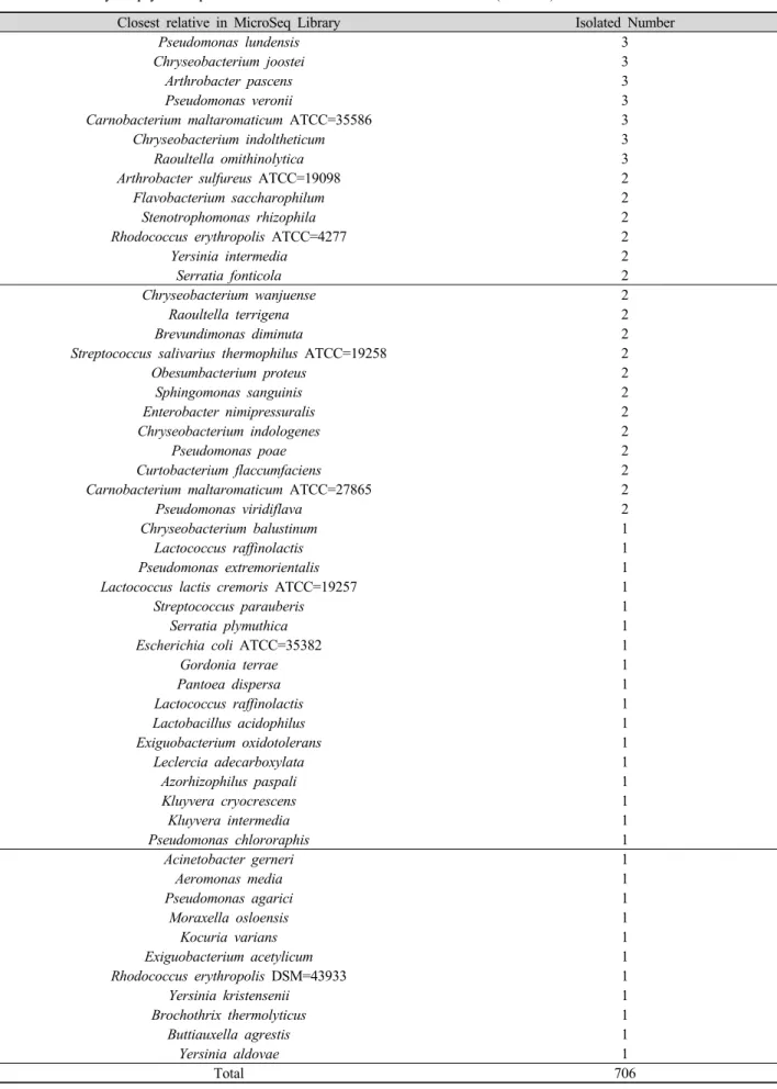
Table 3. Summary of psychrotrophic bacteria from raw milk over four seasons. (continue)
Closest relative in MicroSeq Library Isolated Number
Pseudomonas lundensis 3
Chryseobacterium joostei 3
Arthrobacter pascens 3
Pseudomonas veronii 3
Carnobacterium maltaromaticum ATCC=35586 3
Chryseobacterium indoltheticum 3
Raoultella omithinolytica 3
Arthrobacter sulfureus ATCC=19098 2
Flavobacterium saccharophilum 2
Stenotrophomonas rhizophila 2
Rhodococcus erythropolis ATCC=4277 2
Yersinia intermedia 2
Serratia fonticola 2
Chryseobacterium wanjuense 2
Raoultella terrigena 2
Brevundimonas diminuta 2
Streptococcus salivarius thermophilus ATCC=19258 2
Obesumbacterium proteus 2
Sphingomonas sanguinis 2
Enterobacter nimipressuralis 2
Chryseobacterium indologenes 2
Pseudomonas poae 2
Curtobacterium flaccumfaciens 2
Carnobacterium maltaromaticum ATCC=27865 2
Pseudomonas viridiflava 2
Chryseobacterium balustinum 1
Lactococcus raffinolactis 1
Pseudomonas extremorientalis 1
Lactococcus lactis cremoris ATCC=19257 1
Streptococcus parauberis 1
Serratia plymuthica 1
Escherichia coli ATCC=35382 1
Gordonia terrae 1
Pantoea dispersa 1
Lactococcus raffinolactis 1
Lactobacillus acidophilus 1
Exiguobacterium oxidotolerans 1
Leclercia adecarboxylata 1
Azorhizophilus paspali 1
Kluyvera cryocrescens 1
Kluyvera intermedia 1
Pseudomonas chlororaphis 1
Acinetobacter gerneri 1
Aeromonas media 1
Pseudomonas agarici 1
Moraxella osloensis 1
Kocuria varians 1
Exiguobacterium acetylicum 1
Rhodococcus erythropolis DSM=43933 1
Yersinia kristensenii 1
Brochothrix thermolyticus 1
Buttiauxella agrestis 1
Yersinia aldovae 1
Total 706
Table 4. Predominant psychrotrophic bacteria by regional groups.
Province Predominant isolated bacteria Isolated counts
A Acinetobacter johnsonii 12
B Pseudomonas fluorescens
A(bt)ATCC=17554 21
C Enterobacter amnigenus 13
D Psychrobacter maritimus 19
E Acinetobacter johnsonii 19
F Acinetobacter haemolyticus 8
G Pseudomonas fluorescens
A(bt)ATCC=17554 20
H Acinetobacter johnsonii 9
I Pseudomonas mucidolens 17
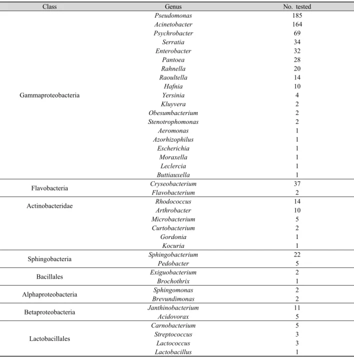
Table 5. Classification of identified psychrotrophic bacteria based on class and genus.
Class Genus No. tested
Gammaproteobacteria
Pseudomonas 185
Acinetobacter 164
Psychrobacter 69
Serratia 34
Enterobacter 32
Pantoea 28
Rahnella 20
Raoultella 14
Hafnia 10
Yersinia 4
Kluyvera 2
Obesumbacterium 2
Stenotrophomonas 2
Aeromonas 1
Azorhizophilus 1
Escherichia 1
Moraxella 1
Leclercia 1
Buttiauxella 1
Flavobacteria Cryseobacterium 37
Flavobacterium 2
Actinobacteridae Rhodococcus 14
Arthrobacter 10
Microbacterium 5
Curtobacterium 2
Gordonia 1
Kocuria 1
Sphingobacteria Sphingobacterium 22
Pedobacter 5
Bacillales Exiguobacterium 2
Brochothrix 1
Alphaproteobacteria Sphingomonas 2
Brevundimonas 2
Betaproteobacteria Janthinobacterium 11
Acidovorax 5
Lactobacillales
Carnobacterium 5
Streptococcus 3
Lactococcus 3
Lactobacillus 1
haemolyticus , G 지역은 20개의 Pseudomonas fluorescens , H 지역은 9개의 Acinetobacter johnsonii , I 지역은 17개 의 Pseudomonas mucidolens 로 나타났다(Table 4). 동정 된 균을 강(class)과 속(genus)을 기준으로 분류하였는데, Gamma-proteobacteria 강이 572개로 가장 우세하였고, 그 중 특히 Pseudomonas 속이 185개로 가장 높은 검출빈 도수를 나타내었으며 Acinetobacter 속이 164개로 그 다음 으로 높게 나타났다(Table 5). Martins 등(2006)은 원유의 내냉성 미생물 오염발생 원인균으로 가장 빈번하게 검출되 는 균이 Pseudomonas 속이라는 연구 결과를 발표하였으 며, Gunasekera 등(2003), Wiedmann 등(2000) 및 Chen 등(2002)도 원유에서 가장 많이 검출되는 내냉성 미생물은 Pseudomonas 속으로 보고하였다. 또한 Touch와 Deeth (2009)의 연구에서도 Pseudomonas 속이 원유에서 내냉성 미생물 중 생장 속도가 다른 균들에 비해 빠르기 때문에 가장 높은 빈도수를 차지하고 있으며, 특히 그 중 Pseudomonas fluorescens 와 Pseudomonas fragi 가 가장 빈번하게 검출 된다고 보고하였다. Lee 등(1990b)에 따르면 경기지방의 농장으로부터 수집한 원유 시료에서 분리한 내냉성 미생물 중 Pseudomonas 속이 44.1%를 차지했다. 또한 Shin 등 (1993)은 원유에서 분리한 총 200개의 내냉성 미생물 중 그램음성균은 160개(80.0%)로 나타났으며 그 중 Pseudomonas 속이 128개(64.0%)로 가장 높은 비율을 나타냈고, 특히 P.
fluorescens 가 85개(66.5%)로 대부분을 차지하였다. So 등(1992)의 보고에서도 원유에서 분리한 300개의 내냉성 미생물 중 49%가 Pseudomonas 속으로 동정되었는데 이 는 본 연구와 동일한 결과임을 알 수 있었다. Kikuchi와 Matsui(1974)의 보고에서도 1년 동안 집유한 원유의 내냉 성 미생물 중 82.1%로 Pseudomonas 속이 가장 높은 비율 을 나타냈다. 또한 Kato 등(1976)은 월별로 원유 내 검출되 는 내냉성 미생물 빈도수를 측정하였는데 6월부터 12월까 지 집유한 원유의 내냉성 미생물의 분리동정 결과는 Streptococcus 균종의 경우 11월에 가장 많이 검출되었고, Pseudomonas 속의 경우 12월에, Achromobacter 속의 경우 7월에, Flavobacterium 속의 경우 10월에 가장 높은 수준 의 균이 검출되었다고 보고하였다.
IV. 요 약
본 연구는 원유의 내냉성 미생물의 계절별 분포에 관해 조사하기 위해서 수행하였다. 내냉성 미생물은 최적 생장
온도가 30℃ 근처이지만, 7℃ 이하에서도 생존가능한 균으 로 Pseudomonas , Achromobacter , Aeromonas 속 등이 있다. 원유로부터 분리한 총 내냉성 미생물 균수는 다른 계절에 비해 겨울철 원유에서 3.0×10
4CFU/mL로 가장 높 은 수준으로 검출되었고, 다른 지역에 비해 B 지역에서 2.4×10
5CFU/mL로 유의적으로 높은 균수가 측정되었다 ( p <0.05). 분리한 총 706개의 균을 동정한 결과는 Gamma- proteobacteria 강이 전체의 81.02%로 가장 우세하였고, 그 중 Pseudomonas 속이 32.34%로 가장 높은 검출 빈도 수를 나타내었으며, 특히 Pseudomonas fluorescens 가 39.46%로 가장 우세하였다. 동정된 내냉성 미생물 의 지역 별 분포는 A 지역은 12개의 Acinetobacter johnsonii , B 지역은 21개의 Pseudomonas fluorescens , C 지역은 13개 의 Enterobacter amnigenus , D 지역은 19개의 Psychrobacter maritimus , E 지역은 19개의 Acinetobacter johnsonii , F 지역은 8개의 Acinetobacter haemolyticus , G 지역은 20개의 Pseudomonas fluorescens , H 지역은 9개의 Acinetobacter johnsonii , I 지역은 17개의 Pseudomonas mucidolens 가 높은 검출 빈도수를 나타내었다.
참 고 문 헌