Original Article
원고 접수일 2011년 7월 27일, 원고 수정일 2011년 8월 23일, 게재 확정일 2011년 8월 24일
책임저자 박용태
(210-702) 강원도 강릉시 강릉대학로 120, 강릉원주대학교 치과대학 구강악안면외 과학교실
Tel: 033-640-3139, Fax: 033-640-3113, E-mail: [email protected]
RECEIVED July 27, 2011, REVISED August 23, 2011, ACCEPTED August 24, 2011
Correspondence to Yong-Tae Park
Department of Oral and Maxillofacial Surgery, College of Dentistry, Gangneung-Wonju National University
120, Gangneung Daehangno, Gangneung 210-702, Korea
Tel: 82-33-640-3139, Fax: 82-33-640-3113, E-mail: [email protected]
CC This is an open access article distributed under the terms of the Creative Commons Attribution Non-Commercial License (http://creativecommons.org/licenses/
by-nc/3.0) which permits unrestricted non-commercial use, distribution, and reproduction in any medium, provided the original work is properly cited.
가토 두개골 결손 모델에서 실크단백과
나노하이드록시아파타이트, 옥수수 녹말 복합물을 이용한 골 이식재 개발
박용태ㆍ권광준ㆍ박영욱ㆍ김성곤ㆍ김찬우ㆍ조유영1ㆍ권해용1ㆍ강석우1
강릉원주대학교 치과대학 구강악안면외과학교실, 1농촌진흥청
Abstract
The Effect of Silk Fibroin/Nano-hydroxyapatite/Corn Starch Composite Porous Scaffold on Bone Regeneration in
the Rabbit Calvarial Defect Model
Yong-Tae Park, Kwang-Jun Kwon, Young-Wook Park, Seong-Gon Kim, Chan-Woo Kim, You-Young Jo
1, Hae-Yong Kweon
1, Seok-Woo Kang
1Department of Oral and Maxillofacial Surgery, College of Dentistry, Gangneung-Wonju National University,
1
Rural Development Administration
Purpose: This study evaluated the capability of bone formation with silk fibroin/nano-hydroxyapatite/corn starch composite scaffold as a bone defect replacement matrix when grafted in a calvarial bone defect of rabbits
in vivo
.Methods: Ten New Zealand white rabbits were used for this study and bilateral round-shaped defects were formed in the parietal bone (diameter: 8.0 mm). The silk fibroin 10% nano-hydroxyapatite/30% corn starch/60% composite scaffold was grafted into the right parietal bone (experimental group). The left side (control group) was grafted with a nano-hydroxyapatite (30%)/corn starch (70%) scaffold. The animals were sacrificed at 4 weeks and 8 weeks. A micro-computerized tomography (μCT) of each specimen was taken. Subsequently, the specimens were decalcified and stained with Masson's trichrome for histological and histomorphometric analysis.
Results: The average μCT and histomorphometric measures of bone formation were higher in the control group than in the experimental group at 4 weeks and 8 weeks after surgery though not statistically significant (
P
>0.05).Conclusion: The rabbit calvarial defect was not successfully repaired by silk fibroin/nano-hydroxyapatite/corn starch composite scaffold and may have been due to an inflammatory reaction caused by silk powder. In the future, the development of composite bone graft material based on various components should be performed with caution.
Key words: Silk fibroin/nano-hydroxyapatite/corn starch composite scaffold, Micro-computed tomography, Bone regeneration
골형성(osteogenesis)이 가능한 유일한 이식재로써 현재 가장 이상적인 재료로 알려져 있다[1]. 하지만 자가골은 몇몇 단점으로 인해 그 사용이 제한적인데, 이는 공여부에서 채취할 수 있는 골량의 한계 및 공여부의 손상에 따른 반흔형성, 이식골편의 흡수, 공여부 수술에 따른 전체 수술시간 및 술후 감염 가능성 증가 등이라 할 수 있다[2-4]. 동종골 및 이종골은 공여자 및 공여 동물이 가지고 있는 질병에 전염될 수 있고 이식 후 감염이 발생할 수 있으며, 스스로 골형성은 불가능하고 골유도(osteoinduction) 및 골전도(osteoconduction)만이 가능하여 그 효과가 자가골 이식에 비해 떨어지는 단점이 있다[5,6]. 하지만 사용할 수 있는 골량의 제한이 없고 공여부 손상 등이 없는 장점이 있어 현재 이들 재료에 대한 많은 연구가 이루어지고 있다. 합성골은 공여자 로부터의 질병 전염 위험이 없고 추가적인 공여부 손상 없이 술자가 원하는 만큼 사용할 수 있다는 장점이 있으나 골형성 능력이 기타 골이식재에 비해 낮다는 단점이 있다. 따라서 합성골 의 골형성 능력을 증가시키기 위한 많은 연구들이 시행되어 왔다.
본 연구에 사용된 hydroxyapatite (Ca
10(PO
4)
6(OH)
2, HA)는 인간 골조직의 주요 무기질 성분이다. HA는 합성 세라믹으로 골전도 성질 및 생체적합성, 생활성이 우수하며 숙주골로 대체되 는 성질이 있다[7-9]. 또한 HA가 nano 형태의 섬유구조로 사용될 경우 micro-size에 비해 단백질 흡수와 세포부착이 증가되는 경향 이 있다. 또한 세포활성도(bioactivity)가 증가하며[10] osteo- blast-like cell의 성장을 촉진한다. 위와 같은 장점들로 인해 HA 는 골이식재의 구성 성분으로 많이 사용되고 있다.
Silk fibroin (SF)은 섬유상의 단백질로써 거미나 누에의 유충 에서 만들어진다[11,12]. 비록 염증 및 면역반응을 유발할 수 있으 나 외층에 존재하는 sericin이라는 당단백질을 제거 시 부작용이 줄어들어 안전한 사용이 가능하다[13]. SF는 강한 인장력을 보이 며 높은 생체친화성, 느린 생분해성, 뛰어난 기계적 성질을 가지며 수용액 상태에서 낮은 용해도를 보인다[14-17]. 이러한 장점은 좋은 scaffold의 조건인 치유기간 동안의 구조 유지, 역할 완수 후 체내에서 분해, 치유에 필요한 세포들이 이주할 공간 제공 등을 가능케 하므로[18] SF는 최근 scaffold로 많이 사용되고
손부에 각각 이식한 후 micro computerized tomography (μ- CT) 분석 및 조직학적, 조직형태학적 분석을 시행하여 SF가 포함 된 복합체가 골 결손부의 신생골 형성을 증대시키는지 여부를 평가하는 것이다.
연구방법
1. 실험 동물
본 연구는 강릉원주대학교 연구윤리위원회의 승인을 얻은 후, 위원회의 감독을 받으며 수행하였다. 실험동물은 충분한 크기의 골 결손부를 형성할 수 있고, 골 형성 능력이 뛰어나며 관리 및 처치가 용이한 생후 10∼11주의 체중 2.0∼2.5 kg New Zealand산 백색 가토(Samtako, Gyeonggi-do, Korea) 10마리 를 강릉원주대학교 창업보육센터 내에 설치되어 있는 동물 사육시 설에서 실험동물용 고형 사료와 물을 자유롭게 공급하며 일정기간 사육한 뒤 실험에 사용했다.
2. 실험 재료
Low molecular weight SF분말(molecular weight 0.5∼1.0 kDa)은 농촌진흥청(Suwon, Korea)에서 제공받은 것을 사용하 였다. Nano-hydroxyapatite분말은 egg shell을 이용하여 제조 하였다. 옥수수 녹말은 분말 형태를 사용하였다(Sigma Aldrich, St. Louis, Missouri, USA). 실험군에 사용될 복합체의 제작방법 은 다음과 같다. SF 0.25 g, nHA 0.75 g, corn starch 1.5 g (silk:nHA:starch 무게비=1:3:6)을 2.5 g 증류수와 섞은 후 이 혼합체를 압력을 가할 수 있는 프레스 장비(ANJEON Hydraulics, Seoul, Korea)에 설치된 disk mold (지름 8 mm, 깊이 5 mm)에 주입한다. 10분간 2,000 psi의 압력을 가한다.
이후 복합체를 20
oC에서 2시간 보관 후 50
oC에서 1시간 동안 건조한다. 그 후 20 psi의 N
2gas에 12시간 동안 노출시킨다.
위 과정을 거쳐 고형의 환상형 복합체를 제조한다(Fig. 1).
대조군에 사용될 복합체의 제조방법은 복합체 재료에서 silk를
Fig. 1. Silk fibroin/nHA/corn starch composite porous scaffold
(Diameter 8 mm, thickness 1∼2 mm). nHA, nano hydroxyapatite.Fig. 2. Silk fibroin/nHA/corn starch composite scaffold was
grafted into the right side of parietal bone (Experimental group:asterisk) and nHA/starch composite scaffold was grafted into left side (Control group). nHA, nano hydroxyapatite.
빼고 nHA 0.75 g과 starch 1.75 g을 증류수 2.5 g에 혼합하는 것을 제외하고는 실험군에서의 복합체 제조방법과 동일하다. 복 합체 제작을 마친 후 ethylene oxide gas sterilizer를 이용하여 멸균을 시행하였다.
3. 동물 실험
전신마취를 유도하기 위해 토끼 대퇴부 근육에 0.5 mL Tiletamine and Zolazepam (125 mg/mL; Zoletil; Bayer Korea, Seoul, Korea)과 0.5 mL xylazine (10 mg/kg body weight; Rompun; Bayer Korea, Seoul, Korea)을 주사한다.
실험동물의 수술 부위인 두정골 상부 피부를 제모하고 10% povi- done-iodine 용액으로 소독한 다음 수술 중 지혈목적으로 1:
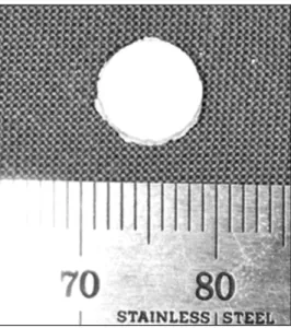
100,000 에피네프린이 함유된 2% lidocaine을 주사하였다. 두피 의 정중부에 시상방향으로 절개를 시행한 후 피부 및 골막을 거상하여 두정골을 노출시킨다(Fig. 2). 토끼 두개골 정중봉합부 에 대칭되도록 두정골상에 trephine bur를 이용하여 두 개의 골결손부를 형성하였다(지름 8 mm, 깊이 약 2 mm). 실험군(우 측 골결손부) 및 대조군(좌측 골결손부)에 각각에 맞게 제작한 복합체를 이식하였다. 골 결손부 상방에 골막을 위치시킨 후 골막 과 근육을 3-0 black silk로 봉합하였다. 술 후 감염 방지를 위해 gentamycin 1 mg/kg (Kookje, Seoul, Korea)을 1일 3회 3일간 근육주사하였다.
수술 4주 후 5마리의 토끼를 희생시킨 후 bur를 이용해 실험군 과 대조군을 포함한 두개골 부위 표본을 채취하였다. 표본은 10%
중성 formalin 용액에 담가 보관했다. 수술 8주 후 나머지 5마리 의 토끼에 대해 4주 후와 같은 과정을 시행하였다.
4. 방사선학적 분석
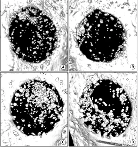
수술 후 4주 및 8주에 가토를 희생시킨 후 채취한 표본은 Explore Locus SP scanner (GE Medical System, Ontario, Canada)를 이용하여 촬영하였다. CT scanner 내부에 있는 turn- table에 표본을 고정한 후 수평 및 수직으로 움직여 위치를 조정하 고 0.05 mm 두께의 절편으로 단층촬영을 시행하였다. 촬영된 이미지는 MicroView software (GE Medical Systems, Ontario, Canada)를 이용하여 삼차원으로 재구성되었다. 수술 시 형성한 결손부가 지름 8 mm, 깊이 약 2 mm 정도의 원통형이 므로 관심영역(ROI, region of interest)은 결손부 크기와 형태에 맞게 100.4 (84×4×3.14×2)로 설정하였다. 각 표본 결손부의 BV (bone volume)를 컴퓨터 소프트웨어를 이용해 분석하였다.
5. 조직학적 검사 및 조직 형태학적 분석
술 후 4주와 8주에 얻은 조직표본들을 10% 중성 포르말린에 고정하고, 5% nitric acid를 이용하여 탈회하고 ethyl alcohol 및 xylene를 이용하여 탈수시킨 후 파라핀 블록에 포매하여 박절 편을 만들어 Masson's trichrome 염색을 시행하였다. 얻어진 조직표본은 광학현미경으로 조직학적 소견을 관찰하였고 디지털 현미경 카메라를 사용하여 광학 현미경상 소견을 디지털 이미지로 취득하였다. 컴퓨터 프로그램인 SigmaScan Pro 5.0 (Systat Software Inc., SJ, CA, USA)을 이용하여 촬영된 이미지상에서 신생골의 면적 비율을 측정하였다.
6. 통계 분석
대조군들과 실험군들간 측정수치 차이의 통계학적 유의성 검사
를 위해 SPSS (SPSS Inc., Chicago, IL, USA)를 이용해 paired
Fig. 3. Microscopic computed to-
mography. (A) Experimental group at 4 weeks. (B) Control group at 4 weeks. (C) Experimental group at 8 weeks. (D) Control group at 8 weeks.Table 1. Micro-computerized tomography analysis
4 weeks 8 weeks
nHA+starch
(control) Silk+nHA+starch
(experiment)
P value
nHA+starch(control) Silk+nHA+starch
(experiment)
P value
Bone volume (mm3) 15.53±7.73 9.68±6.50 0.329 23.47±11.65 17.92±12.50 0.244
nHA, nano-hydroxyapatite.
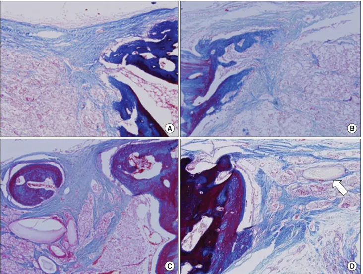
Fig. 4. Histologic section (Masson’s trichrome staining, original magnification ×100). New bone formation of each group was found
at the border area of bone defect. (A) Experimental group at 4 weeks, (B) Control group at 4 weeks, (C) Experimental group at 8 weeks, (D) Control group at 8 weeks. Residual silk fibroin/starch composite were found in this group (arrow).Table 2. Result of measuring area of new bone formation
4 weeks 8 weeks
nHA+starch
(control) Silk+nHA+starch
(experiment)
P value
nHA+starch(control) Silk+nHA+starch
(experiment)
P value
Mean new bone formation (%) 27.20±1.61 23.08±5.17 0.279 36.11±2.74 32.98±5.58 0.139 nHA, nano-hydroxyapatite.지 않고 남은 starch와 SF의 복합체와, 이들의 분해를 위한 면역반 응에 관여하는 lymphocyte와 giant cell 등을 관찰할 수 있었다.
3. 조직형태학적 분석결과
각 시편을 현미경으로 관찰하여 신생골이 형성된 부분의 면적 을 측정한 뒤, 합산 후 실험군과 대조군의 평균치를 각각 구하였다 (Table 2). 4주에서 신생골의 골형성량은 실험군에서 평균 23.08%이며 대조군에서는 평균 27.20%로 대조군에서 높은 양상 을 보였으나 통계적으로 유의하게 높진 않다( P >0.05). 8주에서
는 신생골의 골형성량이 실험군에서 평균 32.98%이고 대조군에 서 평균 36.11%로 대조군에서 높으나 역시 통계적으로 유의할만 한 차이를 보이진 않았다( P >0.05).
고 찰
본 연구에서는 starch와 SF의 복합체를 scaffold로 이용하고
HA를 첨가하여 제조한 골 이식재와 silk는 첨가하지 않고 HA와
starch만을 혼합한 골 이식재를 각각 가토에 이식 후 4주 및
많이 필요하다. 또 흡수되는 동안 염증세포에 의한 염증반응이 필연적으로 생기게 된다[25,26]. 따라서 성공적인 골이식을 위해 서는 염증반응을 최소한으로 하고 신생골 형성을 방해하지 않는 적절한 생분해속도가 필수적이다. 본 연구에서 사용된 SF는 분자 량이 0.5∼1 kDa 정도로 낮아서 빠른 분해양상을 기대할 수 있다[27,28].
본 연구에서는 합성골 이식재 식립 후 골 형성 효과를 평가하기 위해 μ-CT를 이용하였다. 이 방법은 삼차원적으로 시편을 분석 할 수 있고, 관찰하고자 하는 평면을 재구성할 수 있다[29]. 또한 새로이 형성된 골의 부피 및 밀도를 컴퓨터에 수학적으로 계측할 수 있어 보다 정확한 비교가 가능하다. SF과 HA를 재료로 한 복합체가 백서 피하조직 모형에서 12주까지도 분해되지 않는 기 존 연구 결과[30] 등을 고려 시 4주 및 8주 차 표본에서 방사선 사진 상에 중심부에서 보이는 방사선 불투과성 물질은 신생 골조 직과 일부 분해되지 않고 남은 HA가 혼재된 양상으로 생각한다.
실험 결과를 살펴보면, 조직형태학적 분석 및 방사선 분석 모두 에서 silk가 들어가지 않은 대조군의 BV와 평균 신생골 형성량이 silk가 포함된 실험군보다 더 높은 양상을 보인다(Table 1, 2).
이에 대한 원인을 생각해보면 첫째, scaffold로 사용되는 silk의 pore 사이에 starch가 삽입되어 scaffold의 porosity가 감소해 신생골이나 혈관이 자라 들어올 공간이 부족해져 골생성이 원활히 이루어지지 않은 것으로 생각해볼 수 있다. 둘째는 분자량이 낮아 서 체내에서 생분해가 빠르게 이루어져서 골이 자라 들어올 기계 적 지지체 역할을 충분히 수행하지 못했을 것으로 생각한다. 세 번째로는 잔존 이식재 복합체가 8주까지도 완전히 분해되지 않아 신생골 형성을 방해하였을 가능성을 생각해볼 수 있다. 이를 위해 4주와 8주에서 복합체의 흡수율 파악하는 과정이 필요하였을 것 으로 생각한다.
본 연구에서는 아무런 처치도 하지 않은 대조군을 설정하지 않았다. 이전에 이미 보고된 연구 결과들을 참조하여 가토 두개골 에 지름 8 mm의 골 결손부를 형성 후 아무런 처치도 하지 않을 시 골 형성량을 알아본 결과 수술 후 4주가 지났을 때 평균 신생골 형성량은 7.52∼17.11%이고 8주가 지났을 때의 평균 신생골
수 증가를 막기 위해 골 이식재를 사용하지 않은 결손부에 대한 동물실험을 하지 않고 신생골 형성 결과를 이전에 보고된 연구 결과들을 참조하였지만, 아무런 처치를 하지 않은 대조군을 설정 해 함께 동물 실험을 하는 것이 보다 정확한 결과를 산출할 수 있는 방법이라 생각한다. SF를 이용하여 만든 골 이식재를 적용한 이전 연구들을 살펴보면, Kye 등[36]은 전기 방사된 nano silk fiber와 nano-hydroxyapatite를 이용하여 만든 골 이식재를 가 토의 두개골 결손부에 적용하여 아무런 처치를 하지 않은 골 결손부와 비교 시 통계적으로 유의하게 많은 양의 신생골 형성을 확인하였고, Song 등[37]은 실크 단백질과 platelet-rich fibrin (PRF)의 복합체를 이용해 가토의 두개골 결손부에서 신생골 형성 효과가 있음을 보고하였다. 이 외에도 SF에 BMP-2와 nano-hy- droxyapatite를 첨가한 골 이식재 사용 시 골 형성량이 증가하였 으며[38], stem cell을 얇은 필름 형태의 SF와 함께 이식한 경우에 도 신생 골 형성을 관찰 할 수 있었다[39]. 이처럼 silk를 사용하여 만든 골 이식재가 동물실험에서 골 형성에 효과가 있다는 사실이 여러 연구를 통해 입증되었으므로, 향후 추가적인 연구를 통해 silk의 염증반응 정도를 줄이고, 적절한 조성비를 찾는다면 silk는 신생골 형성을 유도하는 골 이식재의 중요하고 유용한 구성 성분이 될 것이라 생각된다.
결 론
본 연구에서는 가토의 두개골에 지름 8 mm, 깊이 약 2 mm의 결손부 2개를 인위적으로 형성한 후 우측에는 실험군인 SF/nHA/
corn starch 복합체를 이식하고, 좌측에는 대조군인 SF를 제외한 nHA/corn starch 복합체를 이식한 후 4주 및 8주에 실험동물을 희생하여 방사선 및 조직형태학적 분석을 시행하여 다음과 같은 결론을 얻었다.
1. Micro-CT 분석에서 4주 및 8주째 대조군의 신생골의 BV 수치가 실험군보다 높았으나 통계적으로 유의한 수준은 아니었다 ( P >0.05).
2. 조직형태학적 분석에서 4주 및 8주째 대조군의 신생골 면적
비율이 실험군보다 높았으나 통계적으로 유의하지 않다( P > 0.05).
위 결과를 바탕으로 SF/nHA/corn starch 복합체가 nHA/
corn starch 복합체에 비하여 통계적으로 유의한 양의 골형성 증대를 야기하지 못함을 확인하였다. 본 연구에서는 보다 다양한 조성 비율에서의 생체 반응 차이는 검토하지 못하였다. 기존의 연구에서 실크 복합체가 일반적으로 골 형성에 유리하다고 보고되 었음을 감안 하면 조성의 차이에 따라서 결과가 달라질 수 있으리 라 생각한다. 이에 대한 추가적인 연구가 검토되어야 할 것이다.
Acknowledgements
이 논문은 농촌진흥청 바이오그린21사업(과제번호:
PJ007170201006)의 지원에 의해 이루어진 것임.
References