Ⅰ. 서 론
조직 결손이 발생하였을 때 연속성을 회복하고 조직을 증대 하기 위하여 다양한 이식재와 대체재가 사용된다. 연조직 결 손부에 사용되는 이식재로는 자가 이식재와 이물성형재를 포 함한 비자가 이식재 등 다양한 재료들이 있으며1,2)
, 결손된 골의
재건을 위한 골 이식재로서 자가골, 동종골, 이종골, 합성골 또는 이들을 혼합하여 사용한다3-6)
. 이러한 이식재 중에서 자가
이식재가 예후가 좋은 것으로 인정되고 있으나 공여부가 한정 되어 있고 부가적인 수술이 필요한 단점이 있다7,8). 반면에 비자
가 이식재는 면역조직적합성과 비경제성 등의 문제점을 가지고 있다9,10)
.
특히 골 이식술의 대용품으로 합성골이나 이종골 등이 연구 개발되어 왔으나 골전도능만을 제공하는 경우가 많을 뿐더러 흡수되지 않고 남아 있는 경우도 있어 이로 인한 감염이나 염 증 반응 때문에 골 유합의 지연과 같은 합병증을 일으킬 수 있
다11)
. 골 치유를 촉진하는 골 생성인자들에 관한 연구와 함께
조직공학의 발달은 이러한 문제점들을 해결할 수 있는 다양한 골 대체재와 조직 활성 인자 등의 사용 가능성을 제시하고 있 으나 아직 이상적인 골간 대체재의 개발이 진행되고 있다12,13)
.
무세포성 진피 기질(AlloDerm�
)은 사체의 피부를 채취하여
유 선 열501-757
광주광역시 동구 학동5
번지전남대학교 치의학전문대학원 구강악안면외과학교실 Sun-Youl Ryu
Dept. of OMFS, School of Dentistry, Chonnam National University 5 Hak-dong, Dong-Ku, Gwangju, 501-757, Korea
Tel: 82-62-220-5439 Fax: 82-62-228-8712 E-mail: [email protected]
가토에서 조직 재생 이식재로서 무세포성 진피 기질의 효용
최종학∙류재영∙유선열
전남대학교 치의학전문대학원 구강악안면외과학교실, 전남대학교 치의학연구소
Abstract (J. Kor. Oral Maxillofac. Surg. 2008;34:220-229)
USEFULNESS OF ACELLULAR DERMAL MATRIX GRAFT ON THE TISSUE REGENERATION IN RABBITS
Jong-Hak Choi, Jae-Young Ryu, Sun-Youl Ryu Department of Oral and Maxillofacial Surgery, School of Dentistry,
Dental Science Research Institute, Chonnam National University
Purpose: The present study was aimed to examine the effect of acellular dermal matrix (AlloDerm
�) grafted to the experimental tissue defect on tis- sue regeneration.
Materials and Methods: Male albino rabbits were used. Soft tissue defects were prepared in the external abdominal oblique muscle. The animals were then divided into 3 groups by the graft material used: no graft, autogenous dermis graft, and AlloDerm
�graft. The healing sites were histologi- cally examined at weeks 4 and 8 after the graft. In another series, critical sized defects with 8-㎜ diameter were prepared in the right and left iliac bones. The animals were then divided into 5 groups: no graft, grafted with autogenous iliac bone, AlloDerm
�graft, AlloDerm
�graft impregnated with rhBMP-2, and AlloDerm
�graft with rhTGF-�
1. The healing sites of bone defect were investigated with radiologic densitometry and histological evaluation at weeks 4 and 8 after the graft.
Results: In the soft tissue defect, normal healing was seen in the group of no graft. Inflammatory cells and foreign body reactions were observed in the group of autogenous dermis graft, and the migration of fibroblasts and the formation of vessels into the collagen fibers were observed in the group of AlloDerm
�graft. In the bone defect, the site of bone defect was healed by fibrous tissues in the group of no graft. The marked radiopacity and good regeneration were seen in the group of autogenous bone graft. There remained the traces of AlloDerm
�with no satisfactory results in the group of AlloDerm
�graft. In the groups of the AlloDerm
�graft with rhBMP-2 or rhTGF-�
1, there were numerous osteoblasts in the boundary of the adjacent bone which was closely approximated to the AlloDerm
�with regeneration features. However, the fibrous capsule also remained as in the group of AlloDerm
�graft, which separated the AlloDerm
�and the adjacent bone.
Conclusions: These results suggest that AlloDerm
�can be useful to substitute the autogenous dermis in the soft tissue defect. However, it may not be useful as a bone graft material or a carrier, since the bone defect was not completely healed by the bony tissue, regardless of the presence of osteogenic factors like rhBMP-2 or rhTGF-�
1.
Key words: Acellular dermal matrix (AlloDerm
�), Experimental tissue defect, Tissue regeneration, Graft material, Carrier
세포성 면역반응의 항원 대상인 주요 조직적합성 복합체 항원
I형과 II형을 가지는 표피와 진피 기질 내의 세포들을 제거하고
급속 동결 건조시켜 진피층의 3차원 구조를 유지한 인체의 동 종진피로서 수혜부 조직과의 친화성이 좋아 진피 기질 내로 섬유세포와 신생 혈관의 성장이 빠른 것으로 알려져 있다14,15).
따라서 비성형술이나 구순증대술 등 악안면 성형 영역을 포함 한 여러 영역에 이용범위가 증가하고 있다16,17). 골 결손부에
AlloDerm
�을 이식한 연구에서 박 등18)은 인접 골로부터AlloDerm
�내로 골 재생이 일어난다고 하였고, Novaes와Souza
19)는 AlloDerm�이 조직유도 재생술에 차단막으로 사용될 수 있다고 하였다. 이러한 관점에서 골 결손부에 AlloDerm�을 이식할 경우 주변 연조직의 내증식은 차단되고 인접 골 절단 면으로부터 신생 골조직이 내성장되어 들어오면서 골재생을 유도할 수 있는지 구명할 필요가 있다.골의 재형성은 호르몬과 골 생성인자에 의해 조절되는 일련 의 과정이다. 골 생성인자로서 인슐린양 성장인자(insulin-like
growth factor), 섬유모세포성장인자(fibroblast growth factor), 혈소
판유래성장인자(platelet-derived growth factor), 골형성 단백(bonemorphogenetic protein, BMP), 및 형질전환 성장인자 베타(trans- forming growth factor �, TGF-�) 등이 있으며, 이들은 차단막이
나 골전도 물질의 사용과는 달리 치유 과정에서 조직 재생에 필수적인 신생 혈관의 형성, 세포의 화학주성 및 분열 촉진 등 을 조절한다20-22). 골 생성인자 사용시에는 운반체(carrier)에 따
라 결과가 변할 수 있는 것으로 알려져 있으며23)특히 운반체로 서 섬유성 콜라겐이 추천되고 있다24). 따라서 이러한 운반체로
서 AlloDerm�의 사용 가능성에 대하여 밝힐 필요가 있다.본 연구는 무세포성 진피 기질이 조직 재생을 위한 이식재로
서 또는 운반체로서 사용 적합한가 구명하고자 하였다. 가토 에서 실험적으로 유발한 조직 결손부에 이식한 AlloDerm�의 치유 양상 및 조직 재생 효과를 알아보고, BMP와 TGF-�를 침 습시킨 AlloDerm�의 골 재생 효과를 비교함으로써 조직 재생 을 위한 이식재로서 또는 운반체로서 AlloDerm�의 사용 가능 성을 조사하였다.
Ⅱ. 실험재료 및 방법 1. 실험재료
실험재료는 전남대학교 의과대학 동물사에서 동일한 조건 에서 사육한 체중 3.0 ㎏ 정도의 건강한 잡종 가토 32 마리를 사 용하였다. 실험 대상으로는 24 군데의 복부와 40 개의 장골을 택하였다. 연조직 및 골 결손부에 이식한 재료로는 실험 중에 채취한 자가진피, 자가장골 및 AlloDerm�
(LifeCell Corp., Branchburg, NJ, USA)(Fig. 1)을 사용하였으며, 골 결손부에 AlloDerm
�과 함께 사용한 골 생성인자로는 rhBMP-2와 rhTGF-�1
(R&D Systems Inc., Minneapolis, MN, USA)을 사용하였다.
2. 실험방법
가토를 0.2 ㎖/㎏의 tiletamine/zolazepam(Zoletil�
, Virbac, France)
과 0.1 ㎖/㎏의 xylazine hydrochloride(Rompun�, Bayer, Korea)를 혼
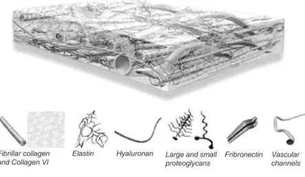
합한 용액을 근육주사하여 마취하였다. 연조직 결손부 실험을 위해 가토를 앙와위로 고정한 다음, 복부와 좌우측 서혜부의 털을 제거하고 베타딘으로 소독하였다. 지혈을 위해 복부와Fig. 1. AlloDerm
�is an acellular dermal matrix derived from donated human skin tissue. The human donor tissue undergoes a multi-step proprietary process that removes both epidermis and the cells responsible for immune response and graft rejection. What remains is a matrix of framework of the natural biological components, ready to enable the body to mount its own tissue regeneration process. [Cited from the webpage of LifeCell corporation].
Fibrillar collagen and Collagen VI
Elastin Hyaluronan Large and small proteoglycans
Fribronectin Vascular channels
서혜부 주변에 1:100,000 에피네프린 함유 2% 리도케인을 국소 주입하였다. 자가진피는 가토의 대퇴 곧은근(rectus femoris
muscle)과 배바깥 경사근(external abdominal oblique muscle) 상방
의 표피를 제거한 후 진피를 8 × 8 ㎜ 크기로 채취하였다. 가토 복부에 정중 절개를 가하여 배바깥 경사근의 근막을 노출시키 고 조직을 박리한 다음, 배바깥 경사근에 8 × 8 ㎜ 크기의 결손 부를 형성하였다. 결손부에 아무 것도 이식하지 않은 S-1군(n=8), 서혜부에서 채취한 자가 진피를 이식한 S-2군(n=8), 그리
고 생리식염수에 30분간 침습시킨 AlloDerm�만을 이식한 S-3 군(n=8)으로 분류하였다. 결손부에 이식편들을 위치시킨 다음,5-0 Vicryl과 5-0 Nylon을 이용하여 층별 봉합하였다. 각 이식편
은 다시 5-0 silk를 이용하여 표피를 관통하는 봉합을 시행하여 고정하였으며, 수술 후 이식편 절취 시 이 봉합을 경계를 확인 하는 표지자로 이용하였다.골 결손부 실험을 위해 가토를 복와위로 위치시키고 둔부의 털을 제거한 다음 베타딘으로 소독하였다. 지혈을 위해 장골 능 주변에 1:100,000 에피네프린 함유 2% 리도케인을 국소 주 입하였다. 장골능 부위를 촉진하고 상방의 피부에 길이 5 ㎝ 정 도의 절개를 가한 후 장골능 부위를 노출시켰다. 식염수를 주 수하면서 trephine bur를 이용하여 내외측 장골을 관통하는 직 경 8 ㎜의 원형 골 결손부를 형성하였다. 골 결손부에 아무 것 도 이식하지 않은 B-1군(n=8), 반대측 자가장골편을 분쇄하여 이식한 B-2군(n=8), 생리식염수에 30분간 침습시킨 AlloDerm� 만을 이식한 B-3군(n=8), 10 ㎍/㎖ 농도의 rhBMP-2가 섞인 생리 식염수에 AlloDerm�을 침습시켜 이식한 B-4군(n=8), 그리고 10
㎍/㎖ 농도의 rhTGF-�1이 섞인 생리식염수에 AlloDerm�을 침 습시켜 이식한 B-5군(n=8)으로 분류하였다. 골막이 골 생성에 미치는 영향을 줄이고자 골 결손부 주변의 골막을 제거하였으 며, 5-0 Vicryl과 5-0 Nylon을 이용하여 층별 봉합하였다. 실험동 물에서 감염 방지를 위하여 술전과 술후 5일 동안 항생제를 투 여하였다.
이식 후 4주 및 8주에 실험동물을 희생시켰다. 희생한 실험동 물의 장골을 채취하여 구내 방사선촬영기(GD-70, Genoray,
Korea)를 이용하여 관전압 70 kVp, 관전류 3 mA, 노출시간 0.15
초로 노출조건을 동일하게 하여 방사선사진을 촬영하였다. 방 사선사진을 이용하여 흑화도(Densitometry)를 측정하였다. 방 사선사진 촬영 후 이식편과 주위 정상 장골을 포함한 조직시 편을 절취하였으며, 복부에서도 이식편과 주위 정상 연조직을 포함한 조직시편을 절취하였다. 절취한 조직편을 10% 포르마 린 용액에 고정한 다음 파라핀에 포매하고 5 μm 두께의 절편을
제작한 다음, H-E 염색하고 광학현미경으로 관찰하였다.3. 통계학적 분석
각 실험군에서 방사선 흑화도를 비교하기 위하여 통계분석 프로그램인 SPSS(version 12.0, Datasolution, Korea)를 이용하였으 며, 일원배치 분산분석을 시행하였다.
Ⅲ. 결 과 1. 육안적 소견
실험동물의 복부 또는 장골 상방의 모든 수술부위에서 뚜렷 한 염증 소견이나 열개 또는 기타 이식 부위와 관련된 합병증 은 관찰되지 않았다. 장골 조직시편에서 육안적 소견으로는 아무 것도 이식하지 않은 군에서만 골 결손부의 섬유성 치유 양상이 관찰되었으며, AlloDerm�만을 사용한 경우에는 일부 골 결손부에서 AlloDerm�의 형태를 확인할 수 있었다. 그 외의 골 결손부에서는 정상적인 치유를 보였으며 각 실험군 간에 별다른 차이를 관찰할 수 없었다.
2. 방사선학적 소견
이식 4주와 8주 후에 희생한 모든 실험군 가운데 자가장골을 이식한 군에서 방사선불투과상이 가장 높게 나타났다(Fig. 2).
아무 것도 이식하지 않은 군과 AlloDerm�만을 이식한 군에서 는 방사선불투과상이 가장 낮게 나타났으며, 실제 방사선사진 상 골 결손부가 거의 그대로 유지되었다(Fig. 3, 4). AlloDerm�에
rhBMP와 rhTGF-�를 침습시켜 이식한 군에서는 중간 정도의
방사선불투과상을 나타냈다(Fig. 5, 6). 자가장골을 이식한 군 에서 다른 실험군에 비해 방사선불투과상의 정도가 유의하게 높았다(p<0.05)(Table 1).3. 조직학적 소견
가. 연조직 결손부
S-1군에서는 이식 4주와 8주 모두 정상적인 치유가 나타났다 (Fig. 7-A, B). S-2군에서는 이식 4주에 자가진피 이식편에서 각
화된 상피로 이장되어 있는 상피내 낭종(epidermal inclusioncyst)이 관찰되었다(Fig. 8-A). 이식 8주에는 자가진피 이식편에
서 상피내 낭종과 함께 낭종의 내강에서는 각질이 관찰되며, 이장된 낭종 벽에서는 모낭이 관찰되었다(Fig. 8-B). S-3군에서 는 이식 4주에 AlloDerm�의 주변에서 염증세포가 관찰되었으 며, AlloDerm�에서 콜라겐 섬유의 주변부로 섬유아세포의 이 주가 관찰되었다(Fig. 9-A). 이식 8주에는 염증 반응이 감소하 면서 AlloDerm�의 콜라겐 섬유 내에서 신생 혈관 및 섬유아세 포의 침윤이 관찰되었다(Fig. 9-B).나. 골 결손부
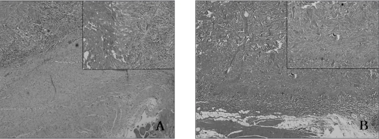
B-1군에서는 이식 4주에 골 결손부가 빈 공간으로 유지되고
있었고, 골 절단면에서 골아세포와 신생골이 관찰되나 결손부 내부에서는 관찰할 만한 조직이 없었다(Fig. 10-A). 이식 8주에 는 골 결손부가 신생 골조직으로 채워지지 못하고 대신 섬유 성 조직이 일부 위치하고 있으며, 빈 공간으로 남아 있는 부분 도 관찰되었다(Fig. 10-B).Fig. 2. Radiographs of the B-2 group at 4 (A) and 8 (B) weeks after the graft. The marked radiopacity of the bone defect is observed at weeks 4 and 8.
Fig. 3. Radiographs of the B-1 group at 4 (A) and 8 (B) weeks after the graft. The radiolucency of the bone defect is observed at weeks 4 and 8.
Fig. 4. Radiographs of the B-3 group at 4 (A) and 8 (B) weeks after the graft. The marked radiolucency of the bone defect is observed at weeks 4 and 8.
Fig. 5. Radiographs of the B-4 group at 4 (A) and 8 (B) weeks after the graft. A, The radiolucency in the bone defect is observed. B, The more increased radiopacity is observed at week 8 than at week 4.
Fig. 6. Radiographs of the B-5 group at 4 (A) and 8 (B)
weeks after the graft. A, The radiolucency of the bone
defect is observed. B, The radiopacity is more increased at
B-2군에서는 이식 4주에 골 결손부에 이식한 자가골과 조혈
세포를 포함하는 골수 조직이 관찰되었다. 이식한 자가골은 골아세포로 둘러싸여 활발한 신생골 형성을 보였다(Fig. 11-A).이식 8주에 골 결손부는 4주에 비해 신생골 형성이 증가하여 이식한 자가골과 신생골이 혼재되어 관찰되었으며 이식한 골
조직과 신생골의 구분이 어려웠다. 골수에서는 대부분 조혈세 포로 구성된 적골수(hemopoietic marrow)를 관찰할 수 있었다
(Fig. 11-B).
B-3군에서는 이식 4주에 AlloDerm
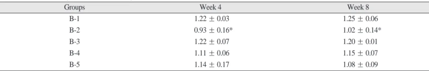
�이 골 결손부에서 잘 유지 되고 있었으며, AlloDerm�을 둘러싸고 있는 섬유성 피막과 인Table 1.
Summary of densitometryGroups Week 4 Week 8
B-1 1.22 ± 0.03 1.25 ± 0.06
B-2 0.93 ± 0.16* 1.02 ± 0.14*
B-3 1.22 ± 0.07 1.20 ± 0.01
B-4 1.11 ± 0.06 1.15 ± 0.07
B-5 1.14 ± 0.17 1.08 ± 0.09
The values are expressed as the mean
± SD. * p<0.05, compared with the B-1.Fig. 7. Photomicrographs of the S-1 group at 4 (A) and 8 (B) weeks after the graft. A, The adjacent tis- sue is closely attached to the muscle tissue (H-E stain, original magnification × 40). B, There is no traces of the tissue defect (H-E stain, original magnification × 40).
Fig. 8. Photomicrographs of the S-2 group at 4 (A) and 8 (B) weeks after the graft. A, The epidermal
inclusion cyst lined by the keratinized epithelia is observed (H-E stain, original magnification × 40 and
inset × 100). B, The keratins are observed in the lumen of the cyst and the hair follicles noted in the
lining wall (H-E stain, original magnification × 40 and inset × 100).
Fig. 9. Photomicrographs of the S-3 group at 4 (A) and 8 (B) weeks after the graft. A, The migration of the fibroblasts into the periphery of collagen fibers is observed (H-E stain, original magnification × 40 and inset × 100). B, The inflammatory cells are decreased and the vascular ingrowth is observed at the collagen fibers of the AlloDerm
�(H-E stain, original magnification × 40 and inset × 100).
Fig. 10. Photomicrographs of the B-1 group at 4 (A) and 8 (B) weeks after the graft. A, The defect site remain empty. The osteoblasts and the new bones are seen at the osteostomy sites of the bone defect (H-E stain, original magnification × 40). B, The fibrous tissue is observed in the defect site (H-E stain, original magnification × 40).
Fig. 11. Photomicrographs of the B-2 group at 4 (A) and 8 (B) weeks after the graft. A, The grafted
autogenous bone and bone marrow containing the hematopoietic cells can be seen in the defect site (H-
E stain, original magnification × 40). B, The increased bone regeneration and the hemopoietic marrow
can be seen in the defect site (H-E stain, original magnification × 40).
Fig. 12. Photomicrographs of the B-3 group at 4 (A) and 8 (B) weeks after the graft. A, The AlloDerm
�is well maintained at the defect site and enclosed by the fibrous capsule (H-E stain, original magnification
× 40). B, The fibrous capsule separate the AlloDerm
�and the adjacent bone (H-E stain, original magnification × 40).
Fig. 13. Photomicrographs of the B-4 group at 4 (A) and 8 (B) weeks after the graft. A, The adjacent bone tissues are closely attached to the AlloDerm
�. There are numerous osteoblasts and blood vessels in the boundary of the adjacent bone (H-E stain, original magnification × 40 and inset × 100). B, There is an active bone formation in the adjacent bone tissue. The adjacent bone tissue is also mature (H-E stain, original magnification × 40 and inset × 100).
Fig. 14. Photomicrographs of the B-5 group at 4 (A) and 8 (B) weeks after the graft. A, The adjacent
tissues are closely attached to the AlloDerm
�. There are numerous osteoblasts in the boundary of
adjacent bone (H-E stain, original magnification × 40 and inset × 100). B, The adjacent tissue is also
closely attached to the AlloDerm
�. There is an active bone regeneration in the adjacent tissue (H-E stain,
original magnification × 40 and inset × 100).
접 골조직 사이에서는 염증 반응이 관찰되지 않고 경계선 없 이 잘 접촉되어 있었다(Fig. 12-A). 이식 8주에 골 결손부 주변의 골조직은 성숙한 층판골 양상을 보였고, AlloDerm�주위의 섬 유성 피막도 성숙되어 인접 골조직과 분리되는 경계선을 보였 다(Fig. 12-B).
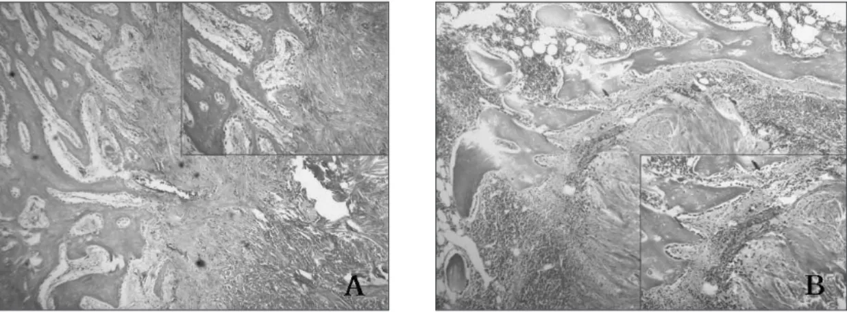
B-4군에서는 이식 4주에 골 결손부에 위치한 AlloDerm
�과 인 접 골조직은 밀접하게 잘 연결되어 있었고 인접 골조직이AlloDerm
�주변부로 이주되는 양상을 보였다(Fig. 13-A). 이식 8 주에는 4주에 비해 AlloDerm�주위의 섬유성 피막이 성숙되어B-3군에서와 같이 경계선을 이루는 양상이 관찰되었으나, 인
접 골조직이 성숙되고 많은 골아세포가 관찰되어 활발한 골 형성 소견을 보였다(Fig. 13-B).골 결손부에 rhTGF-�1을 침습시킨 AlloDerm�을 이식한 B-5 군에서는 이식 4주에 B-4군과 유사하게 AlloDerm�과 인접 골 조직이 밀접해 있고 일부에서는 염증반응이 관찰되었다(Fig.
14-A). 이식 8주에는 4주에 비해 AlloDerm
�주위에 적은 양의 섬유성 피막만이 남아 있었으며 인접 골조직은 AlloDerm�주 변에 밀접해 있으면서 활발한 골 형성 양상을 보였으나, B-3군 에서와 같이 AlloDerm�과 인접 골조직이 구분되어 있었다(Fig.14-B).
Ⅳ. 고 찰
조직 결손의 회복을 촉진하기 위해 여러 가지 이식재가 이용 되고 있으며, 이때 적절한 대체재를 사용하였는지 여부는 치 유의 성공 또는 실패에 결정적인 역할을 하게 된다25)
. 자가 이
식재의 경우 공여부의 수술로 인하여 반흔과 부가적인 결손을 야기하게 되고, 비자가 이식재는 면역조직적합성과 비경제성 등의 문제점을 가지고 있다9,10).
비교적 근래에 상용화된 인체 의 동종진피인 AlloDerm�은 세포성 면역반응을 제거한 무세포 성 진피 기질로서 점차 그 이용범위가 확대되고 있다16,17). 본 연
구에서는 가토에서 실험적으로 유발한 연조직 결손부와 골 결 손부에서 자가진피와 AlloDerm�을 이식했을 때 조직 재생 효 과를 비교함으로써 자가진피의 대체재로서 AlloDerm�의 사용 가능성을 조사하였다. 연조직 결손부에서 자가진피는 표피가 잔존하는 경우 상피내 낭종을 형성하였고 공여부의 반흔과 부 가적인 결손을 야기하였다. 그러나 AlloDerm�이식군에서는 활발한 섬유아세포의 이주 및 신생 혈관의 형성을 관찰할 수 있었다. 이는 연조직 내에 AlloDerm�을 이식하면 AlloDerm�내 로 신생 혈관과 섬유아세포가 성장하여 결손부 치유가 진행된 다고 알려진 기존의 여러 연구 결과14,15,26,27)와 일치하였다. 또한AlloDerm
�이식편에서 다핵거대세포와 같은 이물반응을 유발하는 세포가 관찰되지 않았다. 따라서 AlloDerm�은 연조직 결 손의 회복에 사용할 수 있는 생체적합성이 우수한 이식재라고 생각된다.
박 등18)에 의하면 AlloDerm�을 골 결손부에 이식하였을 때
AlloDerm
�내로 골 재생이 이루어진다고 하였다. 그러나 본 연구에서는 골 결손부에 AlloDerm�을 이식한 경우 AlloDerm�내
에서 골 재생 소견은 관찰되지 않았다. 방사선학적 소견에서 도 방사선불투과상이 가장 낮았고 아무 것도 이식하지 않은 군과 비슷하게 낮은 흑화도를 보였다. AlloDerm�만을 이식한 경우 AlloDerm�주위에 섬유성 피막이 둘러싸고 있어 인접 골 조직이 AlloDerm�내로 침투하기 어려울 것이라 보인다. 이는 인접 골조직의 골아세포가 AlloDerm�의 주성분인 콜라겐 섬유 내로 이주하지 못하기 때문이라 생각된다. 골 조직 대신에 사 람 치아의 백악질 상방에 AlloDerm�을 이식한 연구28)에서 백악 질과 AlloDerm�간의 부착 또는 백악질의 신생 소견은 관찰되 지 않았다. 결국 AlloDerm�과 골조직 간의 완전한 혼재의 가능 성은 적을 것으로 생각된다. 이상적인 이식재의 조건은 이식 재 내로 수혜부 조직의 이주와 신생 혈관이 발생하여 주변의 숙주 조직과 혼재되고 결국 잠재적인 치환이 발생하는 것이라 고 할 수 있다17)
.
본 연구에서 골 결손부에 아무 것도 이식하지 않은 경우 섬 유 조직이 결손부로 함입되어 비정상적인 치유 양상을 나타냈 다. 조직학적 소견에서도 결손부에 정상적인 골 재생 소견은 관찰되지 않았으며 빈 공간으로 남아 있거나 부분적으로 섬유 성 조직이 관찰되었다. 이는 이전 연구들29,30)에서 언급된 것처 럼 형성된 골 결손부의 크기가 결손임계치수임을 의미한다 하 겠다. 한편 골 결손부에 자가장골을 이식한 경우에 방사선사 진상에서 가장 높은 흑화도를 보여 광화작용(mineralization)이 많이 진행되었음을 시사하였다. 자가골은 현재까지 알려진 골 결손부를 회복하기 위한 재료 중 가장 우수한 재료이다29,31)
.
골 이식재와 관련하여 최근에는 조직 공학을 통한 접근이 시 도되고 있다. 골 재생은 호르몬과 골 생성인자에 의해 조절되 는 일련의 과정이므로 골형성 능력을 돕기 위하여 각종 골 생 성인자를 개발하여 임상에 적용하기 위한 연구가 진행되고 있 다. BMP는 골 기질 내에서 높은 수치로 발견되는 골 유도 물질 로서, 골아세포의 분화 및 증식에 작용하여 신생골 형성을 유 도하는 것으로 알려져 있다32,33)
. 특히 BMP는 골수 내에서 유래
된 줄기세포 외에도 전지방세포(preadipocyte)나 섬유아세포, 근세포 등의 세포로 분화된 경우에도 조골세포로의 역분화를 유도할 수 있을 만큼 골 형성에 큰 효과를 갖는 물질이다34-37). 한
편 TGF-�는 골막의 간엽세포로부터 골아세포로의 분화를 유 도하며, 동시에 이 세포들의 증식과 골에 특징적인 세포 기질 단백의 합성을 촉진하는 작용이 있다고 알려져 있다38). 그러나 TGF-�는 고농도에서는 세포 증식을 억제하고, 저농도에서는
촉진시키는 상반된 작용이 있다22). 본 연구에서는 AlloDerm
�에rhBMP와 rhTGF-�를 침습시켜 이식한 경우 치유 소견에서
AlloDerm
�만을 사용하였을 때에 비해 유의한 차이를 관찰할수 없었다.
이와 같이 골 이식재와 골 생성인자들을 혼합 이식하려는 시 도와 함께, 골 생성인자의 운반체에 대해서도 연구가 진행되
고 있다39-41)
. AlloDerm
�은 주위에서 섬유 조직이 함입되는 것을막는 일종의 차단막 역할을 하는 것으로 보고되었으며19)
, 그와
더불어 골 생성인자가 방출된다면 골형성과 골전도에 더욱 도 움이 될 수 있을 것으로 생각된다. 특히 BMP와 같은 골 생성인자는 운반체에 따라 그 효과에 차이가 나타나는 것으로 알려
져 있다23)
. 골 생성인자의 효과적인 운반체로서 섬유성 콜라겐
이 추천되고 있으며, 섬유성 콜라겐은 BMP를 서서히 유리시 켜 보다 지속적인 골형성을 유도한다24)
. AlloDerm
�은 콜라겐 성분으로 되어 있으므로 골 생성인자의 운반체 역할을 수행할 수 있을 것으로 생각되어, AlloDerm�에 골 생성인자를 침습시 킨 경우의 골 재생 효과를 비교하였다.본 연구에서 rhBMP와 rhTGF-�를 침습시킨 AlloDerm�을 이 식한 경우 AlloDerm�만을 이식한 경우에 비해 이식재 주변의 섬유성 피막은 감소하였고, 인접 골조직과 AlloDerm�사이가 더욱 근접해 있으면서 인접 골조직에서 많은 골아세포가 보이 고 보다 활발한 골형성 소견이 관찰되었다. 그러나 AlloDerm� 만을 이식한 경우에서와 같이 AlloDerm�과 인접 골조직이 섬 유성 피막으로 구분되어 있었으며 AlloDerm�내에서 골 재생 소견을 관찰할 수 없어 골 생성인자와 골 재생 소견 간의 직접 적인 연관성은 확인하기 어려웠다. 골 결손부에 AlloDerm�만 을 이식한 경우에는 결손부에 AlloDerm�이 남아 있어 만족할 만한 골 재생 양상을 관찰할 수 없었다. AlloDerm�과 rhBMP-2 또는 rhTGF-�1를 함께 이식한 경우에도 AlloDerm�과 인접 골 조직이 밀접하게 접근해 있었지만 골 결손부가 완전한 골조직 으로 치유되지는 않았다. 이러한 결과는 골 재생을 위한 이식 재로서 또는 운반체로서 AlloDerm�의 사용이 적절하지 않음을 시사한다 하겠다. AlloDerm�이 골 생성인자를 방출하는 운반 체 역할을 수행할 가능성에 대해서는 추후 더 구명되어야 할 것으로 생각된다.
Ⅴ. 결 론
본 연구는 무세포성 진피 기질(AlloDerm�
)이 조직 재생을 위
한 이식재로서 또는 운반체로서 사용 적합한가 구명하고자 하 였다. 연조직 결손부에 아무 것도 이식하지 않은 경우 정상적 인 자가조직으로 치유되었다. 자가진피를 이식한 군에서는 상 피내 낭종이나 모낭 형성이 관찰되었으며, AlloDerm�을 이식 한 군에서는 AlloDerm�의 콜라겐 섬유 내에서 신생 혈관 및 섬 유아세포의 침윤이 관찰되었다. 골 결손부에 아무 것도 이식 하지 않은 군에서는 섬유성 조직으로 치유되었고, 자가장골을 이식한 군에서 가장 높은 방사선 불투과성과 우수한 골 재생 양상을 관찰할 수 있었다. 반면에 AlloDerm�만을 이식한 군에 서는 골 결손부에 AlloDerm�의 흔적이 남아 있어 만족할 만한 골 재생 양상을 관찰할 수 없었다. rhBMP 또는 rhTGF-�를 침 습시킨 AlloDerm�이식군에서는 인접 골조직이 이식재와 밀접 해 있으면서 더 활발한 골아세포들을 관찰할 수 있었으나AlloDerm
�만을 이식한 군에서와 같이 AlloDerm�과 인접 골조 직이 섬유성 피막으로 구분되어 나타났다. 이상의 결과는AlloDerm
�이 연조직 결손부에서는 자가진피의 대체재로 사용가능한 반면, 골 결손부에서는 BMP-2와 TGF-�1과 같은 골 생 성인자의 사용 여부에 무관하게 골 재생을 위한 이식재로서 또는 운반체로서 적절하지 않음을 시사하였다.
참고문헌