Abstract (J. Kor. Oral Maxillofac. Surg. 2009;35:397-402)
Ⅰ.
서 론Bisphosphonates(BPs)는 파골세포의 분화 및 증식을 억제 함으로써 골다공증, Paget’s disease, fibrous dysplasia, can- cer-induced bone disease, osteogenesis imperfecta와 같은 질 병에서 골흡수를 억제하기 위한 치료제로 널리 사용되고 있다
1-5). 그러나 최근 BPs를 장기 복용한 환자들에서 BPs 유
도 악골괴사(Bisphosphonate-related osteonecrosis of the jaws : BRONJ)와 같은 심각한 합병증이 발견되고 있으며, 이는 Marx와 Stern에 의해 2003년 처음 서술된 이래
6)현재 구강 악안면외과 영역의 새로운 질환으로 대두되고 있는 실정 이다
7).
BPs는 phosphate에 연 결 된 산 소 가 탄 소 로 대 체 된 pyrophosphate analogue 계통의 합성물로 hydroxyapatite crystals에 대한 친화성이 매우 크며
8), 탄소와 연결된 측쇄 구조의 종류에 따라 골흡수를 저지하는 능력에 차이가 있 다. 예를 들어, Alendronate(Aln, 4-amino-1-hydroxy-1 phos- phonobutyl phosphonic acid)와 Pamidronate(Pam, 3-amino-1- hydroxy-1-phosphonoprophyl phosphonic acid) 같은 질소를 함유한 BPs는 질소를 함유하지 않은 etidronate나 clo- dronate와 같은 단순한 BPs에 비해 100배 이상의 효과를 가
류 동 목130-701
서울특별시 동대문구 회기동1
번지경희대학교 치의학전문대학원 부속병원 구강악안면외과 Dong-Mok Ryu
Hoegi 1, Dongdaemunku, Seoul, 130-701, Korea Dept. of OMFS, KyungHee University Dental School Tel: 82-2-958-9440
E-mail: [email protected]
Alendronate와 Pamidronate가 인간 골수유래 간엽줄기세포의 증식과 알칼리성 인산분해효소 활성에 미치는 영향
김영란∙류동목∙권용대∙윤영필
1경희대학교 치의학전문대학원 구강악안면외과학 교실,
1경희대학교 치의학전문대학원 구강생물학 교실
EFFECTS OF ALENDRONATE AND PAMIDRONATE ON THE PROLIFERATION AND THE ALKALINE PHOSPHATASE ACTIVITY OF HUMAN BONE MARROW DERIVED MESENCHYMAL STEM CELLS
Young Ran Kim, Dong-Mok Ryu, Yong-Dae Kwon, Yong Pil Yun
1Department of Oral and Maxillofacial Surgery,
1Department of oral biology and institute of oral biology Kyung Hee University Dental School, Seoul, Korea
The purpose of this study is to investigate the effects of alendronate and pamidronate on proliferation and the alkaline phosphatase activity of human bone marrow derived mesenchymal stem cells and to relate the results with bisphosphonate related osteonecrosis of the jaw(BRONJ).
With the consent of patients with no systemic disease and undergoing iliac bone graft, cancellous bone was collected to obtain human bone marrow derived mesenchymal stem cells through cell culture. 96 well plate were prepared with a concentration of 10
4cell/ well. Alendronate and pamidronate were added to each well with the concentration of 10
-6M, 10
-8M and 10
-10M, respectively. Then proliferation capacity of each well was evaluated with the cell counting kit. 24 well plates were prepared with a concentration of 10
5cell/ml/well and with the bone supplement, alendronate and pamidronate were added with the concentration of 10
-6M, 10
-8M and 10
-10M, respectively on each plate. The plates were cultured for either 24 or 72 hours. Then the cells were sonicated to measure the alkaline phosphatase activity and protein assay was done to standardize the data for analysis.
As the concentration of alendronate or pamidronate added to the culture increased, the proliferation capacity of the cells decreased. However, no sta- tistical significance was found between the group with 10
-10M of bisphophonate and the control group. Pamidronate was not capable of increasing the alkaline phosphatase activity in all trials. However, alkaline phosphatase activity increased with 24 hours of 10
-8M of alendronate treatment and with 48 hours of 10
-10M of alendronate treatment.
Cell toxicity increased as the bisphosphonate concentration increased. This seems to be associated with the long half life of bisphosphonate, result- ing in high concentration of bisphosphonate in the jaw and thus displaying delayed healing after surgical procedures. Alendronate has shown to increase the alkaline phophatase activity of human bone marrow derived mesenchymal stem cells. However, this data is insufficient to conclude that alendronate facilitates the differentiation of human bone marrow derived mesenchymal stem cells. Further studies on DNA level and animal studies are required to support these results.
Key words: Bisphosphonate, Cell proliferation, Alkaline phosphatase, Osteonecrosis, Mesenchymal stem cells
[원고접수일 2009. 9. 21 / 1차수정일 2009. 10. 6 / 2차수정일 2009. 10. 15 / 게재확정일 2009. 10. 30]
지며 그에 따른 부작용의 빈도도 높은 것으로 알려져 있다
9,10). BPs는 주로 파골세포에 직접적, 간접적으로 작용하여 골 흡수를 억제하는 것으로 보인다. 즉, 파골세포의 증식과 전 구체 생성을 억제하여
11,12)파골세포의 수를 감소시키며 파 골세포의 ruffled border를 불안정하게 함으로써 골흡수 과 정을 저해시킨다
10). 또한 질소를 함유한 BPs는 farnesyl pyrophosphate(FPP) 합성효소를 억제하여 mevalonate path- way를 방해함으로써
10,13,14)파골세포의 활성과 생존에 필요 한 단백질 활성을 억제하여 결국에는 세포자살을 일으킨 다
15,16).
이와 더불어 최근 연구에 의하면, BPs는 골모세포와 골세 포의 세포자살을 예방하고
17)골모세포의 증식과 분화를 향 상시키는 것으로 보고되고 있으며
18,19)이는 in vivo 상태 연 구에서도 확인되고 있다
20). 그러나 BPs의 종류와 실험 모델 에 따라 골모세포에 대한 효과는 다양하였으며
21,22)아직 확 실히 밝혀지지 않았다.
인간 골수유래 간엽줄기세포(human bone marrow derived mesenchymal stem cells : hMSCs)는 미분화 세포로 자기 증 식능력을 가지며, 중배엽계의 다양한 세포(골아세포, 연골 세포, 지방세포, 골격근, 심근, 인대, 건 등)로 분화할 수 있 는 능력을 가진다
23). 따라서 이는 골대사 균형에 중요한 역 할을 할 것으로 추측되며 BPs에 어떠한 반응을 보이는 지 연구해 보는 것은 상당히 의미있는 일이라 사료된다. 이번 연구에서는 골다공증 치료제로 가장 널리 사용되고 있는 경구용 약물인 Aln(Sigma, St.Louis, MO)와 암과 관련된 골 흡수의 치료제로 많이 사용되고 있는 정맥 내 주사제인 Pam(Sigma, St.Louis, MO)를 이용하여 BPs가 hMSCs의 증 식과 골모세포로의 분화에 어떠한 영향을 미치는 지 알아 보고자 한다. 알칼리성 인산분해효소(alkalaine phosphatase : ALP)는 골형성 전구세포(Osteogenic precursor cell)에 의 해 분비되며 골모세포로의 분화를 뜻하는 초기 징후이기 때문에
19)이 효소의 활성을 측정하여 BPs가 골모세포 분화 에 어떠한 영향을 미치는지 실험하였다.
Ⅱ.
연구 방법 및 재료1. 간엽줄기세포의 분리와 배양
장골 이식이 예정된 전신 질환이 없는 환자에서 서면동 의를 얻어 골 이식 후 남은 약간의 해면골을 채취하였다.
이 골조직을 PBS로 여러 번 세척한 후, 1% 10000u/ml 페니 실린, 10mg/ml streptomycin, 10% fetal bovine serum(FBS : Wellgene, Korea)이 함유된 DMEM (Dulbecco’s Modified Eagle Medium : Wellgene, Korea)에 초대 배양하였다. 통상 의 배양 환경인 37℃, 5% CO
2, 95% 습도를 유지하였으며 배양액은 2-3일 간격으로 교체하였다. 이 후 0.25% trypsin- 10% EDTA (ethylene diaminotetraacetic acid)로 계대 배양을 실시하여 hMSCs를 얻었으며 4-6대의 세포를 이용해 실험
하였다. 이 때 채취된 세포가 hMSCs인지 검사하기 위해 유 세포 분석(flow cytometry)을 시행하였다. 배양된 세포를 anti-CD105-phycoerythrin(PE), anti-CD95, anti-CD34 항체와 반응시킨 후, Cell-Lab Quanta
TMSC (Beckman coulter, USA) 기기를 통해 분석하였다. 그 결과 CD105, CD95와 같은 표 면항원은 발견되었으나 hematopoietic lineage marker인
CD34 표면항원은 발견되지 않았으며, 약 95% 이상이
hMSCs로 확인되었다.
2. 세포 생존능 평가
96-well plate에 hMSCs이 10
4cell/well이 되도록 sample을 형성한 후 Aln, Pam을 여러 농도로 첨가하여 세포 생존에 어떠한 영향을 미치는 지 관찰하였다.
대조군과 실험군은 다음과 같이 분류하였다.
∙대 조 군 : DMEM + 10% FBS + 1% antibiotics
∙실험군 1 : 대조군 배양액 + 10
-10M Aln
∙실험군 2 : 대조군 배양액 + 10
-8M Aln
∙실험군 3 : 대조군 배양액 + 10
-6M Aln
∙실험군 4 : 대조군 배양액 + 10
-10M Pam
∙실험군 5 : 대조군 배양액 + 10
-8M Pam
∙실험군 6 : 대조군 배양액 + 10
-6M Pam
각 배양조건에서 24시간, 48시간을 배양한 뒤, Cell count- ing kit(CCK-8, Dojindo, Japan)를 이 용 하 여 ELISA microplate reader (Bio-Rad, Austrailia)로 450nm 파장에서 흡 광도를 측정하여 세포 생존능을 평가하였다.
3. 알칼리성 인산분해효소 평가
24-well plate에 hMSCs이 10
5cell/ml/well이 되도록 sample 을 형성한 후, 여러 농도의 Aln나 Pam를 첨가하여 배양하 였다. 여기에 골모세포로의 분화를 유도하기 위해 50μg/ml L-ascorbic acid(Sigma, St.Louis, MO), 10nM dexamethason (Sigma, St.Louis, MO), 10μM β-glycerophosphate(Sigma, St.Louis, MO)를 첨가하였다.
대조군과 실험군은 다음과 같이 분류하였으며, 각각 10 개의 sample을 형성하였다
∙대 조 군 : DMEM + 10% FBS + 1% antibiotics + 50μg/ml L-ascorbic acid + 10nM dexamethason + 10μM β-glycerophosphate
∙실험군 1 : 대조군 배양액 + 10
-10M Aln
∙실험군 2 : 대조군 배양액 + 10
-8M Aln
∙실험군 3 : 대조군 배양액 + 10
-6M Aln
∙실험군 4 : 대조군 배양액 + 10
-10M Pam
∙실험군 5 : 대조군 배양액 + 10
-8M Pam
∙실험군 6 : 대조군 배양액 + 10
-6M Pam
각 배양조건에서 24시간, 72시간을 배양한 후, cell을 분 리하여 sonication시켰다. 이 후 ALP의 활성을 분석하였고 (ALP-S, ASAN, Korea) 단백질 정량을 통해(Micro BCA
TMProtein Assay Kit, Thermo, USA) 그 정도를 표준화하여 각 실험군을 비교하였다. 단백질 정량 분석은 2번씩 반복하여 시행하였다.
Ⅲ.
연구 결과1. 세포 생존능 평가
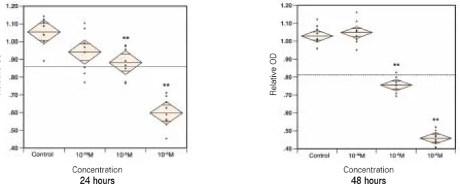
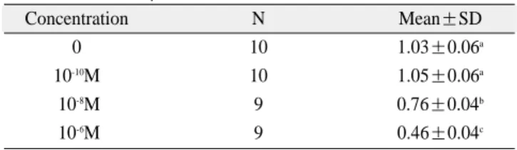
대조군의 중앙값을 기준으로 각 측정치의 상대적인 OD(optical density)값으로 환산한 결과, Aln와 Pam 모두 첨 가된 농도가 높아질수록 세포 생존능이 저하되었다. 대조 군과 10
-10M의 BPs가 첨가된 group간에는 통계적 유의성이
없었으나 10
-6M, 10
-8M의 BPs가 첨가된 group과의 차이는 통계적으로 유의하였다. (Fig. 1-2, Table 1-4, One-Way ANOVA with Tukey-Kramer Posthoc, P < 0.01).
2. 알칼리성 인산분해 효소 평가
단위 단백질 당 ALP 활성을 계산하고 그것을 대조군의 평균값으로 나누어 환산한 결과, 10
-8M의 Aln로 처리한 경 우는 24시간 이내에, 10
-10M의 Aln로 처리한 경우 72시간 내 에 ALP의 활성이 통계적으로 유의성 있게 증가하였다 (Wilcoxon, p<0.001). 반면, Pam는 모든 농도에서 ALP의 양 을 증가시키지 못했다. 10
-6M의 Aln로 처리한 경우에는 세 포 생존률이 낮아 실험에 필요한 만큼의 세포를 얻을 수 없 어 실험이 불가능하였다(Table 5).
Fig. 1. Cellular viability test of hMSCs according to the concentration of alendronate after 24, 48 hours of incubation.
Fig. 2. Cellular viability test of hMSCs according to the concentration of pamidronate after 24, 48 hours of incubation.
Concentration
24 hoursConcentration
48 hoursConcentration
24 hours
Concentration
48 hoursRelative OD
Relative OD Relative OD Relative OD
Ⅳ.
총괄 및 고찰BPs는 임상적으로 골다공증, Paget's disease, fibrous dys- plasia와 같은 질환에서 골소실을 치료하기 위해 널리 사용 되고 있는 약물이다
3-5). 기본적인 작용기전은 BPs가 골 표 면의 파골세포 부착부를 파괴하고 ruffled border의 기능을 방해함으로 골흡수를 저해시키는 것이다. 더 나아가 BPs는 파골세포의 증식과 전구체 생성을 억제하고 세포자살을 유도하는 것으로 보고되고 있다
10-12). 골모세포에 대한 영향 에 대해서도 여러 문헌 보고들이 있으나 그 결과가 BPs 종 류나 처리기간, 실험에 사용된 세포의 종류에 따라 다양하 게 나타나고 있다
18-22).
Im 등은
19)MG-63 cell에 다양한 농도의 Aln와 residronate 를 첨가하여 배양한 뒤 세포증식능을 관찰한 결과, 두 BPs 모두 10
-4M의 고농도에서는 증식이 저하되었으며 10
-7M에
서 10
-9M 사이에서는 대조군과 유의성 있는 차이를 보이며
증가하였고 10
-12M의 저농도에서는 대조군과 비슷해지는 양상을 보였다고 보고하였다. Fromigue 등은
18)여러 농도의 Pam, clodronate, ibandronate, zoledronate를 인간 골모세포
에 첨가하여 24, 48, 72시간 배양한 결과 BPs 종류, 처리 시 간, 농도에 따라 세포증식능이 다양하게 변화하였다고 보 고하였다. 예를 들면 10
-6M의 clodronate를 첨가한 경우에는 24시간 뒤 세포 증식능이 대조군에 비해 60%가량 증가하 였으나, Pam를 첨가한 경우에는 어떠한 농도에서도 증가 되는 양상은 없었고 오히려 10
-4M의 고농도에서는 저하되 는 것을 볼 수 있었다. Moreno 등에
21)의하면 인간 골모세포 에 여러 농도의 Aln를 첨가한 결과, 10
-5M 이하를 첨가하였 을 경우에는 세포 증식능에 어떠한 영향도 미치지 않는 것 으로 나타났지만 10
-4M을 첨가하였을 때는 세포 증식능이 저하되었고 10
-3M이상을 첨가하였을 때는 살아있는 세포 를 발견할 수 없었다고 보고하였다. Reinholz등은
22)다양한 농도의 Pam를 인간 골모세포에 적용해 보았으나 세포 증 식능에 변화가 없었으며, 오히려 농도가 높아질수록 세포 증식능이 감소하였고 10μg/ml보다 높은 농도에서는 세포 가 죽기 시작했다고 보고하였다.
골모세포 외에 다른 세포에 BPs를 첨가하여 세포 증식능 의 변화를 관찰한 연구들도 많이 보고되고 있다. Knoch 등 에
24)의하면 인간 골수기질세포(human bone marrow stromal Table 1.Cellular viability test of hMSCs according to the
concentration of alendronate after 24 hours of incubation.
Concentration N Mean±SD
0 10 0.98±0.05
a10
-10M 10 1.00±0.03
a10
-8M 9 0.95±0.06
a,b10
-6M 9 0.57±0.02
c1)Statistical significances were tested by oneway ANOVA among groups
2)The same letters indicate non-significant difference between groups based on Tukey-Kramer posthoc test.
Table 2.
Cellular viability test of hMSCs according to the concentration of alendronate after 48 hours of incubation.Concentration N Mean±SD
0 9 1.05±0.07
a10
-10M 9 1.11±0.06
a10
-8M 11 0.94±0.11
b10
-6M 12 0.46±0.07
cTable 4.
Cellular viability test of hMSCs according to the concentration of pamidronate after 48 hours of incubation.Concentration N Mean±SD
0 10 1.03±0.06
a10
-10M 10 1.05±0.06
a10
-8M 9 0.76±0.04
b10
-6M 9 0.46±0.04
cTable 3.
Cellular viability test of hMSCs according to the concentration of pamidronate after 24 hours of incubation.Concentration N Mean±SD
0 10 1.05±0.08
a10
-10M 10 0.94±0.11
a,b10
-8M 9 0.88±0.08
b10
-6M 9 0.60
±0.08cTable 5.
Alkaline phosphatase activity in hMSCsAlkaline phosphatase activity is represented as a fraction of untreated cell control and normalized to protein quantity at each time period.
Cells were treated with alendronate and pamidronate at different con- centration as indicated. Data is presented at mean ± standard devia- tion.
Time Concentration Alendronate Pamidronate
10
-10M 0.99±0.06 0.96±0.12
24hrs 10
-8M 1.13±0.09 0.98±0.12
10
-6M 0.95±0.06
Control 1.17±0.14 1.04±0.08
10
-10M 1.23±0.16 0.97±0.06
72hrs 10
-8M 1.02±0.29 0.98±0.05
10
-6M 0.81±0.05
Control 1.03±0.15 1.22±0.08
cells)에 10
-8M의 Aln를 첨가한 결과, 7일과 14일째 세포증 식능이 14%정도 증가했다고 보고하였다. Heino 등은
1)10
-6M 의 Aln 및 Pam를 첨가하여 쥐의 척골을 47일간 배양한 결 과, 연골세포 증식능이 각각 21%, 24% 증가하였다고 보고 하였다.
저자의 실험에서는 Aln와 Pam 모두 고농도에서 hMSCs 에 독성을 가지는 것으로 나타났다. 10
-6M의 BPs를 첨가하 였을 경우, 세포 생존능이 현저하게 떨어졌으나 10
-10M의 BPs를 첨가하였을 때는 대조군과 별다른 차이가 없었다.
이는 in vitro 상태에서, Aln 및 Pam가 골흡수 억제자로서 역할을 할 수 있는 50% 유효량(effective dose 50, ED
50) 2×
10
-9M 및 3×10
-9M
25)근처에서는 hMSCs 생존능에 거의 영 향을 미치지 않는다고 해석할 수 있다. 그러나 in vivo 상태 에서는 흡수된 BPs가 골의 무기질에 강하게 결합하여 대사 가 잘 되지 않기 때문에 오랫동안 골 내에 높은 농도로 잔 존하게 되므로
26)이들이 파골세포 뿐 아니라 hMSCs의 생 존능에도 영향을 미치지 않을까 생각된다.
이처럼 같은 BPs에 대해서 다양한 실험결과를 보일 수 있 는 것은 전술한 바와 같이, 실험에 사용된 세포가 다르기 때문인 것으로 생각되며, 특히 인간 골수기질세포나 hMSCs의 경우에는 얻어낸 개체에 따라서 또는 같은 개체 내에서도 clone에 따라 그 성상이 다를 수 있다
27).
BPs가 골모세포로의 분화에 미치는 영향에 대해서도 다 양한 결과들이 보고되고 있다. Knoch 등은
24)인간 골수기질 세포에 10
-8M의 Aln, risedronate, zoledronate를 첨가한 결과, 모두 세포 증식능은 대조군에 비해 증가하였으나 ALP활 성에는 변화가 없었다고 보고하였다. 그러나 개체가 다른 세포들간, 또는 처리한 BPs의 종류간 ALP 활성에 차이가 있음을 관찰할 수 있었다. Fromigue 등은
18)인간 골모세포 에 10
-6M의 Pam를 첨가하여 배양한 결과, 24, 48시간 내에 는 ALP 활성에 변화가 없었으나 96시간 내에 대조군에 비 해 25% 증가되는 양상을 보였다고 보고하였다. Im 등은
19)인간 골소주 세포에 10
-8M의 Aln를 첨가한 결과, 24시간 내 에 ALP활성이 증가하였으며 이후에는 대조군과 비교하여 차이가 없었다고 보고하였다. Reinholz 등은
22)Pam가 인간 골모세포 증식능을 향상시키지는 못했지만 첨가되는 농도 가 증가할수록 ALP활성이 증가하였다고 보고하였다. 또 한 10μg/ml의 Pam를 첨가하여 기간에 따라 ALP 활성을 측 정한 결과, 2일째부터 증가하기 시작하여 5일째에는 대조 군보다 82% 증가하였으며 이후에는 점차 감소하였다고 보 고하였다.
저자의 실험에서는 비록 Aln가 hMSCs의 생존능을 향상 시키지는 못했지만 ALP활성은 증가시키는 것으로 나타났 다. 10
-10M을 첨가하였을 경우 72시간 이내에, 10
-8M을 첨가 하였을 경우 24시간 이내에 ALP활성이 대조군보다 증가 하였다. 이는 세포 증식이 끝난 뒤, 간질 성숙 단계에서 골 모세포로의 분화를 촉진하는 것이라 생각된다. 이와는 대 조적으로 Pam의 경우에는 어떠한 농도에서도 ALP활성을
증가시키지 못했다. 이는 두 BPs의 약물학적 효능(potency) 차이 때문인 것으로 생각되며 etidronate의 효능을 1로 하여 BPs간 상대적 효능을 비교해 보면, Aln는 1000, Pam는 100 으로 계산해 볼 수 있다
9,10).
본 실험을 통해 BPs가 hMSCs 생존능을 향상시키는 효과 는 없으며 오히려 10
-8M이상의 농도에서는 세포독성을 가 진다는 것을 알 수 있었다. 따라서 in vivo 상태에서는 BPs 의 반감기가 10년 이상으로
5,28)길기 때문에 오랫동안 악골 에 고농도로 축적될 수 있으므로 외과적 수술 시 이러한 점 을 고려하여야 하겠다. 또한 Aln의 경우에는 ALP 활성을 증가시키는 것으로 나타났으나 이것만으로 hMSCs이 골모 세포로 분화하는데 도움을 준다고 단정하기는 어려울 것 으로 생각되며 이를 증명하기 위해서 추가적으로 유전자 단위에서의 연구와 동물실험 등이 필요할 것으로 사료된 다.
Ⅴ.
결 론본 연구에서는 골다공증 치료제로 가장 널리 사용되고 있는 경구용 약물인 Aln와 암과 관련된 골흡수의 치료제로 많이 사용되고 있는 정맥 내 주사제인 Pam를 이용하여 이 들 약물이 hMSCs의 증식과 분화에 미치는 영향을 알아보 고자 하였다. 이를 위해서, 위의 세포 배양 시 10
-10M, 10
-8M, 10
-6M의 Aln와 Pam을 각각 첨가하였으며 Cell counting kit 를 이용하여 흡광도를 측정함으로써 세포 생존능을 평가 하였다. 동일한 조건에서 ALP 활성을 측정하였으며 단백 질 정량 분석을 통해 수치를 표준화하여 비교 분석하였다.
이에 다음과 같은 결론을 얻었다.
1. Aln와 Pam는 10
-6M의 고농도에서는 hMSCs 생존능을 저하시켰으나 10
-10M의 저농도에서는 hMSCs 생존능에 영향을 미치지 않았다.
2. Aln는 10
-10M의 농도에서는 72시간 내에, 10
-8M의 농도 에서는 24시간 내에 ALP의 활성이 증가되었으나 10
-6M 의 고농도에서는 세포수의 감소로 ALP 활성의 측정이 불가능하였다.
3. Pam는 hMSCs의 ALP 활성을 증가시키지 못하였다.
이상의 결과로 BPs 계열의 약물은 10
-10M의 저농도에서 는 hMSCs의 증식에 특별한 영향을 미치지 않으나 10
-6M의 고농도에서는 hMSCs의 생존능을 억제하는 것으로 보아 임상에서 악골 내에 높은 농도로 축적되었을 때에는 손상 된 골조직의 치유에 좋지 않은 영향을 미칠 것으로 생각되 며 근래 구강악안면외과 임상에서 접하게 되는 BRONJ 환 자의 치료 시 고려해야 될 것으로 사료된다.
참고문헌