• 검색 결과가 없습니다.

Clinical Phenotype of a First Unprovoked Acute Pulmonary Embolism Associated with Antiphospholipid Antibody Syndrome

N/A
N/A
Protected

Academic year: 2021

Share "Clinical Phenotype of a First Unprovoked Acute Pulmonary Embolism Associated with Antiphospholipid Antibody Syndrome"

Copied!
9
0
0

로드 중.... (전체 텍스트 보기)

전체 글

(1)

Introduction

In patients with a first symptomatic pulmonary embolism (PE), clinicians should evaluate for predisposing factors. Un- provoked venous thromboembolism (VTE), for which there is the absence of a temporary or reversible risk factor within the 6 weeks to 3 months before diagnosis, has a moderately high risk of recurrence, which is estimated to be 10% within 1 year and 30% within 5 years

1-3

. The following risk factors for recur- rence in patients with an unprovoked PE may help to identify those at higher long-term relative risk of recurrence: (1) one or more previous episodes of VTE, (2) antiphospholipid an- tibody syndrome (APS), (3) hereditary thrombophilia, or (4) residual thrombosis in the proximal veins

3-6

.

One of those risk factors is APS, a systemic autoimmune

Clinical Phenotype of a First Unprovoked Acute Pulmonary Embolism Associated with Antiphospholipid Antibody Syndrome

Yong Sub Na, M.S.

1

, Seongsoo Jang, M.D.

2

, Seokchan Hong, M.D.

3

, Yeon Mok Oh, M.D.

4

, Sang Do Lee, M.D.

4

and Jae Seung Lee, M.D.

5

1

Department of Pulmonary and Critical Care Medicine, Chosun University Hospital, Gwangju, Departments of

2

Laboratory Medicine,

3

Rheumatology, and

4

Pulmonary and Critical Care Medicine, Asan Medical Center, University of Ulsan College of Medicine, Seoul,

5

Department of Pulmonary and Critical Care Medicine, Center for Pulmonary Hypertension and Venous Thrombosis, Asan Medical Center, University of Ulsan College of Medicine, Seoul, Korea

Background: Antiphospholipid antibody syndrome (APS), an important cause of acquired thrombophilia, is diagnosed when vascular thrombosis or pregnancy morbidity occurs with persistently positive antiphospholipid antibodies (aPL).

APS is a risk factor for unprovoked recurrence of pulmonary embolism (PE). Performing laboratory testing for aPL after a first unprovoked acute PE is controversial. We investigated if a specific phenotype existed in patients with unprovoked with acute PE, suggesting the need to evaluate them for APS.

Methods: We retrospectively reviewed patients with PE and APS (n=24) and those with unprovoked PE with aPL negative (n=44), evaluated 2006–2016 at the Asan Medical Center. We compared patient demographics, clinical manifestations, laboratory findings, and radiological findings between the groups.

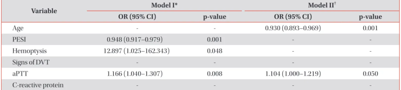
Results: On multivariate logistic regression analysis, two models of independent risk factors for APS-PE were suggested.

Model I included hemoptysis (odds ratio [OR], 12.897; 95% confidence interval [CI], 1.025–162.343), low PE severity index (OR, 0.948; 95% CI, 0.917–0.979), and activated partial thromboplastin time (aPTT; OR, 1.166; 95% CI, 1.040–1.307).

Model II included age (OR, 0.930; 95% CI, 0.893–0.969) and aPTT (OR, 1.104; 95% CI, 1.000–1.217).

Conclusion: We conclude that patients with first unprovoked PE with hemoptysis and are age <40; have a low pulmonary embolism severity index, especially in risk class I–II; and/or prolonged aPTT (above 75th percentile of the reference interval), should be suspected of having APS, and undergo laboratory testing for aPL.

Keywords: Antiphospholipid Syndrome; Antibodies, Antiphospholipid; Pulmonary Embolism; Phenotype; Risk Factors

Address for correspondence: Jae Seung Lee, M.D.

Department of Pulmonary and Critical Care Medicine, Center for Pulmonary Hypertension and Venous Thrombosis, Asan Medical Center, University of Ulsan College of Medicine, 88 Olympic-ro 43-gil, Songpa- gu, Seoul 05505, Korea

Phone: 82-2-3010-3994, Fax: 82-2-3010-6968 E-mail: [email protected]

Received: May. 10, 2018 Revised: Jul. 22, 2018 Accepted: Oct. 16, 2018

cc It is identical to the Creative Commons Attribution Non-Commercial License (http://creativecommons.org/licenses/by-nc/4.0/).

Copyright © 2019

The Korean Academy of Tuberculosis and Respiratory Diseases.

(2)

disease that is diagnosed when arterial and/or venous throm- bosis, and recurrent fetal losses occurs in the presence of persistent antiphospholipid antibodies (aPL)

7

. APS tends to be more common in young to middle aged women. Estimates have indicated an incidence of around five new cases per 100,000 persons per year, with prevalence around 40–50 cas- es per 100,000 persons

8

. PE is the most common pulmonary manifestation and may be the first sign of APS

9,10

. Patients with PE-associated APS are recommended to have extended anti- coagulant therapy because of the risk of recurrent VTE

11

. Pro- thrombotic states and impaired clot dissolution are believed to contribute to the occurrence of chronic thromboembolic pulmonary hypertension in APS

12

.

aPL contributes to the pathogenesis of thrombosis

13

. Galli et al.

14

reported that the odds ratios (OR) of aPL for cerebral stroke and deep vein thrombosis (DVT) ranged between 4.09 and 16.2, all with significant 95% confidence intervals (CI). Re- cently updated British Journal of Haematology guidelines on the investigation and management of APS recommend test- ing for aPL before stopping anticoagulation after unprovoked proximal DVT or PE, as a positive result favours long-term an- ticoagulation

15

. However, laboratory testing for aPL after a first symptomatic unprovoked PE remains controversial.

We hypothesized that there might be a specific clinical phenotype in patients with unprovoked acute PE indicating a need for evaluation for APS. This study investigated the clini- cal characteristics of PE in patients with and without APS who had unprovoked PE, looking for clinical predictors for APS.

Materials and Methods

1. Study design and patients

A retrospective descriptive study was conducted. All clini- cal, radiological, and laboratory data were retrospectively collected from medical records. We reviewed a total of 261 pa- tients diagnosed as having had a first episode of PE between June 2006 and September 2016 at the Asan Medical Center, a 2,700-bed university-affiliated tertiary referral hospital in Seoul, Korea. PE was confirmed in all patients by pulmonary angiography or spiral computed tomography or ventilation- perfusion lung scan indicating a high probability of pulmo- nary embolism. We tested for aPL in patients with a first unprovoked PE. If results were positive for aPL, aPL test was performed again at least 12 weeks apart. Patient records were included if the PE was confirmed to be unprovoked (i.e., idio- pathic) and if laboratory testing for aPL had been performed or the patient was known to have APS. We excluded those with provoked PE in the presence of temporary or reversible risk factors (e.g., surgery, trauma, immobilization, pregnancy, oral contraceptive use, or hormone replacement therapy) within the preceding 6 weeks to 3 months

3

.

The study was approved by the institutional review board (IRB) of Asan Medical Center (IRB No. 2015-0516). The need for informed consent was waived owing to the retrospective nature of the study.

2. Classification criteria for the antiphospholipid syndrome

According to the 2006 revised APS classification criteria, APS requires the combination of at least one clinical and one laboratory criterion. Clinical criteria include vascular throm- bosis (arterial, venous, or small vessel thrombosis in any tissue or organ) and/or pregnancy morbidity (one or more unexplained deaths of a morphologically normal foetus at or beyond the 10th week of gestation, or one or more premature births of a morphologically normal neonate before the 34th week of gestation because of severe pre-eclampsia/eclamp- sia, or recognized features of placental insufficiency, and/or three or more unexplained consecutive spontaneous abor- tions before the 10th week of gestation). Laboratory criteria include positive tests for circulating aPL (lupus anticoagulant [LA]; anticardiolipin antibody [ACA], IgG and/or IgM; anti-β

2

glycoprotein-I antibody [aβ2GPI], IgG and/or IgM) on two or more occasions at least 12 weeks apart

7

.

3. Laboratory diagnosis of APS

LA tests were determined according to the criteria of the Subcommittee for Standardization of LA

16

. The diluted Rus- sell’s Viper Venom Test (dRVVT) was performed using an ACL TOP 750 automated coagulometer (Instrumentation Labora- tory, Milan, Italy) with a commercial assay (HemosIL, dRVVT screen; Instrumentation Laboratory, Milan, Italy). Samples with a prolonged dRVVT that was not corrected by mixing with a normal plasma pool were tested for confirmation using an excess of phospholipids (HemosIL, dRVVT confirm; Instru- mentation Laboratory, Milan, Italy). A commercial silica clot- ting time (SCT) with low and high concentrations of synthetic phospholipids (HemosIL, SCT screen; Instrumentation Labo- ratory, Milan, Italy) was used as an activator in the coagulation test. The SCT was carried out on the ACL TOP 750 automated coagulometer (Instrumentation Laboratory, Milan, Italy).

Samples with a prolonged SCT that was not corrected by mix-

ing with a normal plasma pool were tested for confirmation

using a high phospholipid concentration (HemosIL, SCT con-

firm; Instrumentation Laboratory, Milan, Italy). ACA (IgG and

IgM) and aβ2GPI (IgG and IgM) were tested for using com-

mercially assay kits (HemosIL; Instrumentation Laboratory,

Bedford, MA, USA). Both tests were carried out on automated

immunoassay, the HemosIL AcuStar (Instrumentation Labo-

ratory, Bedford, MA, USA). All tests were performed according

to the manufacturer’s instructions.

(3)

4. Risk stratification

The pulmonary embolism severity index (PESI) is a clinical prediction rule for prognosis of PE developed by Aujesky et al.

17

The PESI correlates with 30-day mortality

3,17,18

. The risk of early mortality in patients with acute PE was classified as high, intermediate-high, intermediate-low, and low. Risk parame- ters and scores included shock or hypotension, PESI class III–

V or a simplified PESI score ≥1, signs of right ventricular (RV) dysfunction on an imaging test, and cardiac laboratory bio- markers. Risk-adjusted therapeutic strategies and algorithms are recommended on the basis of this risk classification

3

. 5. Statistical analysis

We compared patient demographics, clinical manifesta- tions, laboratory findings, and radiological findings between patients with a PE associated with APS (APS-PE) and those with an unprovoked PE but who did not have APS (non-APS- PE). Results are expressed as means with standard deviations or median values with interquartile range (IQR) for continu- ous variables, and as percentages for categorical variables. For comparison of continuous variables between the APS-PE and non-APS-PE groups, parametric data were compared using independent Student’s t-tests, and nonparametric data were compared using a Mann-Whitney U test. Categorical variables were compared using a chi-square or Fisher exact test. To evaluate the clinical predictors of APS-PE, univariate and mul- tivariate logistic regression analysis was performed. Variables with a p-values of <0.05 as determined by univariate logistic

regression analysis in consideration of multicollinearity were entered into multivariate logistic regression analysis using a backward elimination method. Assessing goodness-of-fit in logistic regression models was performed by the Hosmer- Lemeshow goodness-of-fit test. Discrimination ability was assessed using the area under the receiver operating charac- teristic (ROC) curve.

All reported p values were two-sided, and a p-value of <0.05 was considered statistically significant. Statistical analysis was performed using the IBM SPSS Statistics version 21.0 (IBM Corp., Armonk, NY, USA).

Results

1. Patients

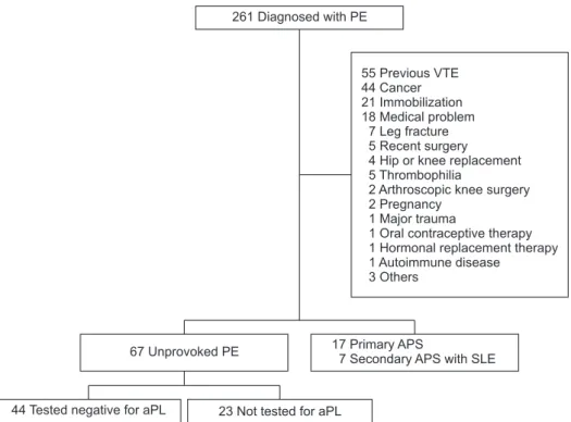
Among 261 patients diagnosed with a first episode of PE, 170 were excluded from analysis because they had a tempo- rary or reversible risk factor (Figure 1). Of 67 patients with an unprovoked PE, 23 were not tested for aPL and were excluded.

So, we compared with 44 patients testing negative for aPL and 24 diagnosed with APS, either primary (n=17) or in associa- tion with systemic lupus erythematous (n=7).

2. Baseline characteristics

Table 1 shows a comparison of the baseline characteristics between the APS-PE and non-APS-PE groups. Individuals in the APS-PE group were significantly younger and had more

261 Diagnosed with PE

55 Previous VTE 44 Cancer 21 Immobilization 18 Medical problem

7 Leg fracture 5 Recent surgery 4 Hip or knee replacement 5 Thrombophilia

2 Arthroscopic knee surgery 2 Pregnancy

1 Major trauma

1 Oral contraceptive therapy 1 Hormonal replacement 1 Autoimmune disease 3 Others

therapy

67 Unprovoked PE 17 Primary APS

7 Secondary APS with SLE

44 Tested negative for aPL 23 Not tested for aPL

Figure 1. Flowchart of patient selection.

One hundred seventy patients with predisposing factors were excluded. An additional 23 patients with unprovoked PE were excluded as they were not tested for APS. PE: pulmonary embolism;

VTE: venous thromboembolism; APS:

antiphospholipid antibody syndrome;

SLE: systemic lupus erythematous; aPL:

antiphospholipid antibody.

(4)

history of arterial thrombosis.

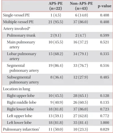
3. Radiological findings

The arteries involved by the modified Boyden classifica- tion

19

, involved lung lobes, and numbers of PEs by pulmonary computed tomography (CT) angiography were similar be- tween the two groups (Table 2), but patients in the APS-PE group were more likely to have a pulmonary infarct than those in the non-APS-PE group.

4. Clinical and laboratory criteria in APS-PE group As shown in Table 3, the types of aPL found in the APS-PE group included LA (in 58.3%), ACA IgM (in 25.0%), ACA IgG (in 29.2%), aβ2GPI IgM (in 20.8%), and aβ2GPI IgG (in 20.8%).

A history of arterial thrombosis was found in three patients (12.5%): coronary artery disease history was found in two and cerebral infarction history in one. Among 11 female patients, one had had eclampsia. Of the 24 patients with APS, seven (29.2%) had systemic lupus erythematosus.

Table 3. Clinical and laboratory criteria in the APS-PE group

APS-PE (n=24) Antiphospholipid antibodies

Lupus anticoagulants 14 (58.3)

Anticardiolipin antibody IgM 6 (25.0) Anticardiolipin antibody IgG 7 (29.2) Anti‒β

2

glycoprotein-I antibody IgM 5 (20.8) Anti‒β

2

glycoprotein-I antibody IgG 5 (20.8)

Arterial thrombosis* 3 (12.5)

Pregnancy complications

1 (9.0)

Secondary APS with SLE 7 (29.2)

Values are presented as number (%).

*Two patients had coronary artery disease. One patient had ce- rebral infarction.

One of eleven women in the APS-PE group had eclampsia.

APS: antiphospholipid antibody syndrome; PE: pulmonary embo- lism; SLE: systemic lupus erythematous.

Table 1. Baseline characteristics of patients with a first unprovoked PE

APS-PE (n=24)

Non- APS-PE

(n=44) p-value

Age, yr 39.7±18.4 60.4±14.0 <0.001

Sex 0.492

Male 13 (54.2) 20 (45.5)

Female 11 (45.8) 15 (54.5)

Body mass index, kg/m

2

23.9 ± 4.4 25.5 ± 3.6 0.112 Deep vein thrombosis* 17 (70.8) 25 (56.8) 0.256 History of arterial thrombosis 3 (12.5) 0 (0) 0.017 History of pregnancy

complications

1 (9.0) 0 (0) 0.176

Presentation 0.175

Acute signs or symptoms 20 (83.3) 42 (95.5) Incidental

4 (16.7) 2 (4.5)

Diagnostic modality 0.283

Computed tomography angiography

22 (91.7) 43 (97.7)

Ventilation-perfusion scan 2 (8.3) 1 (2.3)

Values are presented mean±standard deviation or number (%).

*Deep vein thrombosis was diagnosed by lower extremity com- pression ultrasonography or computed tomography venography.

Incidental PE defined as the finding of PE in situations where it was not suspected.

PE: pulmonary embolism; APS: antiphospholipid antibody syn- drome.

Table 2. Pulmonary CT angiography findings APS-PE

(n=22)

Non-APS-PE

(n=43) p-value Single-vessel PE 1 (4.5) 6 (14.0) 0.408 Multiple-vessel PE 21 (95.5) 37 (86.0) 0.408 Artery involved*

Pulmonary trunk 2 (9.1) 2 (4.7) 0.599 Main pulmonary

artery

10 (45.5) 16 (37.2) 0.521

Lobar pulmonary artery

15 (68.2) 34 (79.1) 0.335

Segmental pulmonary artery

19 (86.4) 33 (76.7) 0.516

Subsegmental pulmonary artery

8 (36.4) 12 (27.9) 0.485

Location in lung

Right upper lobe 10 (45.5) 28 (65.1) 0.128 Right middle lobe 9 (40.9) 26 (60.5) 0.135 Right lower lobe 18 (81.8) 37 (86.0) 0.723 Left upper lobe 13 (59.1) 27 (62.8) 0.772 Left lower lobe 18 (81.8) 35 (81.4) 1.000 Pulmonary infarction

11 (50.0) 10 (23.3) 0.029 Values are presented as number (%).

*Location according to the modified Boyden classification

19

.

De- fined as a wedge-shaped subpleural opacity on pulmonary CT angiography findings.

CT: computed tomography; APS: antiphospholipid antibody syn-

drome; PE: pulmonary embolism.

(5)

5. Clinical manifestations and laboratory findings The clinical manifestations and laboratory findings of the two groups are summarized in Table 4. patients in the APS- PE group had less dyspnea but significantly more hemoptysis, unilateral leg pain, and signs of DVT than the non-APS-PE group. In addition, the APS-PE group had a significantly higher creatinine clearance, prolonged activated partial thrombo- plastin time (aPTT), and lower aspartate transaminase levels.

6. Prognostic assessment

PESI and early mortality risk are shown in Table 5. The non- APS-PE group had significantly higher creatine kinase-MB (0.65, 0.30–1.60 vs. 1.85, 1.00–4.35; p=0.003), PESI, and PESI severity classification than the APS-PE group.

7. Clinical predictors for APS-PE

On univariate logistic regression analysis, significant clinical predictors for APS were age, pulmonary infarction, dyspnea, hemoptysis, unilateral leg pain, signs of DVT, PESI, aPTT, cre- atinine clearance, and C-reactive protein (CRP). The risk of having APS was 11-fold higher for patients with hemoptysis (OR, 11.316; 95% CI, 1.237–103.553) and 3-fold higher for patients with pulmonary infarction (OR, 3.400; 95% CI, 1.170–

9.877), unilateral leg pain (OR, 3.400; 95% CI, 1.170–9.877), or signs of DVT (OR, 3.545; 95% CI, 1.236–10.170) (Table 6). The significant independent risk factors for having APS by multi- variate logistic regression analysis for PESI, hemoptysis, signs of DVT, aPTT (model I) were hemoptysis (OR, 12.897; 95% CI, 1.025–162.343), a low PESI (OR, 0.948; 95% CI, 0.917–0.979), and aPTT (OR, 1.166; 95% CI, 1.040–1.307) (Table 7). When model I was applied to the validation data, the area under the ROC curve was 0.868 and the p-value for the Hosmer‒Leme-

Table 4. Comparison of clinical manifestations and laboratory findings between the APS-PE and non-APS-PE groups APS-PE (n=24) Non-APS-PE (n=44) p-value Clinical manifestations

Dyspnea 14 (58.3) 37 (84.1) 0.019

Pleuritic chest pain 6 (25.0) 5 (11.4) 0.177

Cough 4 (16.7) 2 (4.5) 0.175

Substernal chest pain 5 (20.8) 9 (20.5) 1.000

Fever 3 (12.5) 1 (2.3) 0.122

Hemoptysis 5 (20.8) 1 (2.3) 0.018

Syncope 2 (8.3) 5 (11.4) >0.990

Unilateral leg pain 12 (50.0) 10 (22.7) 0.022

Signs of deep vein thrombosis* 13 (54.2) 11 (25.0) 0.016

Hypotension

1 (4.2) 4 (9.1) 0.649

Heart rate, beats per minute 90.3±14.9 94.1±18.3 0.396

Laboratory findings

White blood cell count, /μL 8,707±4,381 9,792±3,535 0.271

Platelet, ×10

3

/μL 190,125±102,075 205,090±62,744 0.518

Creatinine clearance, mL/min/1.73 m

2

114.4±25.1 82.9±26.2 <0.001

Prothrombin time, sec 11.8±1.2 11.5±1.3 0.366

International normalized ratio 1.05±0.11 1.03±0.11 0.398

Activated partial thromboplastin time, sec 31.8 (27.3–35.2) 27.1 (25.2–28.6) <0.001

Aspartate transaminase, IU/L 22.0 (19.0–29.5) 28.0 (21.5–45.0) 0.029

Alanine transaminase, IU/L 20.5 (11.5–40.5) 27.0 (17.5–47.0) 0.152

C-reactive protein, mg/dL 1.17 (0.45–4.22) 0.55 (0.16–1.79) 0.051

Values are presented as number (%), mean±standard deviation, or median (interquartile range).

*A palpable cord reflecting a thrombosed vein, ipsilateral edema, warmth, and/or superficial venous dilation.

Defined as systolic blood pres-

sure <90 mm Hg or a systolic pressure drop by ≥40 mm Hg for >15 minutes, if not caused by new-onset arrhythmia, hypovolemia or sepsis

3

.

APS: antiphospholipid antibody syndrome; PE: pulmonary embolism.

(6)

show goodness-of-fit test was 0.656. Model II included age, he- moptysis, signs of DVT, aPTT, and CRP. The independent risk factors were age (OR, 0.930; 95% CI, 0.893–0.969) and aPTT (OR, 1.104; 95% CI, 1.000–1.217). When model II was applied to the validation data, the area under the ROC curve was 0.819 and the p-value for the Hosmer–Lemeshow goodness-of-fit test was 0.102.

Discussion

Our study shows which clinical factors in patients with un- provoked PE are predictive of APS, thereby suggesting which

patients need to be evaluated for APS. The factors we found to be associated with APS were (1) younger age, (2) the pres- ence of a pulmonary infarct on CT pulmonary angiography, (3) the absence of dyspnea, (4) the presence of hemoptysis, (5) the presence of unilateral leg pain and signs of DVT, (6) a low PESI, (7) prolonged aPTT, and (8) high creatinine clearance and CRP. Among these factors, the most sensitive predictors for APS were younger age, the presence of hemoptysis, a low PESI, and prolonged aPTT.

The prevalence of APS is approximately 40–50 cases per 100,000 persons

20,21

. APS usually occurs in young patients.

Piette and Cacoub

22

analyzed data on age at first thrombosis in patients with primary APS and found that the first event occurred in patients aged <50 years. This is in contrast to the risk for VTE in the general population which, as Anderson and

Table 6. Clinical predictors of APS on univariate logistic regression

Variable OR (95% CI) p-value

Age 0.929 (0.895–0.964) <0.001

Pulmonary infarction 3.400 (1.170–9.877) 0.025

Dyspnea 0.265 (0.084–0.832) 0.023

Hemoptysis 11.316 (1.237–103.553) 0.032 Unilateral leg pain 3.400 (1.170–9.877) 0.025 Signs of DVT 3.545 (1.236–10.170) 0.019

PESI 0.950 (0.923–0.978) 0.001

aPTT 1.133 (1.025–1.253) 0.015

Creatinine clearance 1.057 (1.027–1.088) <0.001 C-reactive protein 1.150 (1.004–1.318) 0.044 APS: antiphospholipid antibody syndrome; OR: odds ratio; CI:

confidence interval; DVT: deep vein thrombosis; PESI: pulmonary embolism severity index; aPTT: activated partial thromboplastin time.

Table 5. PESI and early mortality risk APS-PE

(n=24)

Non-APS-

PE (n=44) p-value

PESI 0.039

Class I (≤65 points) 14 (58.3) 11 (25.0) Class II (65–85 points) 7 (29.2) 13 (29.5) Class III (86–105 points) 3 (12.5) 14 (31.8) Class IV (106–125 points) 0 (0) 3 (6.8) Class V (>125 points) 0 (0) 3 (6.8)

Average* 58.0±21.6 83.7±24.4 <0.001

Early mortality risk 0.072

High 1 (4.2) 4 (9.1)

Intermediate – high 1 (4.2) 5 (11.4) Intermediate – low 7 (29.2) 22 (50.0)

Low 15 (62.5) 13 (29.5)

Values are presented as number (%) or mean±standard deviation.

*Sum of points.

PESI: pulmonary embolism severity index; APS: antiphospholipid antibody syndrome; PE: pulmonary embolism.

Table 7. Multivariate logistic regression model of independent risk factors for PE associated with APS

Variable Model I* Model II

OR (95% CI) p-value OR (95% CI) p-value

Age - - 0.930 (0.893–0.969) 0.001

PESI 0.948 (0.917–0.979) 0.001 - -

Hemoptysis 12.897 (1.025–162.343) 0.048 - -

Signs of DVT - - - -

aPTT 1.166 (1.040–1.307) 0.008 1.104 (1.000–1.219) 0.050

C-reactive protein - - - -

*Multivariate logistic regression analysis for PESI, hemoptysis, signs of DVT, aPTT, and C-reactive protein.

Multivariate logistic regression analysis for age, hemoptysis, signs ofDVT, aPTT, and C-reactive protein.

PE: pulmonary embolism; APS: antiphospholipid antibody syndrome; OR: odds ratio; CI: confidence interval; PESI: pulmonary embolism

severity index; DVT: deep vein thrombosis; aPTT: activated partial thromboplastin time.

(7)

Spencer

23

reported, increases in patients aged >40 years of age and approximately doubles with each subsequent decade.

In this analysis, the mean age of the APS-PE group was 39.7 years, compared with 60.4 years in the non-APS-PE group.

Therefore, if a patient aged <40 years presents with a first symptomatic unprovoked PE, we recommend testing for aPL.

The presenting symptoms and signs of PE are nonspecific.

Our study showed that the APS-PE group had more hemopty- sis and pulmonary infarction than the non-APS-PE group. The EMPEROR study reported that the most common present- ing signs and symptoms of PE were dyspnea at rest (50.1%), pleuritic chest pain (39.4%), dyspnea on exertion (27%), ex- tremity edema suggestive of DVT (23.5%), and cough without hemoptysis (22.9%). Cough with hemoptysis was present in only 7.6%

24

. He et al.

25

reported that 32% patients with PE had pulmonary infarction. In our analysis, hemoptysis in the APS- PE group was present in 20.8% patients, and half the APS-PE group had pulmonary infarction in pulmonary CT angiogra- phy. Pulmonary infarction occurs more commonly in patients with peripheral than central pulmonary emboli due to the dual blood supply of the lung, i.e., with occlusion of pulmonary artery branches of ≤3 mm in diameter. A wedge-shaped sub- pleural opacity (“Hampton hump”) on X-ray has been thought by many to be diagnostic of pulmonary infarction

25,26

. Miniati et al.

27

reported that hemoptysis in patients with pulmonary infarction was seen in 19 of 105 patients (18%) and was more frequent than in patients without pulmonary infarction. In patients with PE who had APS in our study, pulmonary infarc- tion was more likely to occur, which may be accompanied by hemoptysis. APS is characterized by thrombophilia with systemic hypercoagulability. Because vascular thrombosis in APS involved at any level of the vascular trees, occlusions of pulmonary arteries and bronchial arteries can occur simulta- neously, which can lead to pulmonary infarction

28

. Therefore, hemoptysis and radiological evidence of pulmonary infarc- tion in patients with unprovoked PE are important factors that should increase suspicion for APS.

The most common haematological manifestation in APS is thrombocytopenia, with an incidence ranging 22%–42% in different series. Thrombocytopenia in APS is rarely associ- ated with haemorrhage

29-32

. Many patients with APS have a prolonged aPTT because LA inhibits the formation of the prothrombinase complex

33

. However, the presence of LA is strong risk factors for thrombosis, irrespective of the site and thrombosis type

14

and not for bleeding. In this study, the most common aPL in the APS-PE group was LA, in 14 of 24 patients (58%). The median aPTT value in the APS-PE group was 31.8 seconds (IQR, 27.3–35.2), within the normal range. However, the median value was still significantly higher in the APS-PE than in the non-APS-PE group. On multivariate logistic regres- sion analysis, aPTT was independently associated with APS.

Thus, if patients with an unprovoked PE have an isolated pro- longed aPTT above the 75th percentile of the reference inter-

val, screening for aPL, especially LA, should be considered.

PESI is an accurate and reproducible tool for risk stratifica- tion in acute PE and correlates with 30-day mortality. PESI and classification of early mortality risk may help identify appropriate candidates for outpatient management or hospi- talization. In recently updated guidelines, patients with a con- firmed PE in PESI class I–II or low early mortality risk are rec- ommended to have early discharge and home treatment

3,34,35

. Our study showed that the non-APS-PE group had a higher PESI, with a higher proportion of patients in risk classes III–V, than the APS-PE group. The distribution of early mortality risk between the APS-PE and non-APS-PE groups was not statisti- cally significant, but 70.5% patients in the non-APS-PE group were at intermediate to high mortality risk compared with 37.5% in the APS-PE group. These patients in the non-APS-PE group were generally older than patients in the APS-PE group and had more signs of RV dysfunction. We recommend that laboratory testing for aPL should be considered for patients with unprovoked PE who have a low PESI, especially in risk class I or II.

Our study has limitations. First, it is a retrospective descrip- tive study from a single tertiary centre, with data collected from patients’ medical records, making it susceptible to bias in data selection and analysis. Second, we did not perform a cost- effectiveness analysis of testing for aPL in patients with a first unprovoked PE. Auerbach et al.

36

reported that hypercoagu- lability testing followed by 24 months of anticoagulation was cost effective. Third, we did not analyze treatment outcomes after anticoagulation between the two groups. Fourth, the reason for the high prevalence of APS compared with recent study is unclear

37

.

In conclusion, this study shows that patients with a first un- provoked PE who have hemoptysis, are aged <40 years, had a low PESI (risk class I or II), and/or prolonged aPTT (above the 75th percentile of the reference interval) should be suspected of having APS and should undergo laboratory testing for aPL.

Further research requires cost-effectiveness analysis of testing for aPL in patients with a first unprovoked PE with these fac- tors.

Authors' Contributions

Conceptualization: Oh YM, Lee SD, Lee JS. Methodology:

Lee JS. Investigation: Na YS, Lee JS. Writing - original draft preparation: Na YS. Writing - review and editing: Jang S, Hong S.

Approval of final manuscript: all authors.

Conflicts of Interest

No potential conflict of interest relevant to this article was

reported.

(8)

References

1. Iorio A, Kearon C, Filippucci E, Marcucci M, Macura A, Pengo V, et al. Risk of recurrence after a first episode of symptomatic venous thromboembolism provoked by a transient risk fac- tor: a systematic review. Arch Intern Med 2010;170:1710-6.

2. Kearon C, Akl EA. Duration of anticoagulant therapy for deep vein thrombosis and pulmonary embolism. Blood 2014;123:1794-801.

3. Konstantinides SV, Torbicki A, Agnelli G, Danchin N, Fitzmau- rice D, Galie N, et al. 2014 ESC guidelines on the diagnosis and management of acute pulmonary embolism. Eur Heart J 2014;35:3033-69, 3069a-k.

4. Zhu T, Martinez I, Emmerich J. Venous thromboembolism:

risk factors for recurrence. Arterioscler Thromb Vasc Biol 2009;29:298-310.

5. Kyrle PA, Rosendaal FR, Eichinger S. Risk assessment for re- current venous thrombosis. Lancet 2010;376:2032-9.

6. Heit JA. Predicting the risk of venous thromboembolism re- currence. Am J Hematol 2012;87 Suppl 1:S63-7.

7. Miyakis S, Lockshin MD, Atsumi T, Branch DW, Brey RL, Cer- vera R, et al. International consensus statement on an update of the classification criteria for definite antiphospholipid syn- drome (APS). J Thromb Haemost 2006;4:295-306.

8. Petri M. Epidemiology of the antiphospholipid antibody syn- drome. J Autoimmun 2000;15:145-51.

9. Asherson RA, Cervera R. Review: antiphospholipid antibod- ies and the lung. J Rheumatol 1995;22:62-6.

10. Espinosa G, Cervera R, Font J, Asherson RA. The lung in the antiphospholipid syndrome. Ann Rheum Dis 2002;61:195-8.

11. Kearon C, Akl EA, Ornelas J, Blaivas A, Jimenez D, Bouname- aux H, et al. Antithrombotic therapy for VTE disease: CHEST guideline and expert panel report. Chest 2016;149:315-52.

12. Mirrakhimov AE, Hill NS. Primary antiphospholipid syn- drome and pulmonary hypertension. Curr Pharm Des 2014;20:545-51.

13. Ruiz-Irastorza G, Cuadrado MJ, Ruiz-Arruza I, Brey R, Crowther M, Derksen R, et al. Evidence-based recommen- dations for the prevention and long-term management of thrombosis in antiphospholipid antibody-positive patients:

report of a task force at the 13th International Congress on antiphospholipid antibodies. Lupus 2011;20:206-18.

14. Galli M, Luciani D, Bertolini G, Barbui T. Lupus anticoagu- lants are stronger risk factors for thrombosis than anticardio- lipin antibodies in the antiphospholipid syndrome: a system- atic review of the literature. Blood 2003;101:1827-32.

15. Keeling D, Mackie I, Moore GW, Greer IA, Greaves M; Brit- ish Committee for Standards in Haematology. Guidelines on the investigation and management of antiphospholipid syn- drome. Br J Haematol 2012;157:47-58.

16. Pengo V, Tripodi A, Reber G, Rand JH, Ortel TL, Galli M, et al.

Update of the guidelines for lupus anticoagulant detection.

Subcommittee on Lupus Anticoagulant/Antiphospholipid

Antibody of the Scientific and Standardisation Committee of the International Society on Thrombosis and Haemostasis. J Thromb Haemost 2009;7:1737-40.

17. Aujesky D, Obrosky DS, Stone RA, Auble TE, Perrier A, Cornuz J, et al. Derivation and validation of a prognostic model for pulmonary embolism. Am J Respir Crit Care Med 2005;172:1041-6.

18. Chan CM, Woods C, Shorr AF. The validation and reproduc- ibility of the pulmonary embolism severity index. J Thromb Haemost 2010;8:1509-14.

19. Boyden EA. Segmental anatomy of the lungs. New York:

McGraw-Hill; 1955.

20. Mchrani T, Petri M. Epidemiology of the antiphospholipid syndrome. In: Asherson RA, editor. Handbook of systemic au- toimmune diseases. Vol. 10. Amsterdam: Elsevier; 2009. p. 13- 34.

21. Gomez-Puerta JA, Cervera R. Diagnosis and classification of the antiphospholipid syndrome. J Autoimmun 2014;48-49:20- 5.

22. Piette JC, Cacoub P. Antiphospholipid syndrome in the el- derly: caution. Circulation 1998;97:2195-6.

23. Anderson FA Jr, Spencer FA. Risk factors for venous thrombo- embolism. Circulation 2003;107(23 Suppl 1):I9-16.

24. Pollack CV, Schreiber D, Goldhaber SZ, Slattery D, Fanikos J, O’Neil BJ, et al. Clinical characteristics, management, and out- comes of patients diagnosed with acute pulmonary embo- lism in the emergency department: initial report of EMPER- OR (Multicenter Emergency Medicine Pulmonary Embolism in the Real World Registry). J Am Coll Cardiol 2011;57:700-6.

25. He H, Stein MW, Zalta B, Haramati LB. Pulmonary infarction:

spectrum of findings on multidetector helical CT. J Thorac Imaging 2006;21:1-7.

26. Tsao MS, Schraufnagel D, Wang NS. Pathogenesis of pulmo- nary infarction. Am J Med 1982;72:599-606.

27. Miniati M, Bottai M, Ciccotosto C, Roberto L, Monti S. Pre- dictors of pulmonary infarction. Medicine (Baltimore) 2015;94:e1488.

28. Weng CT, Chung TJ, Liu MF, Weng MY, Lee CH, Chen JY, et al. A retrospective study of pulmonary infarction in patients with systemic lupus erythematosus from southern Taiwan.

Lupus 2011;20:876-85.

29. Thrombosis and thrombocytopenia in antiphospholipid syn- drome (idiopathic and secondary to SLE): first report from the Italian Registry. Italian Registry of Antiphospholipid Anti- bodies (IR-APA). Haematologica 1993;78:313-8.

30. Cuadrado MJ, Mujic F, Munoz E, Khamashta MA, Hughes GR.

Thrombocytopenia in the antiphospholipid syndrome. Ann Rheum Dis 1997;56:194-6.

31. Cervera R, Piette JC, Font J, Khamashta MA, Shoenfeld

Y, Camps MT, et al. Antiphospholipid syndrome: clinical

and immunologic manifestations and patterns of disease

expression in a cohort of 1,000 patients. Arthritis Rheum

2002;46:1019-27.

(9)

32. Uthman I, Godeau B, Taher A, Khamashta M. The hematolog- ic manifestations of the antiphospholipid syndrome. Blood Rev 2008;22:187-94.

33. Abo SM, DeBari VA. Laboratory evaluation of the antiphos- pholipid syndrome. Ann Clin Lab Sci 2007;37:3-14.

34. Donze J, Le Gal G, Fine MJ, Roy PM, Sanchez O, Verschuren F, et al. Prospective validation of the Pulmonary Embolism Severity Index: a clinical prognostic model for pulmonary embolism. Thromb Haemost 2008;100:943-8.

35. Streiff MB, Agnelli G, Connors JM, Crowther M, Eichinger S, Lopes R, et al. Guidance for the treatment of deep vein

thrombosis and pulmonary embolism. J Thromb Thromboly- sis 2016;41:32-67.

36. Auerbach AD, Sanders GD, Hambleton J. Cost-effectiveness of testing for hypercoagulability and effects on treatment strategies in patients with deep vein thrombosis. Am J Med 2004;116:816-28.

37. Kearon C, Parpia S, Spencer FA, Baglin T, Stevens SM, Bauer KA, et al. Antiphospholipid antibodies and recurrent throm- bosis after a first unprovoked venous thromboembolism.

Blood 2018;131:2151-60.

수치

Table 1 shows a comparison of the baseline characteristics  between the APS-PE and non-APS-PE groups
Table 3. Clinical and laboratory criteria in the APS-PE  group
Table 4. Comparison of clinical manifestations and laboratory findings between the APS-PE and non-APS-PE groups APS-PE (n=24) Non-APS-PE (n=44) p-value Clinical manifestations
Table 7. Multivariate logistic regression model of independent risk factors for PE associated with APS

참조

관련 문서