Comparison of Clinical and Imaging Characteristics and
Outcomes between Provoked and Unprovoked Acute Pulmonary Embolism in Koreans
This study was performed to compare clinical and imaging parameters and prognosis of unprovoked pulmonary embolism (PE), provoked PE with reversible risk factors (provoked- rRF), and provoked PE with irreversible risk factors (provoked-iRF) in Koreans. Three hundred consecutive patients (mean age, 63.6 ± 15.0 yr; 42.8% male) diagnosed with acute PE were included. The patients were classified into 3 groups; unprovoked PE, provoked-rRF, and provoked-iRF; 43.7%, 14.7%, and 41.7%, respectively. We followed up the patients for 25.4 ± 33.7 months. Composite endpoint was all-cause mortality and recurrent PE. The provoked-iRF group had significantly higher all-cause mortality, mortality from PE and recurrent PE than the unprovoked and provoked-rRF groups (P < 0.001, P < 0.001, and P = 0.034, respectively). Prognostic factors of composite endpoint in the unprovoked group were high creatinine ( > 1.2 mg/dL; P < 0.001; hazard ratio [HR], 4.735; 95% confidence interval [CI], 1.845-12.152), C-reactive protein (CRP;
> 5 mg/L; P = 0.002; HR, 5.308; 95% CI, 1.824-15.447) and computed tomography (CT) obstruction index (P = 0.034; HR, 1.090; 95% CI, 1.006-1.181). In conclusion, provoked- iRF has a poorer prognosis than unprovoked PE and provoked-rRF. Renal insufficiency, high CRP, and CT obstruction index are poor prognostic factors in unprovoked PE.
Key Words: CT Obstruction Index; Provoked; Pulmonary Embolism; Unprovoked Jae-Sun Uhm, Hae-Ok Jung,
Chan-Joon Kim, Tae-Hoon Kim, Ho-Joong Youn, Sang Hong Baek, Wook-Sung Chung, and Ki Bae Seung Division of Cardiology, Department of Internal Medicine, The Catholic University of Korea College of Medicine, Seoul, Korea
Received: 23 April 2012 Accepted: 14 August 2012 Address for Correspondence:
Hae-Ok Jung, MD
Cardiovascular Center, Seoul St. Mary’s Hospital, The Catholic University of Korea, 222 Banpo-daero, Seocho-gu, Seoul 137-701, Korea
Tel: +82-2.2258-1134, Fax: +82.2-2258-1506 E-mail: [email protected]
http://dx.doi.org/10.3346/jkms.2012.27.11.1347 • J Korean Med Sci 2012; 27: 1347-1353
INTRODUCTION
Acute pulmonary embolism (PE) is a serious, potentially fatal disease in Asians as well as Caucasians, although there are ra- cial differences in the prevalence of PE (1-3). The most common source of embolism is deep vein thrombosis of the lower extrem- ities and pelvis. Virchow’s triad of hemostasis, blood vessel wall alterations, and abnormal blood constituents predisposes to thrombus formation (4). Classic risk factors for PE or deep vein thrombosis are cancer, immobilization, pregnancy, obesity, and estrogen supplementation (5). However, some patients with PE have no identifiable risk factors. PE associated with classic risk factors is categorized as provoked or secondary PE, while PE not associated with classic risk factors is categorized as unpro- voked or spontaneous PE. It has been suggested that PE, partic- ularly unprovoked PE and atherosclerosis may share common risk factors including hypertension, diabetes, smoking, and hy- percholesterolemia (6-9). However, the clinical and prognostic differences between unprovoked PE, provoked PE with revers- ible risk factors and provoked PE with irreversible risk factors
have not been definitely determined. In this study we compared the clinical and imaging characteristics and prognosis of un- provoked PE, provoked PE with reversible risk factors, and pro- voked PE with irreversible risk factors in Koreans.
MATERIALS AND METHODS Patients
Patients hospitalized with acute PE at a single center in Korea from 1998 to 2008 were consecutively included. Patients with acute PE confirmed by chest computed tomography (CT), or lung perfusion/ventilation scans were included. The diagnostic criteria were direct visualization of thrombi in the lumen of the pulmonary arteries on enhanced chest CT, or high probability on lung perfusion/ventilation scans. Patients with chronic or recurrent PE, in addition to patients diagnosed by methods oth- er than chest CT or lung perfusion/ventilation scans were ex- cluded. Patients with PE were classified into 3 groups: unpro- voked, provoked with reversible risk factors (provoked-rRF), and provoked with irreversible risk factors (provoked-iRF). Unpro-
voked PE was defined as PE not related to risk factors including cancer, immobilization (due to neurologic sequelae, fracture of the lower extremities, major surgery, or admission to ICU due to medical disease) for more than a week, pregnancy, or estro- gen supplement within the past 3 months. Provoked PE was defined as PE related to risk factors. Reversible risk factors were defined as a recoverable immobilization state, pregnancy, es- trogen supplementation, and surgically or medically curable cancer. Irreversible risk factors were defined as cancer other than curable state, and an irrecoverable immobilization state.
Data collection and parameters
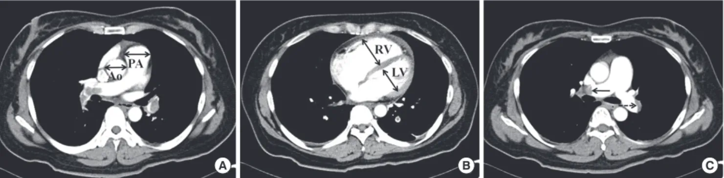
We reviewed patients’ medical records for clinical parameters, imaging findings, and interviewed patients in the outpatient clinic or by phone. Clinical parameters included age, sex, body mass index (BMI), hypertension, diabetes, current smoking, his- tory of significant coronary artery disease (CAD) or cerebrovas- cular accident (CVA), creatinine, total cholesterol, low-density lipoprotein (LDL) cholesterol, high-density lipoprotein (HDL) cholesterol, triglyceride, C-reactive protein (CRP) on the first hospital day, B-type natriuretic peptide (BNP) on the first hos- pital day, D-dimer on the first hospital day, and blood pressure (BP) on admission. Shock was defined as systolic BP < 90 mmHg or signs of poor tissue perfusion. Imaging data included CT and echocardiographic parameters. The dimensions of the right (RV) and left (LV) ventricles at the atrioventricular valvular plane and diameters of the aorta (Ao) and main pulmonary artery (PA) in the axial plane were measured on CT (Fig. 1A, B). RV dimen- sion/LV dimension (CT RV/LV ratio) and PA diameter/Ao diam- eter (CT PA/Ao ratio) were calculated. To define CT obstruction index, arterial trees of each lung were regarded as 10 segmental arteries. The presence of embolism in a segmental artery was scored as 1 or 2 points according the degree of obstruction (Fig.
1C). CT obstruction index (%) was calculated according to the equation: ∑ (n × d)/40 × 100, where n = the number of segmen- tal arteries affected with embolism (1 to 20) and d = degree of obstruction (no = 0, partial = 1, and total = 2) (10, 11). If emboli were present in the proximal pulmonary artery, the score was
equal to the number of segmental arteries arising distally. This index expresses the percentage of pulmonary arterial obstruc- tion by thromboemboli.
Measurement using 2-dimensional Doppler echocardiogra- phy was performed according to the guidelines of the American Society of Echocardiography (12, 13). LV ejection fraction (LVEF) was measured by modified biplane Simpson’s method from apical 4-chamber and 2-chamber views. RV end-diastolic di- mension (RVEDD) was measured at the mid-RV level in apical 4-chamber view. Severity of tricuspid regurgitations (TR) were defined as mild, moderate, and severe according to color TR jet area of < 5 cm2, 5 to 10 cm2, and > 10 cm2 in apical 4-chamber views, respectively (14). Significant TR was defined as moderate to severe TR. Maximal velocity of TR flow was measured by con- tinuous wave Doppler in the apical 4-chamber view. Systolic pulmonary artery pressure (SPAP) was calculated using the con- ventional simplified Bernoulli equation. Composite endpoint was defined as all-cause mortality and recurrence of PE.
Clinical factors including age (> 65 yr), sex, BMI (> 25 kg/m2), hypertension, diabetes, current smoking, history of significant CAD or CVA, creatinine (> 1.2 mg/dL), total cholesterol (> 200 mg/dL), LDL cholesterol (> 120 mg/dL), HDL cholesterol (< 40 mg/dL in men, < 50 mg/dL in women), triglyceride (> 150 mg/
dL) (15), CRP (> 5 mg/L), BNP (> 100 pg/mL), D-dimer (> 20 μg/mL), cancer, metastatic cancer, and shock on admission were analyzed as prognostic factors of the composite endpoint.
As for imaging parameters, CT RV/LV ratio, CT PA/Ao ratio, CT obstruction index, RVEDD, significant TR, SPAP, and LVEF were analyzed as prognostic factors of composite endpoint.
Treatment
Patients were treated by surgery, thrombolytics, unfractionated heparin, or low molecular weight heparin according to vital signs and at the attending physicians’ discretion. The patients in shock on admission were treated by surgery or thrombolyt- ics. At discharge, warfarin was maintained for all patients with- out contraindications. Contraindications of warfarin were bleed- ing diathesis, recent major bleeding, pregnancy, and hypersen-
A B C
Fig. 1. Measurement of CT parameters. Measurement of diameters of the aorta and main pulmonary artery (A), and dimensions of the right and left ventricles (B). (C) An exam- ple of CT obstruction index; the right (solid arrow) and left (dashed arrow) pulmonary arteries are obstructed totally and partially, respectively. CT obstruction index is 75%.
sitivity. Warfarin was maintained with a target prothrombin time (international normalized ratio, INR) between 2.0 and 3.0 for, at least, 3 to 6 months. If patients had contraindications to antico- agulation or recurrent PE during maintenance of warfarin, infe- rior vena cava filtering was performed.
Follow-up
The patients were followed up at the outpatient clinic on a regu- lar basis. If patients had acute symptoms including dyspnea and chest pain, patients were encouraged to visit the emergen- cy room. Chest CT or echocardiography was performed accord- ing to the patients’ complaints. If patients did not visit the out- patient clinic, we interviewed them by phone.
Statistical analysis
Results are expressed as the mean ± standard deviation for data distributed normally. Baseline characteristics and imaging pa- rameters were analyzed by ANOVA and chi-squared test. Ka- plan-Meier method was used for comparison of all-cause mor- tality, mortality from PE and recurrence rate of PE between the groups. Prognostic factors of the composite endpoint were also analyzed by univariate and multivariate Cox proportional haz- ard models. The assumption was assessed by log-minus-log- survival function and found that the proportion hazards assump- tion was reasonable. Parameters of a P value ≤ 0.1 by univari- ate analysis were included for multivariate analysis. Hazard ra- tios (HR) and 95% confidence intervals (CI) were calculated. A P value < 0.05 was considered significant. The data were ana- lyzed using the Statistical Package for the Social Sciences (SPSS, version 15.0; SPSS, Inc., Chicago, IL, USA).
Ethics statement
The study protocol was approved by the institutional review board of the Seoul St. Mary’ Hospital (IRB No. XC10OIMI0120K).
Informed consent was exempted by the board because this was a retrospective study.
RESULTS
Baseline patient characteristics
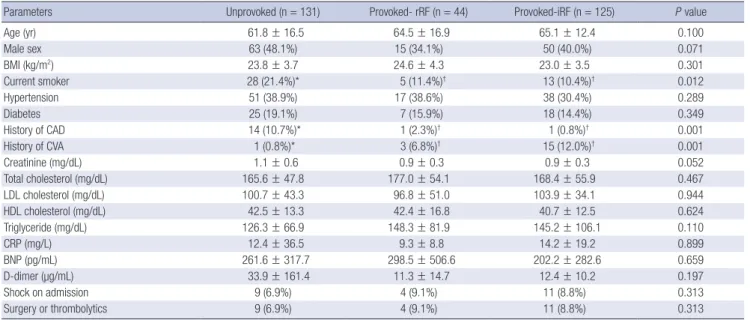
Table 1 reports baseline patient characteristics. Three hundred patients (mean age, 63.6 ± 15.0 yr; 42.8% male) were included.
One hundred thirty-one (43.7%), 44 (14.7%), and 125 (41.7%) patients were in the unprovoked, provoked-rRF, and provoked- iRF groups, respectively. In the provoked group, risk factors in order of frequency were cancer, immobilization, and estrogen supplementation (Table 2). Lung cancer was the most frequent cancer related to PE. Neurologic sequelae were the most fre- quent causes of immobilization related to PE. The number of the patients with metastatic cancer was 65 (58.6% of patients with cancer in the provoked group). Mean follow-up period was 25.4 ± 33.7 months. Twenty-three patients were lost to follow-up.
Clinical factors
The unprovoked group had significantly more current smokers (P = 0.012) and history of CAD (P < 0.001), and less frequent history of CVA (P < 0.001) than the provoked groups (Table 1).
There were no significant differences in age, sex, BMI, hyperten- sion, diabetes, creatinine, cholesterol, triglyceride, CRP, BNP, D-dimer, and frequency of shock on admission between the 3 groups.
Table 1. Baseline patients’ characteristics
Parameters Unprovoked (n = 131) Provoked- rRF (n = 44) Provoked-iRF (n = 125) P value
Age (yr) 61.8 ± 16.5 64.5 ± 16.9 65.1 ± 12.4 0.100
Male sex 63 (48.1%) 15 (34.1%) 50 (40.0%) 0.071
BMI (kg/m2) 23.8 ± 3.7 24.6 ± 4.3 23.0 ± 3.5 0.301
Current smoker 28 (21.4%)* 5 (11.4%)† 13 (10.4%)† 0.012
Hypertension 51 (38.9%) 17 (38.6%) 38 (30.4%) 0.289
Diabetes 25 (19.1%) 7 (15.9%) 18 (14.4%) 0.349
History of CAD 14 (10.7%)* 1 (2.3%)† 1 (0.8%)† 0.001
History of CVA 1 (0.8%)* 3 (6.8%)† 15 (12.0%)† 0.001
Creatinine (mg/dL) 1.1 ± 0.6 0.9 ± 0.3 0.9 ± 0.3 0.052
Total cholesterol (mg/dL) 165.6 ± 47.8 177.0 ± 54.1 168.4 ± 55.9 0.467
LDL cholesterol (mg/dL) 100.7 ± 43.3 96.8 ± 51.0 103.9 ± 34.1 0.944
HDL cholesterol (mg/dL) 42.5 ± 13.3 42.4 ± 16.8 40.7 ± 12.5 0.624
Triglyceride (mg/dL) 126.3 ± 66.9 148.3 ± 81.9 145.2 ± 106.1 0.110
CRP (mg/L) 12.4 ± 36.5 9.3 ± 8.8 14.2 ± 19.2 0.899
BNP (pg/mL) 261.6 ± 317.7 298.5 ± 506.6 202.2 ± 282.6 0.659
D-dimer (µg/mL) 33.9 ± 161.4 11.3 ± 14.7 12.4 ± 10.2 0.197
Shock on admission 9 (6.9%) 4 (9.1%) 11 (8.8%) 0.313
Surgery or thrombolytics 9 (6.9%) 4 (9.1%) 11 (8.8%) 0.313
Statistical significance was evaluated by ANOVA and chi-squared test. *,†The same letters indicate insignificant differences between groups based on Tukey’s multiple compari- son test. rRF, reversible risk factor; iRF, irreversible risk factor; BMI, body mass index; CAD, coronary artery disease; CVA, cerebrovascular accident; LDL, low-density lipoprotein;
HDL, high-density lipoprotein; CRP, C-reactive protein; BNP, B-type natriuretic peptide.
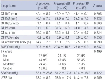
Imaging parameters
The numbers of patients who underwent both CT and echocar- diography were 87, 27, and 80 in the unprovoked, provoked-rRF, and provoked-iRF groups, respectively. There were no signifi- cant differences in imaging parameters between the 3 groups
(Table 3).
Table 2. Risk factors for pulmonary embolism in the provoked group
Risk factors Frequency (%)
Cancer Lung Biliary tract Colon Hematologic Stomach Liver Pancreas Gynecologic Breast Urologic Brain
Oronasopharyngeal Miscellaneous
111 (65.7) 37 (33.3)
9 (8.1) 9 (8.1) 9 (8.1) 7 (6.3) 7 (6.3) 6 (5.4) 6 (5.4) 5 (4.5) 4 (3.6) 3 (2.7) 2 (1.8) 7 (6.3) Immobilization
Neurologic sequelae
Fracture of the lower extremities Admission to ICU due to medical disease Major surgery
57 (33.7) 22 (38.6) 14 (24.6) 12 (21.1) 9 (15.8)
Estrogen supplementation 1 (0.6)
Table 3. Computed tomography and echocardiographic data
Image items Unprovoked
(n = 87)
Provoked-rRF (n = 27)
Provoked-iRF (n = 80) P value CT RVD (mm) 42.1 ± 8.7 42.2 ± 7.9 41.4 ± 9.1 0.706 CT LVD (mm) 40.1 ± 7.9 38.9 ± 7.5 38.3 ± 7.2 0.135 CT RV/LV ratio 1.1 ± 0.4 1.1 ± 0.4 1.1 ± 0.4 0.980 CT PA (mm) 31.0 ± 5.0 31.6 ± 6.0 31.0 ± 4.8 0.833 CT Ao (mm) 36.2 ± 5.0 35.2 ± 4.1 35.4 ± 4.7 0.224 CT PA/Ao ratio 0.9 ± 0.2 0.9 ± 0.1 0.9 ± 0.1 0.358 CT obstruction index (%) 14.2 ± 7.5 15.0 ± 7.5 12.5 ± 6.8 0.290 RVEDD (mmHg) 30.6 ± 9.6 29.8 ± 16.6 27.0 ± 9.9 0.347 TR grade
No Mild Moderate Severe
17.9%
44.9%
24.4%
12.8%
21.1%
47.4%
31.6%
0%
20.9%
55.8%
16.3%
7.0%
0.499
SPAP (mmHg) 53.4 ± 25.8 51.2 ± 17.8 49.4 ± 16.2 0.122 LVEF (%) 63.3 ± 8.6 58.6 ± 17.0 64.2 ± 7.8 0.559 Statistical significance was evaluated by ANOVA and chi-squared test. rRF, reversible risk factor; iRF, irreversible risk factor; CT, computed tomography; RV, right ventricle;
LV, left ventricle; CT RVD, RV dimension from CT; CT LVD, LV dimension from CT; PA, pulmonary artery; Ao, aorta; CT PA, pulmonary artery diameter from CT; CT Ao, aorta diameter from CT; RVEDD, RV end-diastolic dimension from echocardiography; TR, tricuspid regurgitation; SPAP, systolic pulmonary artery pressure; LVEF, LV ejection fraction.
Fig. 2. Cumulative mortality and recurrence rate in each group. (A) Cumulative all- cause mortality, (B) cumulative mortality from pulmonary embolism (PE), and (C) cu- mulative recurrence of PE by Kaplan-Meier method between unprovoked, provoked with reversible risk factors (provoked-rRF), and provoked with irreversible risk factors (provoked-iRF) groups.
Cumulative all-cause mortality Cumulative mortality from PE
Days since PE Days since PE
0 100 200 300 400 500 600 700 0 100 200 300 400 500 600 700
1.0 0.8 0.6 0.4 0.2 0.0
1.0 0.8 0.6 0.4 0.2 0.0 Unprovoked
Provoked-rRF Provoked-iRF
Unprovoked Provoked-rRF Provoked-iRF
P < 0.001 P < 0.001
A B
Cumulative recurrence rate
Days since PE
0 100 200 300 400 500 600 700
1.0 0.8 0.6 0.4 0.2 0.0
Unprovoked Provoked-rRF Provoked-iRF
P = 0.034
C
Treatment
The numbers of patients treated with surgery or thrombolytics were 10 (3.3%) and 16 (5.3%), respectively. The numbers of pa- tients treated with unfractionated heparin only or low molecu- lar weight heparin only were 77 (25.7%), and 197 (65.7%), re- spectively. After discharge, warfarin was maintained for all sur- vivors who had no contraindications of anticoagulation for 3 to 6 months. The proportions of patients with time in therapeutic range of anticoagulation > 60% were 68.7%, 73.5%, and 67.6%
in the unprovoked, provoked-rRF, and provoked-iRF groups, respectively. There were no significant differences in time in therapeutic range of anticoagulation between the groups (P = 0.455). Inferior vena cava filtering was performed for 37 (12.3%) patients; 16 (12.2%), 10 (22.7%), and 11 (8.8%) in the unprovoked, provoked-rRF, and provoked-iRF groups, respectively. There were no significant differences in the numbers of patients who underwent inferior vena cava filtering between the groups (P = 0.054).
Mortality
The all-cause mortality and mortality from PE during the follow- up period was 33.3%, and 18.7%, respectively. The all-cause mortality in the unprovoked, provoked-rRF, and provoked-iRF groups were 15.3%, 18.2%, and 56.8%, respectively. Mortality from PE in the unprovoked, provoked-rRF, and provoked-iRF groups were 9.2%, 11.4%, and 31.2%, respectively. By the Kaplan- Meier method, the provoked-iRF group had significantly higher all-cause mortality, mortality from PE and recurrence of PE than the unprovoked and provoked-rRF groups (P < 0.001, P < 0.001 and P = 0.034, respectively; Fig. 2). However, there were no sig- nificant differences of all-cause mortality, mortality from PE, and recurrence of PE between the unprovoked and provoked- rRF groups (P = 0.377, P = 0.495, and P = 0.410, respectively).
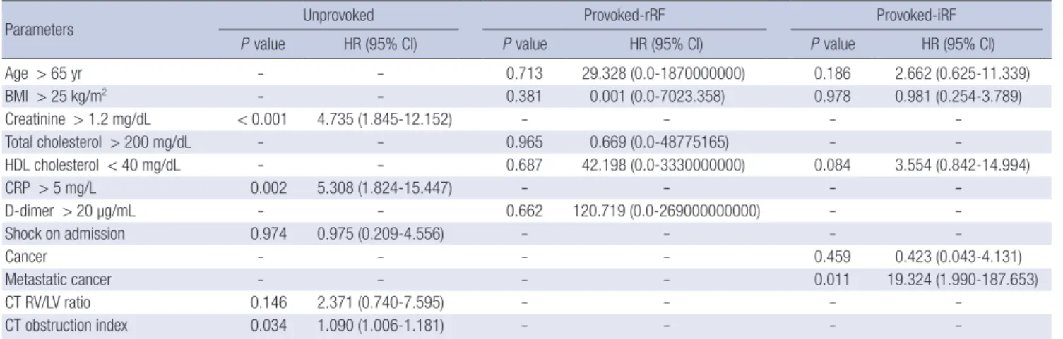
Clinical factors related to the composite endpoint
The rate of composite endpoint was 36.6% in all patients and 19.8%, 20.5%, and 60.0% in the unprovoked, provoked-rRF, and provoked-iRF groups, respectively. By univariate analysis, cre- atinine, CRP, and shock on admission were significantly related to the composite endpoint in the unprovoked group. Age, BMI, HDL cholesterol, cancer, and metastatic cancer were significant- ly related to composite endpoint in the provoked-iRF group.
Age, BMI, total cholesterol, HDL cholesterol, and D-dimer were significantly related to the composite endpoint in the provoked- rRF group. By multivariate analysis, prognostic factors signifi- cantly related to composite endpoint in the unprovoked group were creatinine (> 1.2 mg/dL; P < 0.001), and CRP (> 5 mg/L;
P = 0.002). In the provoked-iRF group, the prognostic factors significantly related to the composite endpoint was metastatic cancer (P = 0.011; Table 4). There were no significant prognostic factors for the composite endpoint in the provoked-rRF group.
Imaging parameters related to the composite endpoint By univariate analysis, CT RV/LV ratio, and CT obstruction in- dex were significantly related to the composite endpoint in the unprovoked group; no imaging parameters were significant in the provoked groups. By multivariate analysis, the CT obstruc- tion index was significantly related to the composite endpoint in the unprovoked group (P = 0.034; Table 4).
DISCUSSION
This was a retrospective study to elucidate the clinical and im- aging characteristics, mortality, and prognostic factors in pa- tients with unprovoked PE, provoked PE with reversible risk fac- tors, and provoked PE with irreversible risk factors. This study showed that the unprovoked group was related to smoking and history of CAD, and that provoked-iRF group had higher mor-
Table 4. Clinical and imaging parameters related to the composite endpoint on multivariate analysis
Parameters Unprovoked Provoked-rRF Provoked-iRF
P value HR (95% CI) P value HR (95% CI) P value HR (95% CI)
Age > 65 yr - - 0.713 29.328 (0.0-1870000000) 0.186 2.662 (0.625-11.339)
BMI > 25 kg/m2 - - 0.381 0.001 (0.0-7023.358) 0.978 0.981 (0.254-3.789)
Creatinine > 1.2 mg/dL < 0.001 4.735 (1.845-12.152) - - - -
Total cholesterol > 200 mg/dL - - 0.965 0.669 (0.0-48775165) - -
HDL cholesterol < 40 mg/dL - - 0.687 42.198 (0.0-3330000000) 0.084 3.554 (0.842-14.994)
CRP > 5 mg/L 0.002 5.308 (1.824-15.447) - - - -
D-dimer > 20 µg/mL - - 0.662 120.719 (0.0-269000000000) - -
Shock on admission 0.974 0.975 (0.209-4.556) - - - -
Cancer - - - - 0.459 0.423 (0.043-4.131)
Metastatic cancer - - - - 0.011 19.324 (1.990-187.653)
CT RV/LV ratio 0.146 2.371 (0.740-7.595) - - - -
CT obstruction index 0.034 1.090 (1.006-1.181) - - - -
Statistical significance was evaluated by Cox proportional hazard model. -, P value > 0.1 on univariate analysis. rRF, reversible risk factor; iRF, irreversible risk factor; HR, haz- ard ratio; CI, confidence interval; BMI, body mass index; HDL, high-density lipoprotein; CRP, C-reactive protein; CT, computed tomography; RV, right ventricle; LV, left ventricle;
CT RV/LV ratio, RV dimension/LV dimension from CT.
tality than the unprovoked and provoked-rRF groups. In addi- tion, this study showed that creatinine, CRP, and CT obstruction index were poor prognostic factors in unprovoked PE.
It is not surprising that CVA is more frequent in the provoked group because patients with neurologic sequelae due to CVA were included in the provoked groups. Recent studies showed strong associations between unprovoked PE and risk factors of atherosclerosis (6, 7, 9). The associations of unprovoked PE with smoking or CAD reported here are consistent with previous studies. However, the lack of associations with other risk factors including age, sex, BMI, hypertension, diabetes, and hyperlipid- emia are not consistent. This may be explained by the fact that although PE and atherosclerosis share the common pathophys- iology of endothelial dysfunction and abnormal blood compo- nents, blood stasis in veins could be different from that in arter- ies (4, 16).
Recent studies reported that shock on admission, high levels of BNP or troponin, RV dilation on CT or echocardiography, RV dysfunction, massive PE and CT obstruction index were signifi- cant predictors of mortality (11, 17-21). In this study, however, only the CT obstruction index was a significant prognostic fac- tor in unprovoked PE; other factors including shock on admis- sion, high BNP, and RV dilation were not significant prognostic factors. This might be due to the influence of hemodynamic in- stability on short-term mortality, but not long-term mortality and recurrence of PE. CT obstruction index was correlated with the amount of embolism. Our data indicate that a large amount of embolism is related to poor prognosis.
In this study, renal insufficiency and a marker of the inflam- matory response were also found to be important prognostic factors in unprovoked PE, in addition to the amount of throm- bus in the pulmonary arteries. It has been reported that chronic kidney disease is associated with increased risk of venous throm- boembolism (22). In this study, poor prognosis in patients with renal insufficiency might be related to risk of venous thrombo- embolism.
It has been reported that high CRP is related to risk of venous thromboembolism (23, 24). However, the mechanisms of asso- ciation of CRP with risk of venous thromboembolism are not clear (25, 26). However, elevated CRP has not been previously reported to be related to PE prognosis. We suggest that CRP is important in predicting prognosis of unprovoked PE. In the provoked-iRF groups, there were no prognostic factors except metastatic cancer; this is likely because the underlying disease, general physical condition, or deconditioning of the patients may be more influential than PE itself on prognosis. It is known that unprovoked PE has high recurrence rate. The reason of no significant differences of recurrence rate between unprovoked and provoked PE with reversible cause in the present study could be because many patients with unprovoked PE were treated with long-term anticoagulation therapy.
There were no studies that evaluated clinical characteristics and mid- to long-term prognosis of PE classified into unpro- voked, provoked-rRF, and provoked-iRF. We demonstrated that provoked PE with irreversible risk factors has higher all-cause mortality, mortality from PE and recurrence of PE than both un- provoked PE and provoked PE with reversible risk factors. This is likely due to the fact that patients with provoked PE and irre- versible risk factors had more serious comorbidities including refractory cancer, and neurologic sequelae. It is not surprising that patients in the provoked-iRF group had a higher recurrence rate than the unprovoked and provoked-rRF groups because they had risk factors for a life-long period after the first episode of PE.
Because we did not perform CT and echocardiography in ev- ery patient, we did not analyze CT and echocardiographic data from the entire study population. Severity of RV dysfunction was not analyzed in this study because quantification methods of RV dysfunction were not consistent for the study period. Cardi- ac markers such as creatine kinase-MB and troponin were not included, because these cardiac markers were not checked rou- tinely in patients with PE at our center. Markers related to inher- ited thrombophilia such as protein C, protein S, and coagula- tion factors are also not included into this study because blood was sampled after heparin injection in some patients.
In conclusion, provoked PE with irreversible risk factors has a poorer prognosis than unprovoked PE and provoked PE with reversible risk factors. Prognosis of unprovoked PE is not differ- ent from that of provoked PE with reversible risk factors. Unpro- voked PE is related to risk factors for atherosclerosis, including smoking, and history of CAD. Renal insufficiency, high CRP, and CT obstruction index are significant prognostic factors of unprovoked PE. We suggest that patients with unprovoked PE combined with renal insufficiency, elevated CRP, or a high CT obstruction index should be monitored more closely for poten- tial adverse outcomes and that aggressive anticoagulation should be performed for patients with provoked PE with irreversible risk factors.
REFERENCES
1. Jang MJ, Bang SM, Oh D. Incidence of venous thromboembolism in Ko- rea: from the health insurance review and assessment service database. J Thromb Haemost 2011; 9: 85-91.
2. In KH. The national survey of acute pulmonary thromboembolism in Korea. Tuberc Respir Dis 2003; 54: 5-14.
3. White RH, Keenan CR. Effects of race and ethnicity on the incidence of venous thromboembolism. Thromb Res 2009; 123: S11-7.
4. Lopez JA, Chen J. Pathophysiology of venous thrombosis. Thromb Res 2009; 123: S30-4.
5. Heit J. Venous thromboembolism epidemiology: implications for preven- tion and management. Semin Thromb Hemost 2002; 28 (Suppl 2): 3-13.
6. Prandoni P, Bilora F, Marchiori A, Bernardi E, Petrobelli F, Lensing AWA,
Prins MH, Girolami A. An association between atherosclerosis and ve- nous thrombosis. N Engl J Med 2003; 348: 1435-41.
7. Prandoni P, Ghirarduzzi A, Prins MH, Pengo V, Davidson BL, Sorensen H, Pesavento R, Iotti M, Casiglia E, Iliceto S, et al. Venous thromboem- bolism and the risk of subsequent symptomatic atherosclerosis. J Thromb Haemost 2006; 4: 1891-6.
8. Ageno W, Becattini C, Brighton T, Selby R, Kamphuisen PW. Cardiovas- cular risk factors and venous thromboembolism; a meta-analysis. Cir- culation 2008; 117: 93-102.
9. Jang MJ, Choi WI, Bang SM, Lee T, Kim YK, Ageno W, Oh D. Metabolic syndrome is associated with venous thromboembolism in the Korean population. Arterioscler Thromb Vasc Biol 2009; 29: 311-5.
10. Qanadli SD, Hajjam ME, Vieillard-Baron A, Joseph T, Mesurolle B, Oli- va VL, Barre O, Bruckert F, Dubourg O, Lacombe P. New CT index to quantify arterial obstruction in pulmonary embolism: comparison with angiographic index and echocardiography. AJR Am J Roentgenol 2001;
176: 1415-20.
11. Van der Meer RW, Pattynama PMT, Van Strijen MJL, Van den Berg-Hui- jsmans AA, Hartmann IJC, Putter H, De Roos A, Huisman MV. Right ventricular dysfunction and pulmonary obstruction index at helical CT:
prediction of clinical outcome during 3-month follow-up in patients with acute pulmonary thromboembolism. Radiology 2005; 235: 798-803.
12. Lang RM, Bierig M, Devereux RB, Flachskampf FA, Foster E, Pellikka PA, Picard MH, Roman MJ, Seward J, Shanewise JS, et al. Recommenda- tions for chamber quantification: a report from the American Society of Echocardiography’s guidelines and standards committee and the cham- ber quantification writing group, developed in conjunction with the Eu- ropean Association of Echocardiography, a branch of the European So- ciety of Cardiology. J Am Soc Echocardiogr 2005; 18: 1440-63.
13. Quinones MA, Otto CM, Stoddard M, Waggoner A, Zoghbi WA. Recom- mendations for quantification of Doppler echocardiography: a report from the Doppler quantification task force of the nomenclature and stan- dards committee of the American Society of Echocardiography. J Am Soc Echocardiogr 2002: 15: 167-84.
14. Zoghbi WA, Enriquez-Sarano M, Foster E, Grayburn PA, Kraft CD, Levine RA, Nihoyannopoulos P, Otto CM, Quinones MA, Rakowski H, et al. Recommendations for evaluation of the severity of native valvular regurgitation with two-dimensional and Doppler echocardiography. J Am Soc Echocardiogr 2003; 16: 777-802.
15. Expert Panel on Detection, Evaluation, and Treatment of High Blood
Cholesterol in Adults. Executive summary of the third report of the na- tional cholesterol education program (NCEP) expert panel on detection, evaluation, and treatment of high blood cholesterol in adults (Adult Treatment Panel III). JAMA 2001; 285: 2486-97.
16. Libby P, Ridker PM, Hansson GK. Inflammation in atherosclerosis: from pathophysiology to practice. J Am Coll Cardiol 2009; 54: 2129-38.
17. Kucher N, Rossi E, De Rosa M, Goldhaber SZ. Massive pulmonary em- bolism. Circulation 2006; 113: 577-82.
18. Sanchez O, Trinquart L, Colombet I, Durieux P, Huisman MV, Chatellier G, Meyer G. Prognostic value of right ventricular dysfunction in patients with haemodynamically stable pulmonary embolism: a systematic re- view. Eur Heart J 2008; 29: 1569-77.
19. Ozsu S, Karaman K, Mentese A, Ozsu A, Karahan SC, Durmus I, Oztu- na F, Kosucu P, Bulbul Y, Ozlu T. Combined risk stratification with com- puterized tomography/echocardiography and biomarkers in patients with normotensive pulmonary embolism. Thromb Res 2010; 126: 486-92.
20. Sanchez O, Trinquart L, Caille V, Couturaud F, Pacouret G, Meneveau N, Verschuren F, Roy PM, Parent F, Righini M, et al. Prognostic factors for pulmonary embolism: the PREP study, a prospective multicenter cohort study. Am J Respir Crit Care Med 2010; 181: 168-73.
21. Stein PD, Janjua M, Matta F, Pathak PK, Jaweesh F, Alrifai A, Chughtai HL. Prognosis based on creatinine kinase isoenzyme MB, cardiac tropo- nin I, and right ventricular size in stable patients with acute pulmonary embolism. Am J Cardiol 2011; 107: 774-7.
22. Wattanakit K, Cushman M, Stehman-Breen C, Heckbert SR, Folsom AR. Chronic kidney disease increases risk for venous thromboembolism.
J Am Soc Nephrol 2008; 19: 135-40.
23. Zacho J, Tybjaerg-Hansen A, Nordestgaard BG. C-reactive protein and risk of venous thromboembolism in the general population. Arterioscler Thromb Vasc Biol 2010; 30: 1672-8.
24. Folsom AR, Lutsey PL, Astor BC, Cushman M. C-reactive protein and venous thromboembolism: a prospective investigation in the ARIC co- hort. Thromb Haemost 2009; 102: 615-9.
25. Fox EA, Kahn SR. The relationship between inflammation and venous thrombosis: a systematic review of clinical studies. Thromb Haemost 2005; 94: 362-5.
26. Lippi G, Favaloro EJ, Montagnana M, Franchini M. C-reactive protein and venous thromboembolism: causal or casual association? Clin Chem Lab Med 2010; 48: 1693-701.