The Effect of Saururus Chinensis Extracts on Antioxidant Activity and Melanin Synthesis
Somi Jung1, Hyejeong Park2, Jaeho Kim3, Yunghee Oh2 and Moon-Moo Kim2*
1Department of Chemistry, Dong-Eui University, 176, Eomgwang-ro, Busanjin-gu, Busan 47340, Korea
2Applied Chemistry, Dong-Eui University, 176, Eomgwang-ro, Busanjin-gu, Busan 47340, Korea
3Department of Chemistry & Biology, Dong-Eui University, 176, Eomgwang-ro, Busanjin-gu, Busan 47340, Korea Received May 27, 2020 /Revised June 22, 2020 /Accepted July 1, 2020
Saururus chinensis has white roots, leaves, and flowers and is known to have antibacterial activity and anti-cancer efficacy. The aim of this study was to investigate the effect of the ethyl acetate fraction of a methanol extract of Saururus chinensis (SCEA) on antioxidant activity and melanin synthesis.
SCEA at 64 μg/ml showed 62% of the DPPH radical scavenging activity of vitamin C, and its re- ducing power was 33% greater than that of vitamin C. Tyrosinase activity was 26% higher and mela- nin synthesis was 44% higher in the presence of SCEA at 64 μg/ml than in a blank group in a dopa oxidation assay. MTT assay showed that SCEA displayed cytotoxicity above 0.5 μg/ml, and SCEA at 1 μg/ml increased melanin synthesis by 69% in live B16F1 cells. SCEA was also separated into 13 frac- tions by silica column chromatography, and fraction 2 (Fr. 2) showed the highest DPPH radical scav- enging activity, reducing power, and melanin synthesis. SCEA also promoted melanin production in live cells. LC-MASS analysis showed that Fr.2 had a molecular weight of 239, and these findings sug- gest that SCEA could be available for the promotion of melanin synthesis in black hair.
Key words : B16F1, melanin synthesis, Saururus chinensis, tyrosinase
*Corresponding author
*Tel : +82-51-890-1511, Fax : +82-51-890-2620 E-mail : [email protected]
This is an Open-Access article distributed under the terms of the Creative Commons Attribution Non-Commercial License (http://creativecommons.org/licenses/by-nc/3.0) which permits unrestricted non-commercial use, distribution, and reproduction in any medium, provided the original work is properly cited.
서 론
한국에서 노인 인구의 증가는 노화관련 질환에 대한 관심을 불러 일으키고 있다. 노화란 연령이 증가함에 따라 신체의 구 조와 기능이 점진적으로 저하되어 결국 질병과 사망을 초래하 는 과정이다. 노화의 원인 중 주요한 요소는 활성산소에 의하 여 세포가 손상되는 것으로 보고되어 있다[25]. 활성산소는 호 흡과정에서 DNA, 단백질, 지질, 탄수화물을 산화시켜 세포를 손상시키는 반응이다[5]. 활성산소를 제거하는 항산화 효소의 활성은 노화됨에 따라 저하된다. 노화가 진행되어 나타나는 현상 중 가장 두드러진 표현형이 백발화 현상이다. 염색을 하 게 된다면 빠르게 흑발로 만들 수 있지만 일시적이어서, 곧 새 백발이 성장하게 되어 다시 염색을 하게 된다. 염색을 반복 하게 되면 화학약품으로 인한 모발 손상이 일어나 부작용이 축적됨으로, 백발화 현상을 억제할 수 있는 멜라닌 생성을 촉 진시킬 수 있는 흑모제의개발이 필요하다. 백발화는 멜라닌의 합성능력이 감소하여 나타나는 현상으로[20], 이를 예방하기 위하여 국내외적으로 멜라닌의 합성 조절에 관한 연구가 드물
게 수행되어 왔으며, 특히 tyrosine을 3,4-dihydroxyphenyl- alanine (DOPA)로 전환하는 tyrosinase에 영향을 주는 요인에 초점이 맞춰 많은 연구가 진행되고 있다. Tyrosinase는 ty- rosine hydroxylase로 작용하여 tyrosine을 DOPA로 전환한다 [15]. 뿐만 아니라 DOPA oxidase로도 작용하여 DOPA를 dop- aquinone으로 전환하는 속도 결정단계의 초기반응에 관여하 여 melanin 합성을 돕는 효소이다[26]. 최근 연구에 의하면 삼백초의 에탄올 추출물이 멜라닌 생성을 억제해 미백효과가 있다고 알려져 있다[23]. 또한, 다양한 삼백초 추출물에 관한 논문에도 미백효과가 나타난다고 알려져 있지만 본 연구에서 발굴된 삼백초 추출물은 멜라닌 생성을 촉진하는 것으로 확인 되었으며, 이와 관련된 연구는 현재까지 보고 된 바가 없다.
최근에 국내외에서는 합성 의약품의 부작용을 막기 위해 천 연물을 식품 보충제 또는 의약 및 화장품 제제의 구성 성분으 로도 사용하는 연구가 활발하다[4]. 본 연구에 사용한 삼백초 (saururus chinensis)는 여러해살이 초본으로 꽃이 필 무렵 윗부 분 잎 2~3개가 백색을 띠고, 뿌리와 꽃도 백색을 띠므로 삼백초 라 일컬어진다[21]. 삼백초 추출물의 성분에는 quercetin quer- cetrin, isoquercitirin, rutin 및 수용성 tannin 등이 있다고 알려 져 있다. 이 중 quercetin과 quercetrin은 flavonoid의 일종이다 [1]. 식물체에 존재하는 flavonoid는 항돌연변이 및 항암활성 을 갖는 것으로 보고되어 있다. Quercetin은 항돌연변이 및 항암활성뿐만 아니라 동맥경화를 유발할 수 있는 인자인 Low Density Lipoprotein (LDL)의 산화와 cytotoxicity 억제 효과도 보고되어 있고[18] 천연 항산화제로 활성산소를 제거해 노화
852 생명과학회지 2020, Vol. 30. No. 10
와 암, 심장병 예방 효과를 나타낸다고 보고되어 있다[2].
따라서 본 연구에서는 삼백초의 유효성분을 분리하기 위하 여 MeOH 추출물을 용매별로 분획하고 칼럼 크로마토그래피 실험을 진행하여 각각의 분획물들의 항산화활성과 멜라닌 합 성능력을 검증해 천연 항산화제 및 흑모제품을 위한 기능성 화장품의 원료로서의 가능성을 규명하고자 한다.
재료 및 방법
재료
본 실험에 사용된 국내산 삼백초(Saururus chinensis)는 서울 동대문구 약령중앙로에 있는 동광종합물산(주)에서 구입하였 으며, 건조시킨 후 실온 보관하여 실험에 사용하였다. 추출 및 분획 용매로 methanol, n-hexane, ethyl acetate, n-butanol 을 사용하였다. 세포배양을 위한 Dulbecco’s Modified Eagle’s Medium (DMEM), Trypsin-EDTA, penicillin (10,000 U/ml)/
streptomycin (10,000 μg/ml)/ amphotericin (2,500 μg/ml), fetal bovine serum (FBS) 시약은 Gibco BRL, Life Technolo- gies (Paisley, Scotland)로 부터 구입하였다. B16F1 cell line은 ATCC (American Type Culture Collection, USA)로 부터 구 입 하였다. MTT reagent, agarose와 기타시약은 Sigma Chem- ical Co. (St. Louis, MO, USA)로 부터 구입하였다. Fraction 2 (Fr. 2)의 정성분석을 위해 High Performance Liquid Chro- matography-Mass Spectrometry (HPLC-MS)의 칼럼은 Pron- tosil Eurobond C18 5um SC-04 (125×4 mm)로 구성된 1260 Infinity HPLC system (Aglilent Corp, USA)를 사용하였다.
시료의 제조
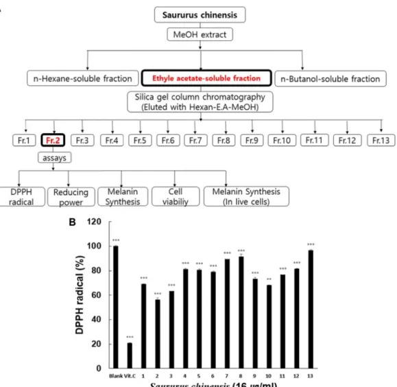
삼백초의 MeOH 추출물은 아래와 같은 방법으로 추출하였 다. Fig. 1A에서 보는 바와 같이 5.5 kg의 삼백초를 완전히 건조시킨 후 분쇄하여, 분쇄된 삼백초를 MeOH 에 3일간 추출 및 여과하고 얻은 165 l의 여과액을 감압 농축 하였다. 농축된 삼백초 MeOH 추출물을 hexane, ethyl acetate, butanol, dis- tilled water (D.W)을 이용하여 용매별로 분획하고 그 중에서 멜라닌 합성 효능이 우수한 결과를 보인 ethyl acetate 분획물 을 실리카겔 칼럼크로마토그래피를 이용해 분리하였다. 또한, Fig 1B에서 보는 바와 같이 칼럼으로 분리한 총 13개의 frac- tion 중 양성대조군에 비해 가장 높은 항산화 효과를 보인 Fr.
2을 본 연구에 이용하였다.
DPPH radical scavenging activity
Brand-Williams [9]의 방법을 변형시켜 DPPH radical에 대 한 Fr. 2의 소거능력을 측정하였다. 최종 부피 500 μl에서 농도 가 1, 2, 4, 8, 16, 32, 64 μg/ml가 되도록 시료를 제조하였고, 각 시료를 시험농도로 처리하고 잘 혼합한 후 실온에서 1시간 동안 반응시켜 517 nm에서 흡광도를 측정하였다. 항산화제로
잘 알려진 vitamin C (100 μg/ml)를 양성대조군으로 사용하여 비교하여 측정하였다. DPPH radical 함량은 시료 첨가군과 대조군의 흡광도 비를 % 값으로 환산하여 나타내었다.
Reducing power assay
Fr. 2 6 μl에 200 mM phosphate buffer (pH 6.5) 20 μl와 1% potassium ferricyanide 20 μl를 순서대로 넣고 교반한 후 50℃에서 20분간 방치하였다. 여기에 10% TCA를 넣어 반응을 종결시킨 뒤 956 xg 10분 동안 원심분리를 한 후 얻은 상층액 25 μl에 증류수 25 μl와 1% ferric chloride를 넣고 교반하였다.
이 과정을 거친 시료를 620 nm에서 흡광도를 측정한다. 양성 대조군으로는 vitamin C (10 μg/ml)를 사용하였다. 시료의 환 원력은 시료 첨가군와 대조군의 흡광도 비를 % 값으로 환산하 여 나타내었다.
멜라닌 합성 측정을 위한 in vitro test
시험관에 0.1 M 인산염완충액(pH 6.5) 45 μl와 L-DOPA 20 μl 그리고 시료액 5 μl을 넣고 2,000 U/ml mushroom tyrosin- ase 20 μl를 순서대로 넣은 다음 37℃에서 1시간 동안 반응시 켰다. 그리고 ELISA reader (ELISA processor II. Behring, Germany)를 이용하여 490 nm에서 흡광도를 측정하였다.
세포독성 측정을 위한 MTT assay
Hansen [7]의 방법에 따라 B16F1 세포에 대한 Fr. 2의 세포 독성을 3-(4,5-dimethyl-2-yl)-2,5-diphenylterrazolium bromide (MTT)를 이용하여 측정하였다.
세포독성평가는 MTT (3-(4,5-dimethythiazol-2-yl)-2,5-di- phenyltetrazolium bromide, Sigma-Aldrich, St. Louis, MO, USA) 시약을 사용하여 실험을 진행하였다. 독성평가에는 mel- anoma cell인 B16F1을 사용하였으며, 7.5×10⁵ cells/well 만큼 의 세포를 96-well plate에 주입한 후 하루 동안 배양하였다.
하루 뒤 배지를 제거하고 적당 농도로 시료를 총 200 μl만큼 투여하고 CO2 배양기에서 다시 한 번 24시간 동안 배양한다.
24시간 후 처리한 시료를 제거하고, MTT 용액을 200 μg/ml의 농도로 하여 96-well plate에 첨가하고 빛을 차단한 채 37℃에 서 3시간 배양한다. 3시간 뒤 PBS buffer로 세척한 뒤 DMSO를 각 well에 200 μl 만큼 주입하고 20분 후 microplate reader로 570 nm의 파장에서 흡광도를 측정하였다
B16F1에서 Melanin 생성 측정
24-well plate에 3×10⁵ cells/well로 세포를 분주하였고, 시 료를 처리하고 1시간 후에 L-DOPA로 melanin생성을 자극 후 48시간 동안 세포를 배양하였다. 세포를 수집하여 125 xg에 서 5분 간 원심 분리하여 침전한 후, 얻은 pellet에 1 N NaOH (10% DMSO) 20 μl를 첨가하고 vortex 후 405 nm에서 흡광도 를 측정하였다. Melanin 표준품(Sigma Chemical Co. (St.
Fig. 1. Screening strategy for the purification of active ingredients from Saururus chinensis (A) Schematic diagram of the extraction procedure and assays for the efficacy of antioxidant and melanin synthesis. (B) The effect of the 13 fractions from Saururus chinensis ethyl acetate extracts on scavenging activity of DPPH radical. Vitamin C at 100 μg/ml was used as a positive control in this experiment. Data are given as means of values ± S.D. from three independent experiments. Level of significance was identified statistically using Student's t test. (**p<0.01, ***p<0.001).
Louis, MO, USA))으로 얻은 표준 검량선을 이용하여 각 well 에서 생성된 멜라닌 양을 산출하였다. 멜라닌은 단위세포(104 cells)에서의 멜라닌 생성량을 비교하였다.
High Performance Liquid Chromatography–Mass Spectrometry spectrum assay
Fr. 2의 정성분석을 위해 High Performance Liquid Chro- matography–Mass Spectrometry (HPLC-MS)를 사용하였다.
Fr. 2 시료를 HPLC에서 순수 분리한 후 HPLC와 연결된 Mass Spectrometer (MS)로 보내어 Positive ion 모드로 각 peak 별 성분에 대해 질량을 분석하였다. 이동상으로 0.1% Formic Acid in ACN과 0.1% Formic Acid in Water 그리고 D.W를 사용하였고, instrument conditions으로 column temperature 25℃, flow rate 0.5 ml/min 조건에서 25분 간 측정하였다.
통계처리
각 실험은 3회 이상 반복 실험을 통하여 그 결과를 얻어 각각의 시료 농도에 대해 평균±표준편차로 나타내었다. 각 시 료 농도 군에 대한 유의 차 검정은 대조군과 비교하여 stu- dent’s test 한 후 p<0.05 값을 통계적으로 유의성 있는 결과로 간주하였다.
결 과
삼백초 추출물의 DPPH radical 소거능
자유 라디칼의 소거 정도를 알아보기 위해 DPPH radical 소거법으로 실험하였다. Fig. 2A에서와 같이 양성대조군으로 사용된 vitamin C와 MeOH 추출물과 ethyl acetate 분획물의 항산화력을 비교한 결과, vitamin C는 100 μg/ml의 농도에서
854 생명과학회지 2020, Vol. 30. No. 10
Fig. 3. The effects of methanol extract, ethyl acetate fraction and fraction 2 on reducing power. (A) The effect of meth- anolic extract and ethyl acetate fraction of Saururus chi- nensis on reducing power. (B) The effect of the fraction 2 on reducing power. The effect of the fraction 2 on re- ducing power. Vitamin C (Vit. C) at 10 μg/ml was used as a positive control. Data are given as means of values
± S.D. from three independent experiments. Level of sig- nificance was identified statistically using Student's t test. (*p<0.05, ***p<0.001)
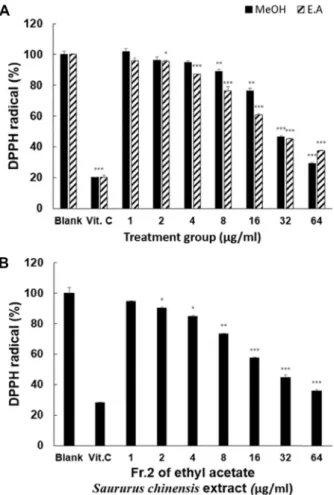
Fig. 2. The effects of methanol extract, ethyl acetate fraction and fraction 2 on scavenging activity of DPPH. (A) The effect of methanolic extract and ethyl acetate fraction of Sauru- rus chinensis on scavenging activity of DPPH radical. (B) The effect of the fraction 2 on scavenging activity of DPPH radical. Vitamin C at 100 μg/ml was used as a positive control in this experiment. Data are given as means of values ± S.D. from three independent experi- ments. Level of significance was identified statistically using Student's t test. (*p<0.05, **p<0.01, ***p<0.001).
80%의 DPPH 소거능을 보여주었고, ethyl acetate 분획물의 경우 4 μg/ml 농도부터 항산화력을 보였으며 64 μg/ml 농도 에서는 62%의 DPPH 소거능을 보였다. MeOH 추출물의 경우 16 μg/ml부터 항산화력을 보였으며 64 μg/ml에서는 70%의 소거능을 보였다. 마지막으로 Fig. 2B는 ethyle acetate층을 칼 럼크로마토그래피를 통해 분획 한 Fr. 2의 DPPH radical 소거 능에 대한 결과로 농도에 비례하여 증가하였으며, 64 μg/ml의 농도에서 64%의 DPPH radical 소거능을 나타났다.
삼백초 추출물의 reducing power
삼백초의 환원력을 측정한 결과, Fig. 3A와 같이 MeOH 추 출물과 ethyl acetate 분획물은 농도에 비례하여 환원력이 증 가하였다. MeOH 추출물은 양성대조군 vitamin C (1,000 μg/
ml)보다 환원력이 낮은 것으로 나타났다. Ethyl acetate 분획물 은 64 μg/ml 의 농도에서 265%로 양성대조군 보다 65% 뛰어 난 환원력을 보였다. 마지막으로 Fig. 3B는 ethyl acetate층을 칼럼크로마토그래피를 이용하여 분획한 Fr. 2의 환원력에 대 한 결과로 농도에 비례하여 환원력이 증가하였고 64 μg/ml 의 농도에서 332%로 양성대조군 vitamin C (1,000 μg/ml) 보 다 108% 높은 환원력을 보였다.
삼백초 추출물의 in vitro tyrosinase 활성에 미치는 영향 Tyrosinase 활성에 대한 삼백초 추출물의 효능을 조사하였 다. Fig. 4A와 같이 삼백초 ethyl acetate 추출물은 8 μg/ml 이상에서 농도가 증가함에 따라 tyrosinase 활성이 증가하였 다. 뿐만 아니라, ethyl acetate 분획물이 MeOH 추출물보다
Fig. 4. The effects of methanol extract, ethyl acetate fraction and fraction 2 on Dopa-oxidation with tyrosinase. (A) The effect of methanolic extract and ethyl acetate fraction of Saururus chinensis on Dopa-oxidation with tyrosinase. (B) The effect of the fraction 2 on Dopa-oxidation with tyros- inase. Vitamin C at 100 μg/ml was used as a positive control in this experiment. Data are given as means of values ± S.D. from three independent experiments. Level of significance was identified statistically using Student's t test. (*p<0.05, ***p<0.001).
Fig. 5. The effects of methanol extract, ethyl acetate fraction and fraction 2 on viability of B16F1 cells. (A) The effect of methanolic extract and ethyl acetate fraction of Saururus chinensis on viability of B16F1 cells. (B) The effect of the fraction 2 on viability of B16F1 cells. The cells were treat- ed with samples at 1, 2, 4, 8 and 16 μg/ml. Cell viability was determined by MTT assay after 24 hr. Data are given as means of values ± S.D. from three independent experiments. Level of significance was identified statisti- cally using Student's t test. (*p<0.05, ***p< 0.001).
tyrosinase 활성이 우수한 것으로 나타났다. MeOH 추출물은 공시험군과 비교하여 64 μg/ml에서 12%의 tyrosinase 활성이 증가하였고, ethyl acetate 분획물은 64 μg/ml 농도에서 44%
의 tyrosinase 활성이 증가하는 것을 확인할 수 있었다. Fig.
4B에서 보는 바와 같이 Fr. 2는 32 μg/ml 농도에서 공시험군과 비교하여, tyrosinase 활성이 46% 증가하였다.
MTT assay
MTT assay를 통해 농도에 따른 세포독성을 측정하였다.
그 결과 Fig. 5A에서 보는 바와 같이 삼백초 추출물 농도에 비례하여 세포 생존력이 감소하여, 세포독성이 증가하는 것으 로 나타났다. 삼백초 MeOH 추출물의 공시험군과 비교하여 4 μg/ml 농도에서 세포생존율이 43% 감소하였고, 삼백초 eth-
yl acetate 추출물은 8 μg/ml의 농도에서 세포생존율이 89%
감소하여 세포독성이 높은 것으로 나타났다. Fig. 5B 와 보는 바와 같이 Fr.2는 농도에 비례하여 세포 생존력이 감소하여 세포독성이 증가하는 것으로 나타났으며, 4 μg/ml의 농도에 서 세포생존율이 64%로 세포독성이 있는 것으로 나타났다.
B16F1에서 Melanin 생성 측정
DOPA 자극을 통하여 삼백초 추출물의 melanin 생성능력 을 조사하기 위해 B16F1 세포를 사용하여 melanin 함량을 측 정하였다. Fig. 6에서 보는 것과 같이 실험의 성공 여부를 확인 하기 위하여 양성대조군으로 사용한 vitamin C (100 μg/ml)는
856 생명과학회지 2020, Vol. 30. No. 10
Fig. 6. The effect of ethyl actate fraction and fraction 2 on mela- nin synthesis in B16F1 cells. (A) The effects of the ethyl acetate fraction of methanolic Saururus chinensis extract on melanin synthesis in B16F1 cells. (B) The effects of the fraction 2 on melanin synthesis in B16F1 cells. Vita- min.C at 100 μg/ml was used as a positive control. The cells were treated with the samples at 0.25, 0.5,1,2 and 4 μg/ml for 8 hr. Data are given as means of values
± S.D from three independent experiments. Level of sig- nificance was identified statistically using Student's t test. (*p<0.05, **p<0.01).
멜라닌 합성을 억제하여 이 실험이 멜라닌 생성을 조사하는 실험으로 유효하다는 것을 확인하였다. Fig. 6A에서 보는 바와 같이 1 μg/ml 이하의 세포독성이 없는 삼백초 ethyl acetate 추출물은 공시험군에 비하여 멜라닌 생성이 증가되는 것으로 나타났다. 마찬가지로 Fig. 6B와 같이 Fr. 2는 세포독성이 없는 0.25 μg/ml에서 공시험군과 비교하였을 때 멜라닌 합성이 증가하였다.
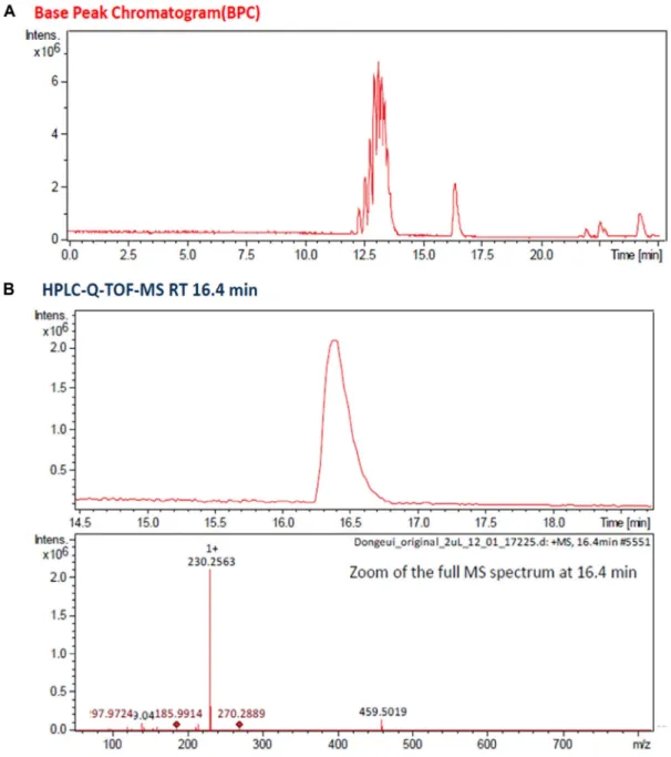
HPLC-MS spectrum에 대한 Fr. 2의 정성분석 삼백초 추출물로부터 항산화 효능과 멜라닌 합성능력이 우 수한 Fr. 2를 HPLC-MS spectrum을 이용하여 Fig. 7A와 같이 분리하였다. Fig. 7B에서 보는 바와 같이 삼백초 추출물의 Fr.
2는 HPLC-MS를 이용한 분석에서 16.4 min에서 관찰된 분자 량이 239인 물질로 확인되었다.
고 찰
스트레스는 신체에서 다량의 활성산소(ROS, reactive oxy- gen species)를 생성하여 노화를 촉진시킨다[24]. 활성산소는 주름, 흰머리, 검버섯의 발생에 관여하는 것으로 보고되고 있 다[8, 11, 17]. 몸 속 해로운 활성산소로부터 인체를 보호하기 위해 최근 국내외적으로 천연 항산화 물질에 대한 관심이 높 아졌다[19]. 항산화력이 우수하다고 알려진 천연물에는 해바 라기 씨앗, 석류, 일일초 등이 있다[3, 10, 27].
본 연구에서는 국내에서 서식하는 식물 중 하나인 삼백초 잎을 이용하여 노화의 진행과정과 관련된 ROS 생성 억제와 멜라닌 합성 촉진 효과를 항산화 능력 실험, 멜라닌 합성 능력 실험을 통해 확인하였다. 삼백초 MeOH 추출물을 hexane, ethyl acetate, butanol, distilled water (D.W)순으로 용매 별 항산화 실험한 결과 ethyl acetate 분획물에서 가장 높은 항산 화력이 나타났다. 따라서 ethyl acetate fraction of methanol extract of Saururus chinensis (SCEA) 분획물을 대상으로 항산 화 능력을 알아보기 위해서 DPPH radical scavenging activity 와 Reducing power activity를 진행하였다. DPPH radical scavenging activity는 양성 대조군인 Vitamin C와 유사한 효 능을 보였으며, Reducing power activity는 양성 대조군인 Vitamin C보다 항산화 효능이 더 높게 나타났다. 이 결과는 Lee 등[16]이 보고한 삼백초 추출물의 DPPH radical scaveng- ing activity (DRSA) 결과와 유사하였다. 또한, SCEA의 멜라닌 합성 능력을 알아보기 위해 tyrosinase 활성 측정실험을 수행 하였다. SCEA 분획물이 tyrosinase 활성이 있는 것으로 나타 났으나 본 실험 조건에서 얻은 이 연구 결과는 흥미롭게도 이미 발표된 멜라닌 생성을 억제하는 연구결과[13]와 다르게 멜라닌 합성을 촉진하는 것으로 나타났다. 본 연구 결과의 신 뢰성을 확보하기 위하여 재현성 실험을 반복한 결과 삼백초 추출물은 tyrosinase 활성을 감소시키는 것이 아니라 증가시 키는 것으로 확인되었다. SCEA 분획물 속에 있는 성분 중 멜 라닌 합성을 증가시키는 단일성분을 확인하기 위해 칼럼 크로 마토그래피로 분리 실험을 진행하였다. 그 결과 13개의 Frac- tion으로 분획하여 항산화 효능과 tyrosinase 활성을 조사한 결과 Fr. 2가 가장 우수하였다. 이전 연구에서는 삼백초 추출물 중 ethyl acetate 분획물 중 하나인 lignans이 항염증제 및 항암 효능이 있다고 보고되었다[14]. 이 후 SCEA 분획물 Fr.2로 DPPH radical scavenging activity, reducing power activity, tyrosinase 활성 측정실험을 재 진행하였다.
다음으로 삼백초 추출물은 세포독성이 있는 것으로 나타나 저 농도에서 살아있는 세포에서 멜라닌 생성촉진 실험을 진행 한 결과 Fr. 2는 멜라닌 생성 촉진 효과가 있는 것으로 확인되
Fig. 7. HPLC-MS spectrum and HD Q-TOF Mass Spectrometry of Fr. 2. (A) HPLC-MS spectrum was analyzed by the 1,260 Infinity HPLC system. (B) The molecular weight of peak was analyzed by Impact HD Q-TOF Mass Spectrometry. Peak was confirmed that the molecular weight is 239.
었다. 이전 연구에서도 간암 및 전립선암 세포에서 독성이 있 는 것으로 보고되어 삼백초 추출물은 세포독성이 있는 것으로 확인되었다[6]. Fr. 2의 유효성분을 분석하기 위하여 HPLC- MS를 이용하여 단일성분을 확인한 결과 분자량이 239인 성분 이 존재하는 것으로 확인되었다. 이전 연구에서는 삼백초 추 출물의 HPLC 분석 실험 결과 분자량은 다르지만 리그난 성분 을 함유한 것으로 보고 되었다[12]. 앞으로 이 성분에 대한 분 리정제와 구조분석에 대한 후속 연구가 필요한 실정이다. 뿐 만 아니라 정제한 유효성분의 멜라닌 생성촉진 효과에 대한 연구도 계속 되어야 한다.
현재까지의 결과를 바탕으로 본 연구에 사용된 삼백초 추출 물은 저농도에서항산화 효능과 멜라닌 합성 능력이 우수한 것으로 나타나, 기능성 흑모 관련 화장품의 원료로 사용 가능 성이 기대된다.
감사의 글
이 논문은 2020년도 정부(교육부)의 재원으로 한국연구재 단의 지원을 받아 수행된 기초연구사업임(No.2017R1D1A3 B06028000).
858 생명과학회지 2020, Vol. 30. No. 10
The Conflict of Interest Statement
The authors declare that they have no conflicts of interest with the contents of this article.
References
1. Alaklabi, A., Arif, I. A., Ahamed, A., Kumar, R. S. and Idhayadhulla, A. 2018. Evaluation of antioxidant and anti- cancer activities of chemical constituents of the saururus chi- nensis root extracts. Saudi. J. Biol. Sci. 25, 1387-1392.
2. Baghel, S. S., Shrivastava, N., Baghel, R. S., Agrawal, P. and Rajput, S. 2012. A review of quercetin: antioxidant and anti- cancer properties. World J. Pharm. Pharm. Sci. 1, 146-160.
3. Derakhshan, Z., Ferrante, M., Tadi, M., Ansari, F., Heydari, A., Hosseini, M. S., Conti, G. O. and Sadrabad, E. K. 2018.
Antioxidant activity and total phenolic content of ethanolic extract of pomegranate peels, juice and seeds. Food Chem.
Toxicol. 114, 108-111.
4. Donno, D., Boggia, R., Zunin, P., Cerutti, A., Guido, M., Mellano, M. G., Prgomet, Z. and Beccaro, G. L. 2016. Phyto- chemical fingerprint and chemometrics for natural food preparation pattern recognition: an innovative technique in food supplement quality control. J. Food Sci. Technol. 53, 1071-1083.
5. Gill, S. S. and Tuteja, N. 2010. Reactive oxygen species and antioxidant machinery in abiotic stress tolerance in crop plants. J. Plant Biochem. Physiol. 48, 909-930.
6. Hahm, J. C., Lee, I. K., Kang, W. K., Kim, S. U. and Ahn, Y. J. 2005. Cytotoxicity of neolignans identified in Saururus chinensis towards human cancer cell lines. Planta Med. 71, 464-469.
7. Hansen, M. B., Nielsen, S. E. and Berg, K. 1989. Re-examina- tion and further development of a precise and rapid dye method for measuring cell growth/cell kill. J. Immunol.
Methods 119, 203-210.
8. Helfrich, Y. R., Sachs, D. L. and Voorhees, J. J. 2008. Overview of skin aging and photoaging. Dermatol. Nurs. 20, 177.
9. Imai, J., Ide, N., Nagae, S., Moriguchi, T., Matsuura, H. and Itakura, Y. 1994. Antioxidant and radical scavenging effects of aged garlic extract and its constituents. Planta Med. 60, 417-420.
10. Islam, R. T., Ahmed, T. and Kishor, M. 2016. In vitro anti- oxidant activity of methanolic extract of Helianthus annuus seeds. J. Med. Plants Studies 4, 15-17.
11. Jo, S. J., Shin, H., Paik, S. H., Choi, J. W., Lee, J. H., Cho, S. and Kwon, O. 2013. The pattern of hair dyeing in Koreans with gray hair. Ann. Dermatol. 25, 401-404.
12. Jung, Y. W., Lee, B. M., Ha, M. T., Tran, M. H., Kim, J.
A., Lee, S., Lee, J. H., Woo, M. H. and Min, B. S. 2019.
Lignans from Saururus chinensis exhibit anti-inflammatory activity by influencing the Nrf2/HO-1 activation pathway.
Arch. Pharm. Res. 42, 332-343.
13. Kim, S. K., Kim, D. S., Woo, W. H. and Mun, Y. J. 2012.
Effect of Saururus chinensis BAILL extract for pharmacopuncture on the melanogenesis in B16F10 cells.
Kor. J. Acupuncture 29, 117-130.
14. Lee, J., Huh, M. S., Kim, Y. C., Hattori, M. and Otake, T.
2010. Lignan, sesquilignans and dilignans, novel HIV-1 pro- tease and cytopathic effect inhibitors purified from the rhi- zomes of Saururus chinensis. Antiviral Res. 85, 425-428.
15. Lee, S. Y., Baek, N. and Nam, T. G. 2016. Natural, semi- synthetic and synthetic tyrosinase inhibitors. J. Enzyme Inhib.
Med. Chem. 31, 1-13.
16. Lee, W. S., Baek, Y. I., Kim, J. R., Cho, K. H., Sok, D. E.
and Jeong, T. S. 2004. Antioxidant activities of a new lignan and a neolignan from Saururus chinensis. Bioorg. Med. Chem.
Lett. 14, 5623-5628.
17. Lephart, E. D. 2016. Skin aging and oxidative stress: Equol’s anti-aging effects via biochemical and molecular mecha- nisms. Ageing Res. Rev. 31, 36-54.
18. Li, Y., Yao, J., Han, C., Yang, J., Chaudhry, M., Wang, S., Liu, H. and Yin, Y. 2016. Quercetin, inflammation and im- munity. Nutrients 8, 167.
19. Lobo, V., Patil, A., Phatak, A. and Chandra, N. 2010. Free radicals, antioxidants and functional foods: Impact on hu- man health. Pharma. Rev. 4, 118.
20. Martin, P. 2015. Parthenogenesis: mechanisms, evolution, and its relevance to the role of marbled crayfish as model organism and potential invader. Freshwater Crayfish: A Global Overview, 63-82.
21. Meng, X., Kim, I., Jeong, Y. J., Cho, Y. M. and Kang, S. C.
2016. Anti-inflammatory effects of Saururus chinensis aerial parts in murine macrophages via induction of heme oxygen- ase-1. Exp. Biol. Med. 241, 396-408.
22. Oyaizu, M. 1986. Studies on products of browning reaction:
antioxidative activity of products of browning reaction. Jpn.
J. Nutr. 44, 307-315.
23. Park, D. J. and Lee, J. C. 2008. A study on the antioxidative and depigmentation activities of the ethanol extract of Saururus herba. Kor. J. Herbol. 23, 193-202.
24. Poprac, P., Jomova, K., Simunkova, M., Kollar, V., Rhodes, C. J. and Valko, M. 2017. Targeting free radicals in oxidative stress-related human diseases. Trends Pharmacol. Sci. 38, 592- 607.
25. Redza-Dutordoir, M. and Averill-Bates, D. A. 2016. Activa- tion of apoptosis signalling pathways by reactive oxygen species. BBA-Mol. Cell Res. 1863, 2977-2992.
26. Sugimoto, K., Nishimura, T., Nomura, K., Sugimoto, K. and Kuriki, T. 2004. Inhibitory effects of α-arbutin on melanin synthesis in cultured human melanoma cells and a three-di- mensional human skin model. Biol. Pharm. Bull. 27, 510-514.
27. Velioglu, Y., Mazza, G., Gao, L. and Oomah, B. 1998. Antiox- idant activity and total phenolics in selected fruits, vegeta- bles, and grain products. J. Agric. Food Chem. 46, 4113-4117.
초록:삼백초 추출물이 항산화활성과 멜라닌 합성에 미치는 영향
정소미1․박혜정2․김재호3․오영희2․김문무2*
(1동의대학교 화학과, 2동의대학교 응용화학과, 3동의대학교 화학생물과)
뿌리, 잎, 꽃이 하얀색인 삼백초는 항균 활성 및 항암 효과를 갖는 것으로 알려져 있다. 본 연구에서는 SCEA의 항산화 활성과 멜라닌 합성에 미치는 영향을 조사하고자 하였다. SCEA를 헥산, 에틸 아세테이트, 부탄올 및 증류 수로 분획 하였다. 용매 분획 중 가장 높은 항산화 활성을 갖는 ethyl acetate 분획물이 본 연구에 사용되었다.
DPPH radical 소거능에서는 64 μg/ml의 SCEA는 양성 대조군과 비교하여 62% 라디칼 소거능을 보였다. 환원력 시험에서 64 μg/ml의 SCEA는 양성 대조군으로 사용된 비타민 C보다 33% 더 낮은 감소력을 보였다. Tyrosinase 활성은 공시험군에 비해 64 μg/ml의 SCEA 처리군에서 26% 증가했다. 또한, 64 μg/ml의 SCEA는 Dopa 산화력 분석에서 공시험군과 비교하여 44%의 멜라닌 합성을 증가시켰다. MTT 분석 결과 SCEA는 0.5 μg/ml 이상의 세 포 독성을 나타냈다. 1 μg/ml의 SCEA는 멜라닌 합성을 살아있는 세포에서 69% 증가시켰다. 또한 SCEA 분획을 실리카 칼럼 크로마토그래피로 13 개의 분획으로 분리 하였다. 특히, Fr. 2는 DPPH 라디칼 소거 활성이 가장 높았 으며, 멜라닌 합성이 감소되었다. 또한 SCEA는 살아있는 세포인 B16F1에서 멜라닌 생성을 촉진시켰다. LC- MASS는 Fr. 2의 분자량이 239임을 보여 주었다. 따라서 위의 결과는 SCEA가 멜라닌 합성 촉진과 관련된 천연 흑모 모발화장품 개발에 사용될 수 있음을 시사한다.