Establishment of Cell Suspension Cultures and Plant Regeneration in White Dandelion (Taraxacum coreanum NAKAI.)
Yan-Lin Sun1, Jae-Hak Kim1 and Soon-Kwan Hong1,2*
1Department of Bio-Health Technology, Kangwon National University, Chuncheon 200-701, Korea
2Institute of Bioscience and Biotechnology, Kangwon National University, Chuncheon 200-701, Korea
Abstract - In this study, we established a novel somatic embryogenesis and plant regeneration system through cell suspension culture of white dandelion (Taraxacum coreanum NAKAI.). Embryogenic calli could be initiated from leaf and root explants of sterile seedlings on solid Murashige and Skoog (MS) medium supplemented with 1.0 mg/L 2,4- dichlorophenoxyacetic acid (2,4-D) after 3-week cultures. To proliferate embryogenic calli rapidly, cell suspension culture was performed with transferred to liquid MS medium with various combinations of plant growth regulators (PGRs) including 2,4-D, α-naphthalene acetic acid (NAA), indole-3-acetic acid (IAA), N6-benzylamino purine (BAP), thidiazuron (TDZ), and kinetin. During suspension cultures, embryogenic calli not only greatly proliferated, but shoot organogenesis also simultaneously occurred from the surface of somatic embryos. Among them, TDZ at lower concentration, 0.1 mg/L produced the highest efficiency of somatic embryo formation and shoot organogenesis. Rooting of embryogenic calli with adventitious shoots was done on solid MS medium containing 0.1 mg/L NAA and 0.3% activated carbon. Nearly 80% of embryogenic calli with shoot organogenesis could be rooted normal. Well-rooted plantlets were transferred into pots under a greenhouse condition, and plants derived from this system appeared phenotypically normal.
Key words - Taraxacum coreanum, Plant regeneration, Cell suspension culture
*Corresponding author. E-mail : [email protected]
Introduction
The genus Taraxacum is perennial, herbaceous plants, a member of the family Asteraceae, subfamily Cichorioideae, tribe Lactuceae and is widely distributed in the warmer temperate zones of the Northern Hemisphere, and mostly inhabited fields, roadsides and ruderal sites (Schütz et al., 2006; Lee et al., 2004). This genus has long been used as medicinal herbs to treat arthritic and rheumatic complaints (Bisset et al., 1994), diabetes mellitus (Hernandez-Galicia et al., 2002), hepatitis (Leu et al., 2005) and some choleretic, diuretic, and anti-inflammatory diseases (Sweeney et al., 2005;
Yang et al., 1996). Apart from being used for therapeutic purposes, dandelion inflorescences, leaves and roots are processed into different healthy food products. Young leaves of this species are consumed fresh as salad and kimchi, while roasted roots are utilized as a coffee substitute (Kang and Kim, 2001; Schütz et al., 2006).
Among nearly 400 plant species of the genus Taraxacum, T. coreanum is one of main plant species in Korea and Japan.
In Traditional Korean Medicine, T. coreanum has long been used for medicinal purposes due to its diuretic and anti- inflammatory activities (Ahn, 1998, Koo et al., 2004).
However, due to its low reproduction capacity to be lost in bioecological competition with other dandelion species, T. coreanum is rare and nearly extinct in natural fields of Korea. Although dandelion is a well-known traditional herbal remedy with a long history, until recently few scientific information is available to improve the low-reproduction status. To meet the needs of plant resource conservation and commercial production, further trait improvements in Taraxacum cultivars are currently required. Previous researches on Taraxacum are mainly focused on its apomictic charac- teristics, and isolation of effective components and their activity detection (Akashi et al., 1994; Schütz et al., 2006;
Wolbis et al., 1993; Paul et al., 2006). Few studies about in vitro tissue culture and plant regeneration of T. coreanum have also been reported (Lee et al., 2002; Lee et al., 2007).
However, tissue culture systems through callus induction, somatic embryogenesis, and plant regeneration is very low- yield and time-consuming (Hall, 1991).
To satisfy the demand of mass production, cell suspension culture was investigated in this work. In comparison to the routine tissue culture, cell suspension culture facilitates more rapid cell division and cell generations of various cell lines and more stable and direct somatic embryogenesis (Sun and Hong, 2010). Moreover, due to a large surface for exposure to culture medium with transforming agents and effective selection of transformants, embryogenic cells from suspension culture are also efficient for gene transformation and gene manipulation (Hall, 1991).
The objectives of this study were to establish reliable plant regeneration system from cell suspension culture with T.
coreanum. In an attempt to demonstrate the application of suspension culture techniques to enhance reproductibility, an efficient plant regeneration system through cell suspension culture has been established. This system would help further application of biotechnology to Taraxacum breeding programs through genetic modification.
Materials and Methods
Plant materials
Seeds of T. coreanum used in this study were collected from Kangwon-Do, Korea from May to June in 2009. The seeds were sterilized by immersion in 4% sodium hypochlorite for 10 min followed in 70% ethanol for 30 sec. The surface- sterilized seeds were washed for four times with sterilized distilled water and dried on sterilized filter paper. Then, the surface-sterilized seeds were inoculated on Murashige and Skoog (MS) medium (Murashige and Skoog, 1962) supple- mented with 3% sucrose and solidified with 3.0 g/L gelrite.
Cultures were maintained at 28℃ under 16/8 h (light/darkness) photoperiod provided by fluorescent white light 24 µmol m-2 s-1. Within 1-week culture in above conditions, seeds germinated. The seedlings were prepared for leaf and root explants for callus induction.
Callus induction
The leaf blades and roots of seedlings were cut into 1 cm
length segment under an aseptic condition. The leaf and root segments were inoculated on 25 ml MS basal medium supple- mented with 1.0 mg/L 2,4-D, solidified with 3.0 g/L gelrite in 90 × 20 mm Petri dishes under light conditions. With 1-week culture, primary calli were induced from the surface of different explants. The 3-week-old embryogenic calli were used for cell suspension culture.
Cell suspension culture
Approximately 500 mg embryonic calli were transferred into 250 ml Erlenmeyer flask containing 100 ml liquid MS medium supplemented with various kinds of plant growth regulators (PGRs) individually, including three auxins, two cytokinins and kinetin. Three auxins were 2,4-dichlorphenoxy- acetic acid (2,4-D), α-naphthalene acetic acid (NAA), and indole-3-acetic acid (IAA), while two cytokinins were N6- benzylamino purine (BAP), and thidiazuron (TDZ). All media contained 30 g/L sucrose. The cultures were maintained on a rotary platform shaker at 100 rpm at 26℃ under light conditions.
Plant regeneration
After 1 month suspension culture, embryogenic calli produced adventitious shoots. To induce shoot organogenesis further and rooting, the embryogenic calli were transferred onto solid half-strength or full MS media supplemented with various concentrations of NAA and 0.3% activated carbon. Regenerated shoots with elongated leaves were transferred on a PGRs-free medium for rooting. Well-rooted plantlets were taken out of the solid culture medium, and washed carefully to remove the gelrite. Plantlets were cultured in a pot containing a mixture of sterilized soil and vermiculite (v/v, 3:1) at 27℃ under light conditions. After 3-week culture under above conditions, plantlets were transferred to a greenhouse condition.
Results and Discussion
To achieve efficient production of in vitro plantlets of white dandelion, many investigations have been reported.
Lee et al. (2002) investigated various explants such as roots, hypocotyls, and cotyledons, and various kinds of growth regulators to optimize callus induction and plant regeneration
Fig. 1. Callus induction, shoot organogenesis and plant regeneration from cell suspension cultures derived from leaf and root explants of T. coreanum. A. T. coreanum plantlets grown in an aseptic condition used for the explants supporter.
B. Callus formation from root explants. C. The proliferation of induced calli. D. Embryogenic calli in cell suspension culture cultured on liquid MS medium containing various PGRs after 4-week suspension culture. E. Induced embryogenic calli after 4-week suspension culture. F. Induced non-embryogenic calli after 4-week suspension culture. G. Embryogenic calli with adventitious shoots during shoot organogenesis. H. Rooting of embryogenic calli with adventitious shoots on the half- strength solid MS medium. I. Plantlets derived from the plant regeneration system through cell suspension culture, grown in a pot. Bars = 2 mm.
Fig. 2. Effects of various PGRs on the proliferation of embryogeic calli with 4-week suspension culture. Data represent the mean of three replications.
conditions, suggesting that roots was considered to be the best explants for efficient plant regeneration. Lee et al. (2007) attempted leaf explants to induce callus induction and plant regeneration of T. coreanum, suggesting that 2 mg/L BAP could cause the highest shoot regeneration frequency. To authors’ knowledge, this work is the first report about embryogenic callus and plant regeneration through cell suspension culture of white dandelion. To determine the effects of various PRGs and their concentrations, three auxins, two cytokinins, and kinetin with both concentrations (0.1 mg/L and 1.0 mg/L) were investigated in this work.
In this study, the sterile plantlets were used to prepare leaf and root explants (Fig. 1A). The leaf and root from sterile plantlets were cut into 1 cm length segments as explants
for callus induction, and inoculated on solid MS medium supplemented 1.0 mg/L 2,4-D, 30 g/L sucrose. Within 1-week culture, primary calli were induced from the surface of leaf and root explants (Fig. 1B). To maintain production of primary calli, 1-month-old primary calli were cut into 2~3 parts, and went on to subculture onto the same MS medium (Fig. 1C).
To satisfy the demand of the growth capacity, 3-week-old primary calli only were used for cell suspension culture in this study.
With 4-week suspension culture in a rotary shaker, embryogenic and non-embryogenic calli were found to be induced from the surface of cultured primary calli (Fig. 1D, E). Callus type was evaluated by morphological characteristics of the microscopical image. Synchronously, the proliferation of suspension-cultured calli was evaluated by fresh weight of embryogenic calli (Fig. 2). From the results of weight changes of embryogenic calli after 4-week suspension culture (Fig. 2), it was suggested that cytokines such as BAP and TDZ, played more important roles than kinetin and some auxins such as NAA, and IAA. With the addition of 2,4-D, NAA, IAA or kinetin, the weights of embryogenic calli showed 5~14-fold higher than those of initial inoculated calli. With the addition of BAP and TDZ, the weights increased more largely compared to the addition of 2,4-D, NAA, IAA or kinetin, with 13.71 g and 17.07 g with 0.1 mg/L and 1.0 mg/L BAP, and 23.04 g and 19.79 g with 0.1 mg/L and 1.0 mg/L TDZ, respectively. Based on above results, 0.1 mg/L TDZ was
Fig. 3. Morphological characteristics of cell suspension cultures on various liquid media after 6-week culture. A. MS. B. MS + 0.1 mg/L 2,4-D. C. MS + 0.1 mg/L NAA. D. MS + 0.1 mg/L IAA. E. MS + 0.1 mg/L BAP. F. MS + 0.1mg/L Kinetin. G. MS + 0.1 mg/L TDZ. H. MS + 1.0 mg/L 2,4-D. I. MS + 1.0 mg/L NAA. J. MS + 1.0 mg/L IAA. K. MS + 1.0 mg/L BAP. L. MS + 1.0 mg/L Kinetin.
M. MS + 1.0 mg/L TDZ.
Fig. 4. The effect of activated charocoal on the vitrification of adventitious shoots. A. Embryogenic calli with vitrified leaves.
B. Threadlike adventitious leaves induced from embryogenic calli. C. Normal rooting of embryogenic calli with the removal of vitrified leaves. Bars = 2 mm.
considered to be the most efficient in the proliferation of embryogenic calli in cell suspension culture of T. coreanum.
Combined with the morphological characteristics of suspension cultured embryogenic calli (Fig. 3), the effects of various PGRs were further evaluated. As known, cytokinins promote cell division, shoot multiplication and axillary bud proliferation, while auxins cause cell enlargement, root initiation and adventitious bud formation (Lian et al., 2009). In this study, both the proliferation rates and sizes of embryogenic calli varied depending on PGR type and concentration (Fig.
3). Liquid media with cytokinin produced greater amounts and larger sizes of embryogenic calli (Fig. 3E-G, K-M) compared to those with kinetin or/and auxins (Fig. 3B-D, H-J). And the control hormone-free medium could not cause the proliferation of embryogenic calli (Fig. 3A).
With further 2-month suspension culture conducted in identical liquid MS medium with relevant PGRs, 100%
somatic embryos would achieve the conversion to shoot organogenesis (Fig. 1F, G). Adventitious shoots with elongated leaves were transferred onto solid half-strength MS media
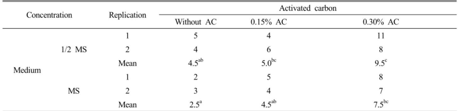
supplemented 1.0 mg/L NAA for rooting (Fig. 1H, I). However, rooting of adventitious shoots did not appear rapidly, on the contrary, browning of callus and vitrification of leaves occurred frequently (Fig. 4A, B). To improve these phenomena, lowering MS basic salt accession and adding activated charocoal in the solid culture media (Table 1). From the results of normal rooting formation, it was suggested that 1/2 MS basic salt medium and higher concentration (0.30%) of activated charocoal were more efficient in the prevention
Table 1. Root formation after 8-week culture on various solid media for rooting.
Concentration Replication Activated carbon
Without AC 0.15% AC 0.30% AC
Medium
1/2 MS
1 5 4 11
2 4 6 8
Mean 4.5ab 5.0bc 9.5c
MS
1 2 5 8
2 3 4 7
Mean 2.5a 4.5ab 7.5bc
All solid media contained 1.5 mg/L NAA. Twenty explants per replication were used in this experiment. Means followed by the same letter are not significantly different at P < 0.05 based on the multiple range test (Duncan, 1955).
from browning of callus and vitrification of leaves. In addition, the removal of vitrified leaves was also considered to treat the adventitious shoots prior to being transferred onto solid culture medium for rooting (Fig. 4C). Well-rooted plantlets were transferred to a pot containing a mixture of sterilized soil and vermiculite (v:v, 3:1) for acclimation at 27℃ in a 16/8 h (light/darkness) photoperiod provided by fluorescent white light (Fig. 1H). The 100% plantlets showing pheno- typically normal were transferred to a greenhouse condition (Fig. 1I).
At present, T. coreanum being worth as commercial, aesthetic and medicinal values is paid more and more attentions. This report has established an efficient, direct shoot organogenesis and plant regeneration system from somatic embryos through cell suspension culture in T.
coreanum. This study provides to develop novel tissue culture techniques for application to T. coreanum. This system would help further application of biotechnology to Taraxacum breeding programs through genetic modification.
Acknowledgement
Following are results of a study on the “Human Resource Development center for Economic Region Leading Industry”
Project, supported by the Ministry of Education , Science &
Technology (MEST) and the Nutraceutical Bio Brain Korea 21 Project Group.
Literature Cited
Ahn, D.K. 1998. Illustrated book of Korean medicinal herbs.
Kyohak Publishing Co. Ltd., Korea. p. 134.
Akashi, T., T. Furuno, T. Takahashi and S.I. Ayabe. 1994.
Biosynthesis of triterpenoids in cultured cells, and regenerated and wild plant organs of Taraxacum officinale. Phyto- chemistry 36:303-308.
Bisset, N.G., J.D. Phillipson, F.C. Czygan, D. Frohne, D.
Höltzel, A. Nagell, H.J. Pfander, G. Willuhn and W. Buff (eds.). 1994. Herbal drugs and phytopharmaceuticals: A Handbook for Practice on a Scientific Basis. CRC Press, Boca Raton, Ann Arbor, London, Tokyo. pp. 486-489.
Duncan, D.B. 1955. Multiple range and multiple F tests.
Biometrics 11:1-42.
Hall, R.D. 1991. The initiation and maintenance of plant cell suspension cultures. Plant Tiss. Cult. Manual. A3:1-21.
Hernandez-Galicia, E., A. Aguilar-Contreras, L. Aguilar- Santamaria, R. Roman-Ramos, A.A. Chavez-Miranda, L.M.
Garcia-Vega, J.L. Flores-Saenz and F.J. Alarcon-Aguilar.
2002. Studies on hypoglycemic activity of Mexican medicinal plants. Proc. West Pharmacol. Soc. 45:118-124.
Kang, M.J. and K.S. Kim. 2001. Current trends of research and biological activities of dandelion. Food Industry Nutr.
6:60-67.
Koo, H.N., S.H. Hong, B.K. Song, C.H. Kim, Y.H. Yoo and H.M. Kim. 2004. Taraxacum offcinale induces cytotoxicity through TNF-α and IL-1α secretion in Hep G2 cells. Life Sci. 74:1149-1157. Doi: 10.1016/j.lfs.2003.07.030.
Lee, H.H., Y.S. Kim and H.Y. Park. 2007. Plant regeneration via organogenesis from leaf explant culture of Taraxacum coreanum Nakai. Korean J. Medicinal Crop Sci. 15(1):62-66 (in Korean).
Lee, M.H., E.S. Yoon, J.H. Jeong and Y.E. Choi. 2004.
Agrobacterium rhizogenes-mediated transformation of Taraxacum platycarpum and changes of morphological characters. Plant Cell Rep. 22:822-827. Doi:10.1007/s00299- 004-0763-5.
Lee, M.H., E.S. Yoon, S.J. Jung, K.H. Bae, J.W. Seo and Y.E.
Choi. 2002. Plant regeneration and effect of auxin and cytokinin on adventitious shoot formation from seedling explant of Taraxacum platycarpum. Korean J. Plant Biotechnol.
29(2):111-115 (in Korean).
Leu, Y.L., Y.L. Wang, S.C. Huang and L.S. Shi. 2005. Chemical constituents from roots of Taraxacum formosanum. Chem.
Pharm. Bull. 53(7):853-855. Doi:10.1248/cpb.53.853.
Lian, Y.J., G.Z. Lin, D.I. Yoo, W.B. Kim and X.M. Zhao. 2009.
Effect of cytokinin on adventitious shoot formation and plant regeneration from explants of Pulsatilla koreana NAKAI. J. Plant Biotechnol. 36(2):170-173 (in Korean).
Murashige, T. and F. Skoog. 1962. A revised medium for rapid growth and bio assays with tobacco tissue culture. Physiol.
Plant. 15:473-497.
Paul, N.C., W.K. Kim, S.K. Woo, M.S. Park and S.H. Yu. 2006.
Diversity of endophytic fungi associated with Taraxacum coreanum and their antifungal activity. Mycobiology 34(4):
185-190.
Schütz, K., R. Carlea and A. Schieber. 2006. Taraxacum--A review on its phytochemical and pharmacological profile. J.
Ethnopharmacol. 107:313-323. Doi:10.1016/j.jep.2006.07.021.
Sun, Y.L. and S.K. Hong. 2010. Establishment of a novel plant regeneration system from suspension-derived callus in the halophytic Leymus chinensis (Trin.). J. Plant Biotechnol.
37(2):228-235. Doi: 10.5010/JPB.2010.37.2.228.
Sweeney, B., M. Vora, C. Ulbricht and E. Basch. 2005.
Evidence-based systematic review of dandelion (Taraxacum officinale) by natural standard research collaboration. J.
Herb. Pharmacother. 5(1):79-93.
Wolbis, M., M. Królikowska and P. Bednarek. 1993.
Polyphenolic compounds in Taraxacum officinale. Acta Pol. Pharm. Drug Res. 50:153-158.
Yang, D.S., W.K. Whang and I.H. Kim. 1996. The constituents of Taraxacum hallaisanensis roots. Arch. Pharm. Res.
19(6):507-513. Doi:10.1007/BF02986020.
(Received 11 November 2010 ; Accepted 31 May 2011)