149 임플란트를 식립하려 할 경우 골열개 또는 골천공이 발생
하는 경우가 많으며, 다양한 골이식재를 사용한 골유도재생 술은 보편적인 술식이 되었다. 골결손량이 큰 부위의 재건이 나 치조능증대술과 같은 많은 양의 골증대가 필요할 경우에 는 자가골을 통한 이식이 가장 좋은 결과를 보인다. 자가골은 장골, 능골, 경골과 같은 연골내골(endochondral bone)과 두개 골, 안면골과 같은 막내골(intramembranous bone)을 이용해 서 시행할 수 있다. 치조능증대술은 수직적, 수평적으로 골량 이 부족한 경우에 치조능의 상방 혹은 협측에 입자나 혹은 블 록형의 골이식재를 이식하여 치조능의 폭경이나 높이를 증가
하는 술식을 말한다. 이것은 일종의 온레이 이식으로써 이식 후에 골흡수가 많이 발생되며 상부 연조직의 열개가 일어날 가능성이 높다(Kim MJ et al., 2004).
그러나 자가골이식은 채취량이 제한되어있고 공여부의 합 병증이 크며 이식 후 흡수가 많이 발생하는 등의 문제점이 있 다. 따라서 동종골, 이종골, 합성골과 같은 골 대체재료가 개발 되어 임상에서 사용하고 있으나 각각의 정점을 공유하기 위 해 자가골과 다른 골 대체재료를 혼합하여 사용한다. 골유도 재생술의 범위가 작을 경우에는 이러한 대체재료의 사용만으 로 충분하다. 그러나 광범위한 치조능증대술이나, 1벽성 혹은 2벽성의 부위에 골증대술을 시행할 경우에는 반드시 자가골 이식을 선택해야 할 것이다. 그러나 동종골이나 이종골을 사 용할 경우에 환자에 따라서는 골재료로부터의 감염 등의 불 안감으로 인하여 거부반응을 보이기도 한다. 합성골은 가격 은 저렴하지만 골형성능과 골유도능이 전혀 없어 생활력있는 골조직 형성을 목적으로 골증대술을 시행할 경우에는 사용에 제한을 받는다(Kim YK, 1998; Kim MJ et al., 2004).
ORAL BIOLOGY RESEARCH 2012; 36(2): 149-152
자가치아골이식재를 이용한 발치창 재건술: 증례보고
김문섭ᆞ김수관*ᆞ오지수
조선대학교 치의학전문대학원 구강악안면외과학교실
Guided bone regeneration on extraction socket using autogenous tooth bone graft materials: Case report
Moon-Seob Kim, Su-Gwan Kim*, Ji-Su Oh
Department of Oral and Maxillofacial Surgery, School of Dentistry, Chosun University, Gwangju, Korea
ABSTRACT
Implants were installed in conjunction with guided bony regeneration (GBR) using an autogenous tooth bone graft material in the patients. Guided Bone regeneration was performed using an autogenous tooth bone graft block and powder in a 54-year old male patient. Excellent osteoconductive bony healing was observed in the 6 month histology examination after the bone graft.
Socket grafting is common method for preservation of alveolar bone to prevent resorption of the socket as inevitable remodeling.
And autogenous tooth bone graft material has excellent biocompatibility and osteoinduction as well as osteoconduction.
Key Words: Autogenous tooth bone, Osteoconductive bony healing, Guided bony regeneration, Tooth socket Case Report
Received Jan 23, 2012; Revised version received Feb 20, 2012 Accepted Mar 18, 2012
Corresponding author: Su-Gwan Kim
Department of Oral and Maxillofacial Surgery, School of Dentistry, Chosun University, 375 Seosuk-dong, Dong-gu, Gwangju 501-759, Korea
Tel: 82-62-220-3815, Fax: 82-62-228-7316 E-mail: [email protected]
This is an open-access article distributed under the terms of the Creative Commons Attribution Non-Commercial License (http://creativecommons.org/licenses/by-nc/3.0) which permits unrestricted non-commercial use, distribution, and reproduction in any medium, provided the original work is properly cited.
150
Guided bone regeneration on extraction socket using autogenous tooth bone graft materials: Case report
저자들은 최근 개발된 입자 및 블록형 자가치아골이식재를 사용하여 골유도증례술을 병행한 증례를 소개하고 조직학적 소견과 함께 자가치아를 이용한 골이식재의 우수한 골치유 능력을 언급하고자 한다.
증례보고
54세 남자 환자가 좌측 제1대구치 부위의 심한 수직적 골결 손으로 임플란트 수복을 위해 내원하였다. 임상 및 방사선 검 사 결과 전반적인 치조골 소실 및 하악 좌측 제1대구치의 부위 의 치근단 농양 소견을 보였다. 하악 좌측 제1대구치를 발치한 후 자가치아골이식재로 처리하여 치조능증대술 및 골유도재 생술에 사용하기로 하였다(Fig. 1). 2010년 7월 5일 #36을 발치 한 후 분말 형태의 자가치아이식재로 가공하여 보관하였다.
2010년 8월 11일 피판을 거상한 후 자가치아골이식재를 이용 한 치조능증대술 및 임플란트식립을 시행하였으나 임플란트 는 초기고정을 얻지 못하여 제거하였으며 자가치아골이식재 를 이용한 치조능증대술만 시행하였다. 피판을 거상한 후 분 말형 자가치아이식재를 치조와에 적용한 후 피판을 이용하여
피개한 후 창상을 봉합하였다(Figs. 2, 3).
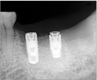
2010년 12월 13일 하악 좌측 제2대구치의 수직파절로 인해 발치 시행 후 골유도 재생술 시행하였던 제1대구치 부위와 제 2대구치 부위에 임플란트를 식립하였다. 제1대구치 부위에는 직경 4.1 mm, 길이 10 mm의 Zimmer 임플란트 (TSV; Zimmer Dental, Carlsbad, CA, USA)를 식립하였으며 제2대구치 부위 에는 직경 4.1 mm, 길이 8 mm의 Zimmer 임플란트를 식립하 였다. Osstell Mentor (Integration Diagnostics AB, Savedalen, Sweden)로 측정한 초기 안정성은 제1대구치 72, 제2대구치 74 implant stability quotient (ISQ)값을 보였다. 식립 시 환자의 동 의 하에 골증대술을 시행했던 부위에 생검을 시행하였다. 임 플란트 식립하기 전 피판 거상시 골증대술 부위에는 골과 유 사한 양상의 조직이 육안으로 관찰되었으며 생검하여 관찰한 결과 자가치아골이식재 근처에 신생골조직이 생성되는 것이 관찰되었다(Figs. 4-6).
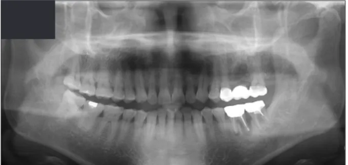
7개월 후 2차 수술을 시행하였다. 이때 Osstell Mentor로 측 Fig. 1. Initaial panoramic radiogrphy. There were generalized alveolar
bone resorption and severe vertical bone resorption on #36 area.
Fig. 2. Preoperative intraoral view.
Fig. 3. Vertical ridge augmentation was performed using autogenous tooth block at #36 area.
Fig. 4. Flap elevation view after 4 month later.
Moon-Seob Kim et al.
151 정한 2차 안정성은 제1대구치가 77 ISQ, 제2대구치가 80 ISQ
값을 보였으며 치유지대주를 연결하고 창상을 봉합하였다 (Figs. 7, 8).
한 달 후 성공적으로 보철물을 수복하였으며, 2차 수술을 한 지 4개월 후 Ostell Mentor로 측정한 안정성은 제1대구치와 제 2대구치 모두 80 ISQ 값을 보였다.
고 찰
치아를 이용한 골이식재의 무기질은 주로 수산화인회석 으로 구성되어 있으며 골대체재료로 사용할 수 있는 가능성 이 지금까지의 다양한 실험연구에서 입증되었다(Kim et al., 1993; Kim et al., 1999; Kim et al., 2002; Kim SY et al., 2004). 최
근 환자들에서 발치된 치아들을 최첨단 의료 공법으로 처리 하여 자가치아골이식재로 제조한 후 동일 환자의 골이식술 에 이용하는 방법이 개발되었다. 자가치아골이식재(AutoBT, Korea Tooth Bank, Seoul, Korea)는 이식 후 골유도 및 골전도 에 의한 치유를 보이며 생체적합성이 매우 좋은 것으로 보고 되었다(Kim et al., 2010).
치아와 치조골의 화학적 조성은 매우 유사하다. 치아의 화 학적 조성을 살펴보면 법랑질의 성분은 총 무기성분 95%, 유 기성분 0.6%, 물 4%이고 상아질의 성분은 무기성분 70-75%, 유기성분 20%, 물 10%로 구성되어 있다. 치조골의 성분은 무기성분 65%, 유기성분 25%, 물 10%로 알려져 있다(Min, 2007). Orban’s Oral histology and embryology에서는 법랑질 의 성분은 무기성분 96%, 유기성분과 물 4%, 상아질은 무기성 분 65%, 유기성분과 물 35%, 백악질은 무기성분 45-50%, 유기 Fig. 5. Periapical radiography after implant installation.
Fig. 6. The remodeling of new bones formed in the vicity of graft materials is obsereved (H&E staining, original magnifi cation ×100).
Fig. 7. Secondary surgery was performed at mandibular 1st, 2nd mo- lar area.
Fig. 8. Periapical radipgraphy after secondary surgery.
152
Guided bone regeneration on extraction socket using autogenous tooth bone graft materials: Case report
성분과 물 50-55%, 치조골은 무기성분 65%, 유기성분 35%로 기술되어 있다(Bhaskar, 1980). 상아질에 존재하는 유기성분 의 약 90% 정도는 콜라겐이며 석회화에 중요한 역할을 담당 한다. 콜라겐은 주로 Type I으로 구성되어 있고 그 외의 유기 성분은 비콜라겐성 단백질(non-collagenous proteins), 탄수화 물(carbohydrate), 지질(lipid), citrate, lactate 등으로 구성되어 있다(Min, 2007). 단백질에는 bone morphogenetic protein를 포함한 다양한 골성장 요소들이 존재하는 것으로 알려져 있 다.
자가치아골이식재 분말과 탈회시킨 블록은 점착성이 우 수하고 결손부에 잘 부착되는 특성을 가지고 있으며 조작이 매우 편리하다. 또한 자가치아를 이용한 골이식재는 치조골 과 매우 유사한 무기질과 유기질 성분을 모두 포함하고 있어 서 골유도 및 골전도에 의해서 매우 빠르고 우수한 골치유를 보이며, 창상이 일부 벌어지거나 노출되더라도 감염에 대한 저항성이 우수하고 2차치유가 잘 되는 장점이 있다(Choung, 2002). 따라서 발치창에 대하여 재건술을 시행할 경우, 자가치 아골이식재는 자가골 이식의 대체 수단이 될 수 있으며 양이 모자랄 경우에는 타 골이식재와 혼합하여 사용한다면 임상에 서 매우 유용하게 사용할 수 있을 것으로 보인다. 본 증례에서 는 하악 구치부의 발치창 재건술을 위해 블록과 분말형 자가 치아골이식재를 이식하였다. 4개월 후 이차수술을 시행하였 는데, 이때 피판 거상시 이식재가 잘 생착되어 있는 것을 확인 할 수 있었고, 조직학적 치유과정을 볼 때 이식재가 서서히 흡 수되면서 신생골로 대체되었으며, 신생골은 자가치아골이식 재들과 직접적인 유합을 보이는 것을 관찰할 수 있었다. 임플 란트 상부 보철물이 완성된 후 경과관찰 기간 중 임상적, 방사 선학적으로 안정적인 상태가 유지되었다.
이에 자가치아골이식재는 골전도에 의한 골치유가 이루어 지는 생체적합성이 우수한 재료로 임플란트 주변 골유도재생 술에 이용할 경우 좋은 결과를 얻을 수 있을 것으로 보인다.
감사의 글
이 논문은 2011년도 조선대학교 치과대학 교육문화재단의 지원을 받아 연구되었음.
참 고 문 헌
Bhaskar SN: Orban's oral histology and embryology. 9th ed. Mosby, St. Louis, 1980.
Choung PH: Inventor; Method for extracting tooth protein from extracted tooth. Korean patent 1020020008789 2002.
Kim MJ, Kim YK, Kim SG: A variety of biomaterial used in dental surgery. Narae Publishing Co., Seoul, 2004.
Kim SG, Yeo HH, Kim YK: Grafting of large defects of the jaws with a particulate dentin-plaster of paris combination. Oral Surg Oral Med Oral Pathol Oral Radiol Endod 88:22-25, 1999.
Kim SG, Chung CH, Kim YK, Park JC, Lim SC: Use of particulate dentin-plaster of Paris combination with/without platelet-rich plasma in the treatment of bone defects around implants. Int J Oral Maxillofac Implants 17:86-94, 2002.
Kim SY, Kim SG, Lim SC, Bae CS: Effects on bone formation in ovariectomized rats aft er implantation of tooth ash and plaster of Paris mixture. J Oral Maxillofac Surg 62:852-857, 2004.
Kim YK, Yeo HH, Ryu CH, Lee HB, Byun UR, Cho JO: An experi- mental study on the tissue reaction of toothash implanted in mandible body of the mature dog. J Korean Assoc Maxillofac Plast Reconstr Surg 15:129-136, 1993.
Kim YK: Inventor; Tooth plaster and manufacturing method thereof. Korean patent 1019980008980 1998.
Kim YK, Kim SG, Byeon JH, Lee HJ, Um IU, Lim SC, Kim SY: Development of a novel bone grafting material using autogenous teeth. Oral Surg Oral Med Oral Pathol Oral Radiol Endod 109:496-503, 2010.
Min BM: Oral biochemistry. Daehan Narae Publishing, Seoul, 2007.