Gossypiboma는 수술 후에 남겨진 거즈나 실 또는 지혈제 등에 의해 이물 반응이 일어나 생긴 육아종을 말한다.
Gossypiboma는 라틴어의 솜을 의미하는“gossypium”과 스 와힐리어의 은폐장소를 의미하는“boma”가 합성되어 만들어 진 용어이다(1). 다른 용어로는 textlioma나 cottonnoid 그리 고 gauzeoma로 불린다(2). 이러한 gossypiboma는 주로 복 부나 흉부 수술 후에 보고된 예가 종종 있으나 두개내 gossypiboma는 매우 드물며 그 영상 소견에 대해서도 매우 드물게 보고되고 있다(3, 4). 저자들은 뇌의 저등급 별아교세 포종의 외과적 절제 수술 이후 시행한 뇌 자기공명영상에서 조 영증강되는 병변으로 보여 별아교세포종의 재발로 오인되었던 gossypiboma를 경험하였기에 문헌검토와 함께 보고하는 바 이다.
증례 보고
34세 남자 환자가 전신 강직간대발작을 주소로 내원하였다.
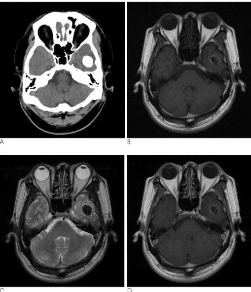
내원하여 시행한 뇌 전산화단층촬영술 상 왼쪽 측두엽에 2×
2 cm 크기의 비교적 치밀한 석회화를 보이는 난원형 종괴가 발견되었다(Fig. 1A). 뇌 자기공명영상에서 상기 종괴는 석회 화로 인해 T1 강조영상에서 저신호강도를 보이고 있었으며 주 변부위에 비균질한 고신호강도를 보이고 있었다(Fig. 1B).
T2 강조영상에서도 종괴는 저신호강도로 보였으며 종괴 주변 에 미약한 부종을 보이고 있었다(Fig. 1C). 조영 증강 T1 강 조영상에서 종괴는 조영증강을 보이지 않았다(Fig. 1D).
환자는 이 종괴에 대해 두개골 절개와 종괴 절제술을 시행받
았다. 병리학적 소견상 종괴는 저등급의 별아교세포종으로 진 단되었다. 수술 한 달 후 환자의 증상은 호전되었고 특별한 합 병증도 관찰되지 않아 환자는 퇴원하였다.
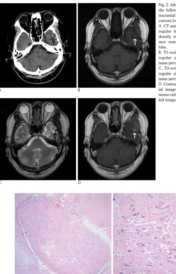
6개월 후 시행한 추적 뇌 자기공명영상에서 측두엽에 종괴가 절제된 자리에 약 1 × 1.4 cm 크기의 불규칙한 모양의 종괴가 발견되었다. 이 종괴는 전산화단층촬영에서 경계가 불명확하고 불규칙한 모양을 보이고 있었으며, 약간 높은 음영을 보이고 있었다(Fig. 2A). 또한, 이 종괴는 T1 강조영상에서 주변 뇌실 질과 동등한 정도의 신호강도를 보이고 있었고(Fig. 2B), T2 강조영상에서는 저신호강도를 보이고 있었다(Fig. 2C). 조영 증강 T1 강조영상에서는 조영증강을 보였다(Fig. 2D). 상기 영상 소견으로 이 종괴는 저등급 별아교세포종의 재발을 배제 할 수가 없었다. 따라서 환자는 이 종괴에 대해 다시 두개골 절 개와 종괴 절제술을 시행받았다. 병리학적 소견상 뇌실질 내에 이물질을 포함하고 있는 다핵거대세포(multinucleated giant cells)와 간질의 섬유화와 염증(interstitial fibrosis and inflammation) 소견이 관찰되어 textiloma(gossypiboma) 으로 진단되었으며 별아교세포종은 관찰되지 않았다(Figs.
3A, B).
고 찰
수술 후 잔류된 거즈나 솜 또는 지혈 물질로 인해 gossypi- boma가 발생한다. 매우 다양한 종류의 지혈물질이 두개내 수 술에 사용되며 이러한 물질은 흡수성과 비흡수성 물질로 나눌 수 있다(5). 결과적으로 비흡수성 물질이 두개내에 남아 염증 반응을 일으키게 되며 비흡수성 물질은 두 가지 타입의 신체반 응을 일으킨다. 첫 번째 반응은 무균성 이물육아종 형성과 함
─ 217 ─ 대한영상의학회지 2011;64:217-220
뇌의 저등급 별아교세포종의 재발로 오인된 Gossypiboma:
증례 보고1
이 진 영∙구 준 범
Gossypiboma는 수술 과정에서 잔류된 거즈나 스펀지와 같은 다양한 종류의 이물질에 의해 염증반응을 일으키는 종물을 말한다. Gossypiboma는 복부나 흉부 수술과 관련하여서 많은 증례 보고가 있었다. 그러나 뇌수술 후에 발생한 gossypiboma는 매우 드문 병변이다. 저자들 은 뇌의 별아교세포종의 외과적 절제 수술 이후 시행한 뇌 자기공명영상에서 조영증강되는 병 변으로 보여 별아교세포종의 재발로 오인되었던 gossypiboma를 경험하였기에 이를 보고하는 바이다.
1동국대학교 일산병원 영상의학과
이 논문은 2010년 11월 15일 접수하여 2011년 1월 6일에 채택되었음.
께 섬유화 반응과 완전한 피막화 반응으로 대부분 증상을 일으 키지 않는다(6). 두 번째 반응은 삼출성 반응으로 종종 농양을 형성할 수 있다. 이 염증성 반응은 조기에 더 심한 증상을 일으 킨다(7).
수술 후 남겨진 이물질의 발생률은 0.01%에서 0.001%로 알려져 있다. 이 중 80%가 gossypiboma이며, 그 중 75%가 복부나 골반 수술로 인한 것들이다(6). 두개내 gossypiboma 는 매우 드물게 보고 있으며 정확한 발생률은 알 수 없다. 두개 내 gossypiboma의 영상소견도 정립된 바는 없으며 다양하게 보고되고 있다. Martins 등(5)은 후두와의 뇌내 신경낭미충증 에 대해 수술을 받은 이후 발생한 두개내 gossypiboma의 자기 공명영상소견에 대해 증례보고를 하였다. 이 증례에서 gossypiboma는 네 번째 뇌실을 막는 종괴로 관찰되었으며 T1 강조영상에서 저신호강도를 보이고 T2 강조영상에서 비균질한 고신호강도에 저신호강도의 점들을 동반하고 있었다. 조영증강 T1 강조영상에서 종괴의 가장자리와 두개골 절개한 자리 주변 의 뇌막에 조영증강을 보였다. Djindjian 등(8)도 뇌내 동정맥 기형 절제 수술 후 발생한 두개내 이물육아종에 증례보고를 하 였다. 이 증례에서 두개내 이물육아종은 뇌 전산화단층촬영에
서 난원형의 저신호강도 종괴로 보였고 조영증강 후 원형 조영 증강을 보였다. 본 증례의 두개 내 gossypiboma는 앞서 기술 한 증례와 다르게 원형 조영증강이 아닌 비교적 균질한 조영증 강을 보였다. 그 외 Jang 등(3)은 두개내 이물육아종의 자기공 명영상소견에 대해 문헌 고찰을 하였다. 고찰 소견상 두개내 이물육아종은 T1 강조영상에서 저신호강도를 보이고, T2 강조 영상에서 저신호 또는 고신호강도를 보이며, 조영증강 영상에 서 결절성 또는 원형 조영증강을 보인다고 한다. 이러한 두개 내 gossypiboma는 환자의 병력을 고려할 때, 영상소견에서 다 양한 종류의 질환과 혼동될 수 있다. 두개 내 gossypiboma와 감별해야 하는 질환으로 혈종, 뇌농양, 종양의 재발, 방사선 괴 사, 뇌경색 등이 있다(9). 본 증례에서도 자기공명영상에서 두 개내 gossypiboma는 종양의 재발로 혼동되었다.
결론적으로 종양의 재발로 혼동되었던 두개내 gossypiboma 의 영상소견에 대해 보고한다. 드물게 보고되고 있는 두개내 gossypiboma의 영상소견에 대해 좀 더 많은 증례의 보고와 이 에 대한 고찰을 통해 다른 질환과의 감별점에 대해 논의가 필 요할 것으로 생각한다.
─ 218 ─
이진영 외: 뇌의 저등급 별아교세포종의 재발로 오인된 Gossypiboma
A B
Fig. 1. CT and MR imaging show low grade astrocytoma in left temporal lobe.
A. CT axial scan shows 2 × 2 cm ovoid dense calcified mass in left temporal lobe.
B. T1-weighted axial image shows low signal intensity mass in left temporal lobe.
C. T2-weighted axial image shows low signal intensity mass in left temporal lobe with minimal peritumoral edema.
D. Contrast enhanced T1-weighted ax- ial image shows no definite enhance- ment in mass.
C D
─ 219 ─ 대한영상의학회지 2011;64:217-220
A B
Fig. 3. Microscopic finding shows intracranial gossypiboma.
A. The low-power magnification shows a well-defined foreign body reaction within the brain parenchyme (×40).
B. Higher magnification reveals multinucleated giant cells engulfing foreign body material surrounded by interstitial fibrosis and inflammation (× 200).
A B
C D
Fig. 2. After 6 months of the operation, the follow up CT and MR show in- tracranial gossypiboma mimicking re- current low grade astrocytoma.
A. CT axial scan shows 1 × 1.4 cm ir- regular heterogeneous slightly high density mass (arrow) in previous tu- mor resection site of left temporal lobe.
B. T1-weighted axial image shows ir- regular shaped iso signal intensity mass (arrow) in left temporal lobe.
C. T2-weighted axial image shows ir- regular shaped low signal intensity mass (arrow) in left temporal lobe.
D. Contrast enhanced T1-weighted ax-
ial image shows relatively homoge-
neous enhancement in mass (arrow) in
left temporal lobe.
참 고 문 헌
1. Rajput A, Loud PA, Gibbs JF, Kraybill WG. Diagnostic challenges in patients with tumors: case 1. Gossypiboma(foreign body) mani- festing 30 years after laparotomy. J Clin Oncol 2003;21:3700-3701 2. Erdem G, Ates O, Koçak A, Alkan A. Lumbar gossypiboma Diagn
Interv Radiol 2010;16:10-12
3. Jang SW, Kim SJ, Kim SM, Lee JH, Choi CG, Lee DH, et al. MR spectroscopy and perfusion MR imaging findings of intracranial foreign body granuloma: a case report. Korean J Radiol 2010;11:
359-363
4. Kim AK, Lee EB, Bagley LJ, Loevner LA. Retained surgical sponges after craniotomies: imaging appearances and complica- tions. AJNR Am J Neuroradiol 2009;30:1270-1272
5. Martins MCB, Amaral RPG, Andrade CS, Lucato LT, Leite CC.
Magnetic resonance imaging findings of intracranial gossypiboma:
a case report and literature review. Radiol Bras 2009;42:407-409 6. Kopka L, Fischer U, Gross AJ, Funke M, Oestmann JW, Grabbe E.
CT of retained surgical sponges (textilomas): pitfalls in detection and evaluation. J Comput Assist Tomogr 1996;20:919-923
7. Choi BI, Kim SH, Yu ES, Chung HS, Han MC, Kim CW. Retained surgical sponge: diagnosis with CT and sonography. AJR Am J
Roentgenol 1988;150:1047-1050
8. Djindjian M, Brugieres P, Razavi-Encha F, Allegret C, Poirier J.
Post-operative intracranial foreign body granuloma: a case report.
Neuroradiology 1987;29:497-499
9. Chater-Cure G, Fonnegra-Caballero A, Baldio′n-Elorza AM, Jime′nez- Hakim E. Gossypiboma in neurosurgery. Case report and litera- ture review. Neurocirugia (Astur) 2009;20:44-49
─ 220 ─
이진영 외: 뇌의 저등급 별아교세포종의 재발로 오인된 Gossypiboma
J Korean Soc Radiol 2011;64:217-220
Address reprint requests to : Joon Bum Koo, M.D., Department of Radiology, Dongguk University Il-San Hospital, Dongguk University, 814 Siksa-dong, Ilsandong-gu, Goyang-si, Gyeonggio 410-773, Korea.
Tel. 82-31-961-8281 Fax. 82-31-961-7826 E-mail: [email protected]
Intracranial Gossypiboma Mimicking a Recurrent Low Grade Astrocytoma : Case Report1
Jin Young Lee, M.D., Joon Bum Koo, M.D.
1
Department of Radiology, Dongguk University Il-San Hospital
Gossypiboma is an inflammatory pseudomass formed by a retained surgical sponge or gauze with reactive tissue after surgery. Gossypiboma has been reported most frequently after abdominal or thoracic surgery. As such, gossypiboma following brain surgery is very rare. We report a case of gossypiboma mimicking tumor re- currence in the brain after a craniotomy and surgical excision of a low grade astrocytoma.
Index words :
BrainGossypiboma
Magnetic Resonance Imaging Tomography, X-ray Computed