Case Report
Obstet Gynecol Sci 2016;59(4):342-345 http://dx.doi.org/10.5468/ogs.2016.59.4.342 pISSN 2287-8572 · eISSN 2287-8580
www.ogscience.org 342
Introduction
Mayer-Rokitansky-Kuster-Hauser (MRKH) syndrome is a mal- formation of the female genitals characterized by normal female phenotype, vaginal agenesis, a rudimentary or absent uterus and normal ovaries [1]. Oppelt et al. [2] further classi- fied MRKH into typical, atypical, and MURCS (Müllerian duct aplasia, renal aplasia, and cervicothoracic somite dysplasia) associated types. The prevalence of MRKH is 1 in 4,000 to 1 in 5,000 women [3], and its association with inguinal ovaries is extremely rare. There are only few case reports of ingui- nal ovaries in MRKH syndrome and accompanying primary ovarian insufficiency is even more rarer [4]. The ideal man- agement consists of surgical reduction of the gonads into pelvic cavity for the ectopic gonads and neovaginoplasty for vaginal agenesis. We present a case of MRKH syndrome with inguinal ovaries and ovarian dysgenesis in a young woman and review the clinicopathological findings in MRKH syn- drome.
Case report
A 21-year-old woman was referred to out center for the evaluation of amenorrhea. She was 161 cm tall and weighs 50 kg. Physical examination showed normal secondary sexual characteristics except for the blind pouch of vagina. A tran- srectal ultrasonography and magnetic resonance imaging revealed atrophic inguinal ovaries (Fig. 1A-C), a single kidney and absence of uterus and cervix. The karyotype was 46, XX
A successful laparoscopic neovaginoplasty using
peritoneum in Müllerian agenesis with inguinal ovaries accompanied by primary ovarian insufficiency
Seonghye Gweon, Jisun Lee, Suna Hwang, Kyoung Joo Hwang, Miran Kim
Department of Obstetrics and Gynecology, Ajou University School of Medicine, Suwon, Korea
The combination of Müllerian agenesis with inguinal ovaries accompanied by primary ovarian insufficiency is extremely rare. A 21-year-old Korean woman was referred to our center with primary amenorrhea. The patient was diagnosed with Müllerian agenesis with inguinal ovaries. Her hormonal profile showed hypergonadotrophic hypogonadism suggesting primary ovarian insufficiency. We performed laparoscopic neovaginoplasty using modified Davydov’s procedure and reposition inguinal ovaries in the pelvic cavity. Oral estrogen replacement was applied for the treatment of primary ovarian insufficiency. This is a rare case report on Mayer-Rokitansky-Kuster-Hauser syndrome accompanied not only by inguinal ovaries but also with primary ovarian insufficiency. We present our first experience on the laparoscopic neovaginoplasty performed on the patient with müllerian agenesis accompanied by inguinal ovaries and primary ovarian insufficiency.
Keywords: Inguinal ovary; Laparoscopy; Müllerian agenesis; Neovaginoplasty; Primary ovarian insufficiency
Articles published in Obstet Gynecol Sci are open-access, distributed under the terms of the Creative Commons Attribution Non-Commercial License (http://creativecommons.
org/licenses/by-nc/3.0/) which permits unrestricted non-commercial use, distribution, and reproduction in any medium, provided the original work is properly cited.
Copyright © 2016 Korean Society of Obstetrics and Gynecology Received: 2015.10.26. Revised: 2016.2.20. Accepted: 2016.3.14.
Corresponding author: Miran Kim
Department of Obstetrics and Gynecology, Ajou University School of Medicine, 164 World cup-ro, Yeongtong-gu, Suwon 16499, Korea Tel: +82-31-219-5300 Fax: +82-31-219-5245
E-mail: [email protected]
http://orcid.org/0000-0001-5553-5334
www.ogscience.org 343 Seonghye Gweon, et al. Neovagina using peritoneum with inguinal ovaries and POI
and serum basal hormone tests showed hypergonadotrophic hypogonadism status (follicle stimulating hormone 72.2 mIU/mL, luteinizing hormone 39.9 mIU/mL, estradiol 5 pg/
mL, prolactin 0.1 ng/mL, antimüllerian hormone 0.08 ng/
mL). Collectively, the diagnosis was MRKH associated with inguinal ovaries and primary ovarian insufficiency.
We performed neovaginoplasty using modified Davydov’s procedure. After making a 1-cm vertical intra-umbilical skin incision, 10 mm trocar was inserted into the abdominal cavity for laparoscopic camera. A 10-mm and two 5-mm trocars were inserted into the lower abdomen. Laparoscopic exploration revealed two rudimental uterine horns and in- guinal ovaries. A probe was inserted in the rectum to secure enough space between sigmoid colon and uterine horns.
Meanwhile, an 8-cm-long vaginal mold (vagina dilator set;
Vaginismus, Vaginismus.com, CA, USA) was inserted and pushed through the vaginal blind pouch to mark the vaginal opening.
About 4-cm transverse incision was made laparoscopically on the peritoneum below the connected rudimental uterine horns until the vaginal mold was exposed into the pelvic cav- ity to create vaginal opening. The pelvic peritoneum in the supravesical fossa was separated from the bladder. We then dissected both round ligaments and pulled the separated pelvic peritoneum out through the newly created vaginal opening. A purse string suture was performed through the
bladder peritoneum, bilateral round ligaments, inguinal ca- nals, tubo-ovarian ligaments, lateral sides of mesorectum, and rectosigmoid junction under the rectal mucosa using non-absorbable prolene 1-0 to make vaginal vault (Fig. 1D).
Concomitantly we pulled the isthmus of the fallopian tube and tubo-ovarian ligament which was herniated in the ingui- nal canal to the pelvic cavity for making vaginal upper fornix The remaining operation was completed vaginally. The pelvic peritoneum superior and inferior to the vaginal open- ing was pulled out into the vaginal cavity and it was repaired using monofilament 2-0 synthetic suture. At the completion of the surgery, the length of newly created vagina was 7 cm and the width was 3 cm. The vaginal mold inserted into the vaginal cavity thereafter until discharge.
We had the patient to keep the vaginal mold inserted for 24 hours at least for 3 days following the surgery. The patient was discharged from the hospital on the 7th day after the surgery without complications. We then limited the mold insertion only for 6 to 8 hours during the daytime and during the sleep. For the next first three months after the surgery, we asked the patient to keep the vaginal mold for 1 hour at least twice a day and maintain inserted over- night. After this period, we informed the patient to keep the mold only overnight until she is able to experience sexual intercourse without difficulties. Up to 12 months following the surgery, the patient continued to use vaginal mold and A B C
Fig. 1. Bilateral inguinal ovaries (arrows). Magnetic resonance imag- ing (A) and laparoscopic finding (B,C). Vaginal vault (D) was made through middle purstring suture to prevent vaginal prolapse and maintain of vaginal sufficient length after inguinal ovaries reposition.
D
www.ogscience.org 344
Vol. 59, No. 4, 2016
neovagina was shown to be intact (Fig. 2). Oral estrogen re- placement was applied for the treatment of primary ovarian insufficiency.
Discussion
Müllerian agenesis or MRKH syndrome is a congenital mal- formation characterized by the failure of uterus and vagina to develop [3]. Most common clinical manifestations include uterovaginal aplasia or hypoplasia [5], and renal or skeletal malformation occurs only in 10% [6].
Genital hernia involving inguinal ovary is uncommon [7]
and the co-occurrence of inguinal ovary with MRKH is even scarcer. Since the first case report by McDonough et al. in 1970 [8], the association between inguinal ovaries with mül- lerian agenesis was well documented by a few case reports [9].
Strubbe et al. [3] reviewed 91 cases of MRKH syndrome and reported that only six patients had an inguinal ovaries. In rare incidences the hernia sac contained ovaries, fallopian tubes as well as uterus in MRKH syndrome [1,7,10].
At 7 to 8 weeks’ gestations, the developing ovary is located on mesentery which forms the cranial suspensory ligament and the genitoinguinal ligament, or gubernaculum. The intra- abdominal portion of the gubernaculum ovary becomes at- tached to the lateral border of the uterus, evolving as the liga- ment of the ovary and the round ligament. As the müllerian duct develops into the uterus, vagina and fallopian tubes, the ovaries remain close to the internal ring as an intraabdominal organ [11].
In inguinal genital hernia, the gubernaculum is proximally attached to the isolated uterine horn and distally to the exter- nal ring [11]. The incomplete closure of the processus vaginalis
formed inside the gubernaculum contributes to the formation of inguinal genital hernia. The processus vaginalis and the round ligament can pass through the inguinal canal, causing inguinal hernia of ovaries, fallopian tubes, and even the uterus [1]. The indirect hernia in MRKH is considered to result from müllerian abnormality, which comprises fusion defects of mül- lerian ducts [10]. The hernia is suggested by an excess length and mobility of suspensory ligaments caused by the lack of fu- sion of the müllerian ducts during the second month of fetal development [12].
Although inguinal ovary is well documented in MRKH, the ovary function in such cases has been underreported. In fact, most MRKH cases were presented with normal ovarian func- tions. Our patient however, showed hypergonadotrophic hy- pogonadism status (follicle stimulating hormone 72.2 mIU/mL, luteinizing hormone 39.9 mIU/mL, estradiol 5 pg/mL, prolactin 0.1 ng/mL, and anti-müllerian hormone 0.08 ng/mL) suggest- ing primary ovarian insufficiency. Ovarian dysgenesis with 46, XX is defined as a primary ovarian defect leading to primary ovarian insufficiency due to failure of development and resis- tance to gonadotropin stimulation [13].
The ovary in the inguinal canal is not at risk of compression of its blood supply but rather is at a significant risk of tor- sion and infarction [14]. The vulnerability of inguinal ovary to infarction suggests a possible explanation for the ovarian in- sufficiency observed in our patient. Therefore, surgical reposi- tioning of the inguinal ovaries into the pelvic cavity should be undertaken as soon as possible to preserve ovarian function.
Despite the ovarian dysgenesis, our patient showed second- ary sexual characteristics. The laparoscopy revealed small yet morphologically otherwise normal ovaries. We postulated that the ovary function may gradually deteriorate to allow second- ary sexual characteristics to develop.
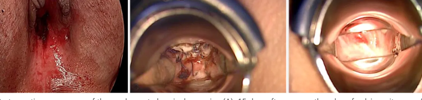
Fig. 2. Postoperative appearance of the newly created vaginal openning (A), 15 days after surgery, the edge of pelvic peritoneum (B), and vaginal vault after surgery at 12 months (C).
A B C
www.ogscience.org 345 Seonghye Gweon, et al. Neovagina using peritoneum with inguinal ovaries and POI
In this article, we reported a rare case of MRKH with in- guinal ovary and primary ovarian insufficiency. Although an indirect ovary hernia in young girls is not a rare condition, it is rare in adults. In this patient, the müllerian system failed to fuse and ovaries were descended into the inguinal ring.
The anatomical anomaly in MRKH is corrected by surgical procedure. We successfully performed neovaginoplasty us- ing modified Davydov’s procedure and repositioned bilateral inguinal ovaries in the pelvic cavity. The modified Davydov’s technique consisted of the laparoscopic procedure followed by vaginal approach [15]. Oral estrogen replacement was also applied for the treatment of primary ovarian insufficiency.
In conclusion, MRKH is a complex syndrome involving uterus, fallopian tubes and other organs such as skeleton or renal system. The treatment must be individualized consider- ing patient’s age, and should be aimed to correct not only the anatomical anomaly but also hormonal dysfunction in cases of ovarian insufficiency.
Conflict of interest
No potential conflict of interest relevant to this article was reported.
References
1. Al Omari W, Hashimi H, Al Bassam MK. Inguinal uter- us, fallopian tube, and ovary associated with adult Mayer-Rokitansky-Kuster-Hauser syndrome. Fertil Steril 2011;95:1119.e1-4.
2. Oppelt P, Renner SP, Kellermann A, Brucker S, Hauser GA, Ludwig KS, et al. Clinical aspects of Mayer-Rokitansky- Kuester-Hauser syndrome: recommendations for clinical diagnosis and staging. Hum Reprod 2006;21:792-7.
3. Strubbe EH, Willemsen WN, Lemmens JA, Thijn CJ, Rol- land R. Mayer-Rokitansky-Kuster-Hauser syndrome:
distinction between two forms based on excretory uro-
graphic, sonographic, and laparoscopic findings. AJR Am J Roentgenol 1993;160:331-4.
4. Bazi T, Berjawi G, Seoud M. Inguinal ovaries associated with Mullerian agenesis: case report and review. Fertil Steril 2006;85:1510.e5-8.
5. Evans TN, Poland ML, Boving RL. Vaginal malformations.
Am J Obstet Gynecol 1981;141:910-20.
6. Willemsen WN. Renal-skeletal-ear- and facial-anomalies in combination with the Mayer-Rokitansky-Kuster (MRK) syndrome. Eur J Obstet Gynecol Reprod Biol 1982;14:121- 30.
7. Kokcu A, Malazgirt Z, Cetinkaya MB, Tosun M. Presence of a uterine horn and fallopian tube within an indirect hernial sac: report of a rare case. Hernia 2010;14:325-7.
8. McDonough PG, Byrd JR, Freedman MA. Complete du- plication and underdevelopment of the Mullerian system in association with gonadal dysgenesis: report of a case.
Obstet Gynecol 1970;35:875-7.
9. Kriplani A, Banerjee N, Aminni AC, Kucheria K, Takkar D.
Hernia uterus inguinale in a 46,XX female: a case report. J Reprod Med 2000;45:48-50.
10. Kamio M, Nagata T, Yamasaki H, Yoshinaga M, Douchi T.
Inguinal hernia containing functioning, rudimentary uter- ine horn and endometriosis. Obstet Gynecol 2009;113(2 Pt 2):563-6.
11. Hutson JM, Kearsey I. Is the ovary in an inguinal hernia
‘descended’ like a testis or not? J Pediatr Surg 2015 Sep 25 [Epub]. http://dx.doi.org/10.1016/j.jpedsurg.2015.09.014.
12. Ming YC, Luo CC, Chao HC, Chu SM. Inguinal hernia containing uterus and uterine adnexa in female infants:
report of two cases. Pediatr Neonatol 2011;52:103-5.
13. Gorgojo JJ, Almodovar F, Lopez E, Donnay S. Gonadal agenesis 46,XX associated with the atypical form of Roki- tansky syndrome. Fertil Steril 2002;77:185-7.
14. Merriman TE, Auldist AW. Ovarian torsion in inguinal her- nias. Pediatr Surg Int 2000;16:383-5.
15. Fedele L, Bianchi S, Zanconato G, Raffaelli R. Laparoscopic creation of a neovagina in patients with Rokitansky syn- drome: analysis of 52 cases. Fertil Steril 2000;74:384-9.