CASE REPORT
Copyright © 2017 The Korean Retina Society
This is an Open Access article distributed under the terms of the Creative Commons Attribution Non-Commercial License (http://creativecommons.org/licenses/by-nc/3.0/) which permits unrestricted non-commercial use, distribution, and reproduction in any medium, provided the original work is properly cited.
pISSN 2508-1926 eISSN 2508-3589 Journal of Retina 2017;2(2):97-100
https://doi.org/10.21561/jor.2017.2.2.97
미만성 거대 B 세포 림프종 환자에서 발병한 거대세포바이러스 망막염
Cytomegalovirus Retinitis in Patients with Diffuse Large B-cell Lymphoma
강혜민 Hae Min Kang
가톨릭관동대학교 국제성모병원 안과
Department of Ophthalmology, Catholic Kwandong University International St. Mary’s Hospital, Incheon, Korea
Purpose: To report two cases of cytomegalovirus (CMV) retinitis in patients with diffuse large B cell lymphoma (DLBCL).
Case summary: A 59-year-old man (patient 1) visited our ophthalmology clinic due to decreased vision in the left eye. The patient had a history of right nephrectomy due to renal cell tumor, which showed complete remission. Best-corrected visual acuity (BCVA) of the left eye was 20/100 according to the Snellen visual acuity chart. Fundus examination revealed yellowish retinal infiltration with retinal hemorrhages, and diagnostic vitrectomy confirmed CMV retinitis. Further systemic evaluation indicated pancytopenia and bone marrow suppression. Positron emission tomography-computed tomography imaging suggested malignant lymphoma, and subsequent biopsy of lymph nodes confirmed DLBCL. In the second case, a 55-year-old woman (patient 2) presented with a sudden decrease in vision in the left eye during systemic chemotherapy for DLBCL. BCVA in the left eye was 20/125 by Snellen visual acuity chart, and the fundus demon- strated diffuse yellowish retinal infiltration with some grainy opacification. After a diagnosis of CMV retinitis, ganciclovir was administered to both patients, leading to improvement of retinal lesions.
Conclusions: CMV retinitis can occur in patients with DLBCL; thus, when patients with DLBCL complain of visual deterioration, CMV reti- nitis should be suspected.
Keywords: Cytomgalovirus (CMV); CMV retinitis; Diffuse large B-cell lymphoma
Introduction
Cytomegalovirus (CMV) retinitis is the most common op- portunistic infection of the eye and can cause profound visual loss [1,2]. Acquired immunodeficiency syndrome caused by human immunodeficiency virus (HIV) is a common underly- ing disease in CMV retinitis, and was particularly prevalent before the era of highly active antiretroviral therapy [2-4].
CMV retinitis also can occur in the absence of HIV when other conditions lead to systemic immunosuppression, such as after chemotherapy for any malignancy, organ transplan- tation, or in cases of inherited immunodeficiency [1,5-7].
CMV retinitis has been also reported in various hematologic malignancies including leukemia and lymphoma [5,8-12].
Diffuse large B cell lymphoma (DLBCL) is the most com- mon lymphoid malignancy in adults, and the most common
Address reprint requests to Hae Min Kang, MD, PhD
Department of Ophthalmology, Catholic Kwandong University International St. Mary’s Hospital, Catholic Kwan- dong University College of Medicine, #25 Simgok-ro 100beon-gil, Seo-gu, Incheon 22711, Korea
Tel: 82-32-290-3888; Fax: 82-32-290-3879 E-mail: [email protected]
Received: 2017. 7. 26 Revised: 2017. 8. 27 Accepted: 2017. 9. 4
98
JOURNAL OF RETINA
https://doi.org/10.21561/jor.2017.2.2.97 subtype of non-Hodgkin’s lymphoma (representing approxi-
mately one-third of all cases) [13]. DLBCL is most prevalent in middle-aged or elderly subjects, with a mean age at diag- nosis in the sixth decade of life. Men are slightly more prone to developing DLBCL than women [13].
Recently, we treated two cases of CMV retinitis in patients with DLBCL, which represent the first cases reported in Korea. In one patient, ophthalmologic confirmation of CMV retinitis led to diagnosis of underlying DLBCL, and the other patient developed CMV retinitis during chemotherapy for DLBCL.
Case Report
Case 1
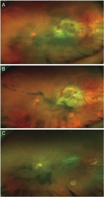
A 59-year-old male patient visited our ophthalmology clinic due to visual deterioration in the left eye that had progressed over 1 week. The best-corrected visual acuity (BCVA) was 20/20 in the right eye, and 20/100 in the left eye according to the Snellen visual acuity chart. Detailed fundus examination after dilation detected no significant findings in the right eye, whereas in the left eye there was moderate vitreous opacity with retinal infiltration and hemorrhages temporal to the fovea, extending to the temporal periphery (Fig. 1A).
The patient had a history of right nephrectomy due to a renal tumor that had been treated at another hospital 15 years pri- or, which remained in complete remission. The patient did not report experiencing any other diseases. However, base- line blood tests showed generalized hematologic problems including anemia, neutropenia, and decreased platelet cell counts. Other laboratory findings were normal, and an HIV test was negative. The patient’s clinical features were sugges- tive of CMV retinitis and his immune status was not conclu- sive for immunosuppression, therefore diagnostic vitrectomy was performed on the same day. After diagnostic vitrectomy, systemic administration of ganciclovir was initiated, but due to further deterioration of renal function, treatment was switched to intravitreal ganciclovir injections. PCR analysis of vitreous fluid confirmed CMV retinitis in this patient, and further systemic assessments including bone marrow biopsy were performed. Bone marrow biopsy revealed hypocellular marrow. Subsequent positron emission tomography-comput- ed tomography revealed multiple enlarged lymph nodes with increased fluorodeoxyglucose (FDG) uptake in the bilateral
parotid glands, neck, supraclavicular area, axillary area, mediastinum, paraspinal area, cardiophrenic area, abdomen, retroperitoneum, bilateral iliac changes, and inguinal area.
Figure 1. Representative image of patient 1. (A) Wide fundus pho- tography of a 59-year-old man who visited the ophthalmology clinic with a sudden decrease in vision in the left eye. Dilated fundus examination revealed moderate vitreous opacity with retinal infil- tration and hemorrhages temporal to the fovea, extending to the temporal periphery. (B) Wide photography on treatment day 1. After diagnostic vitrectomy, the vitreous opacity was removed. A central shadow due to a posterior subcapsular opacity of the lens could be observed. Retinal infiltration with hemorrhages temporal to the fovea was noted, leading to a diagnosis of cytomegalovirus retinitis.
(C) After treatment via intravitreal ganciclovir injection, retinal infiltra- tion subsided, and granular consolidation was achieved.
A
B
C
99
Kang HM. CMV retinitis in DLBCL
https://doi.org/10.21561/jor.2017.2.2.97
In addition, the spleen, right nasopharyngeal wall, sternum, thoracic spines, bilateral acetabulum, left pubic bone, and thighs showed increased FDG uptake, suggesting lympho- ma. Lymph node biopsy led to a diagnosis of DLBCL stage IVB, IPI 3 in this patient, and systemic chemotherapy was started (rituximab, cyclophosphamide, hydroxydaunoru- bicin, oncovin and prednisone or prednisolone; R-CHOP).
After intravitreal cymevene injections in the left eye, BCVA in the left eye was maintained at 20/100 on the Snellen visual acuity chart, although CMV retinitis itself exhibited repeat- ed stabilization and reactivation over the next 8 months (Fig.
1B, C). Despite systemic chemotherapy, the patient expired 8 months after CMV retinitis diagnosis.
Case 2
A 55-year-old female patient was referred from the hema- tology department due to a sudden decrease in vision in the left eye. BCVA was 20/25 in the right eye, and 20/125 in the left eye on the Snellen visual acuity chart. The patient had a 7-year history of diabetes mellitus, and had undergone oph- thalmologic evaluation in our clinic 7 months prior. BCVA was 20/25 in each eye on the Snellen visual acuity chart.
Numerous fine drusen were present, and further assessment showed mild nonproliferative diabetic retinopathy in both eyes (Fig. 2A). Five months prior to visual symptoms, the patient was diagnosed with DLBCL via nasopharyngeal bi- opsy (stage II EA, IPI 1), and started systemic chemotherapy (R-CHOP). Concurrent with sudden visual deterioration, moderate anterior chamber reaction, mild vitreous haze, dif- fuse yellowish retinal infiltration, and grainy opacification with some retinal hemorrhages were noted in the left eye (Fig. 2B). Based on the patient’s clinical features and sys- temic condition, she was diagnosed with CMV retinitis and systemic intravenous ganciclovir administration followed by oral administration was started. One month after the initia- tion of systemic treatment, the anterior chamber reaction was resolved, retinal lesions had stabilized, and BCVA improved to 20/40 in the left eye according to the Snellen visual acuity chart (Fig. 2C). However, the patient expired 7 months after the diagnosis of CMV retinitis due to DLBCL.
Discussion
CMV infection causes retinal whitening or opacification at- tributable to retinal necrosis and variable edema [14,15]. An Figure 2. Representative image of patient 2. (A) Wide fundus pho- tography of a 55-year-old woman before the development of cyto- megalovirus (CMV) retinitis. Due to underlying diabetes mellitus, the patient was referred for routine evaluation for diabetic retinopathy.
At that time, fundus examination revealed some drusen over the macula. (B) After diagnosis of diffuse large B-cell lymphoma and development of CMV retinitis, fundus examination demonstrated a mild vitreous haze, diffuse yellowish retinal infiltration, and grainy opacification with some retinal hemorrhages in the left eye. (C) After treatment for CMV retinitis with systemic ganciclovir, retinal infiltra- tion was greatly improved.
A
B
C
100
JOURNAL OF RETINA
https://doi.org/10.21561/jor.2017.2.2.97 irregular, dry-appearing, granular border, often with charac-
teristic small, isolated satellites at the advancing edge, is the hallmark of untreated CMV retinitis [14,15]. CMV retinitis can be further classified into two variants: a fulminant/edem- atous type showing marked edema with dense, confluent areas of retinal whitening, which is usually accompanied by moderate-to-severe retinal hemorrhage and vascular sheath- ing, and an indolent/granular type characterized by less ede- ma, only faint, grainy opacification, and little or no retinal hemorrhage without vascular sheathing [14,15]. CMV retini- tis and its complications such as retinal detachment, branch retinal vessel occlusion, and retinal neovascularization can lead to profound vision loss in these patients [14,15]. Because the diagnosis of CMV retinitis is primarily clinical, relying on identification of typical features, clinical suspicion is im- portant for the diagnosis and treatment of CMV retinitis.
This represents the first Korean case report of CMV ret- initis, and suggests the importance of clinical suspicion in the diagnosis of this disease for both ophthalmologists and hemato-oncologists. The case of patient 1, clinical suspicion and confirmation of CMV retinitis led to the diagnosis of underlying DLBCL. With this case, we have revalidated the importance of clinical suspicion and a thorough systemic work-up in CMV retinitis patients, even in those who deny the presence of any underlying diseases. By identifying systemic diseases, life-saving treatment in addition to sight-preserving treatment can be administered in a timely manner. In the case of patient 2, we were aware that she was already immunocompromised due to treatment for DLBCL, and timely consultation with the hematology department led to diagnosis of CMV retinitis. CMV retinitis should be suspected when immunocompromised patients report visual disturbances.
We report two cases of CMV retinitis in patients with DLBCL in Korea, and encourage clinical suspicion, timely evaluation and appropriate treatment in such patients.
Conflicts of interest
The authors have no conflicts to disclose.
References
1. Scott WJ, Giangiacomo J, Hodges KE. Accelerated cytomegalo-
virus retinitis secondary to immunosuppressive therapy. Arch Ophthalmol 1986;104:1117-8, 1124.
2. Jabs DA. Cytomegalovirus retinitis and the acquired immuno- deficiency syndrome--bench to bedside: LXVII Edward Jackson Memorial Lecture. Am J Ophthalmol 2011;151:198-216.e1.
3. Pertel P, Hirschtick R, Phair J, et al. Risk of developing cytomega- lovirus retinitis in persons infected with the human immunode- ficiency virus. J Acquir Immune Defic Syndr 1992;5:1069-74.
4. Sugar EA, Jabs DA, Ahuja A, et al. Incidence of cytomegalovirus retinitis in the era of highly active antiretroviral therapy. Am J Ophthalmol 2012;153:1016-24.e5.
5. Fiala M, Payne JE, Berne TV, et al. Epidemiology of cytomegalo- virus infection after transplantation and immunosuppression. J Infect Dis 1975;132:421-33.
6. Wagle AM, Biswas J, Gopal L, Madhavan H. Clinical profile and immunological status of cytomegalovirus retinitis in organ transplant recipients. Indian J Ophthalmol 2002;50:115-21.
7. Pathanapitoon K, Tesavibul N, Choopong P, et al. Clinical mani- festations of cytomegalovirus-associated posterior uveitis and panuveitis in patients without human Immunodeficiency virus infection. JAMA Ophthalmol 2013;131:638-45.
8. Derzko-Dzulynsky LA, Berger AR, Berinstein NL. Cytomegalovirus retinitis and low-grade non-Hodgkin’s lymphoma: case report and review of the literature. Am J Hematol 1998;57:228-32.
9. Chawla R, Venkatesh P, Garg SP, et al. Cytomegalovirus retinitis in a patient with non-Hodgkin’s lymphoma: a diagnostic dilemma.
Eur J Ophthalmol 2005;15:153-7.
10. Nasir MA, Jaffe GJ. Cytomegalovirus retinitis associated with Hodgkin’s disease. Retina 1996;16:324-7.
11. Han SB, Lee JH, Lee JW, et al. Cytomegalovirus retinitis diag- nosed after completion of chemotherapy for acute lympho- blastic leukemia in an adolescent. J Pediatr Hematol Oncol 2015;37:e128-30.
12. Kobayashi R, Takanashi K, Suzuki D, et al. Retinitis from cytomeg- alovirus during maintenance treatment for acute lymphoblastic leukemia. Pediatr Int 2012;54:288-90.
13. Jaffe ES. The 2008 WHO classification of lymphomas: implica- tions for clinical practice and translational research. Hematology Am Soc Hematol Educ Program 2009:523-31. doi: 10.1182/ashed- ucation-2009.1.523.
14. Barrett L, Walmsley S. CMV retinopathy in the antiretroviral ther- apy era: prevention, diagnosis, and management. Curr Infect Dis Rep 2012;14:435-44.
15. Bloom JN, Palestine AG. The diagnosis of cytomegalovirus reti- nitis. Ann Intern Med 1988;109:963-9.