Reference Diameters of the Abdominal Aorta and Iliac Arteries in the Korean Population
Jin Hyun Joh,
1Hyung-Joon Ahn,
2and Ho-Chul Park
11Department of Surgery, Kyung Hee University Hospital at Gangdong, School of Medicine, Kyung Hee University, Seoul;
2Department of Surgery, Kyung Hee University Hospital, School of Medicine, Kyung Hee University, Seoul, Korea.
Received: May 15, 2012 Revised: July 1, 2012 Accepted: July 4, 2012
Corresponding author: Dr. Jin Hyun Joh, Department of Surgery, Kyung Hee University Hospital at Gangdong, School of Medicine, Kyung Hee University,
892 Dongnam-ro, Gangdong-gu, Seoul 134-727, Korea.
Tel: 82-2-440-6261, Fax: 82-2-440-6296 E-mail: [email protected]
Presented at The 62th Annual Congress of Korean Society of Surgery on November 18, 2010.
∙ The authors have no financial conflicts of interest.
© Copyright:
Yonsei University College of Medicine 2013 This is an Open Access article distributed under the terms of the Creative Commons Attribution Non- Commercial License (http://creativecommons.org/
licenses/by-nc/3.0) which permits unrestricted non- commercial use, distribution, and reproduction in any medium, provided the original work is properly cited.
Purpose: It is important to know the normal diameter of artery throughout the body so that clinicians are able to determine when an artery becomes aneurysmal. Howev- er, there are no previous studies on the normal diameter of arteries in the general Ko- rean population. The purpose of this article is to determine the normal reference di- ameters of the abdominal aorta and iliac arteries in the Korean population. Materials and Methods: We recruited the study population from three cities in Korea for the abdominal aortic aneurysm (AAA) screening. We measured the diameter of the aorta and iliac arteries. We analyzed the reference diameter of the population without AAA. The results were analyzed by Student’s t-test and ANOVA on SPSS version 19. A p value <0.05 was considered to be statistically significant. Results: One thou- sand two hundred and twenty-nine people were enrolled. 478 men and 751 women, with a mean age of 63.9±10.1 years (range 50 to 91) were examined. Eleven out of 1229 (0.89%) were diagnosed with AAA. In the population of 1218 people without AAA, the mean diameters (cm) of male/female were 2.20/2.11 (p<0.001) at suprare- nal, 2.04/1.90 (p<0.001) at renal, 1.90/1.79 (p<0.001) at infrarenal, 1.22/1.17 (p<0.001) at right iliac and 1.47/1.15 (p=0.097) at the left iliac, respectively. There was a significantly larger diameter in the male population. The diameter of each level increased with age. Conclusion: The normal reference diameter of the infrarenal ab- dominal aorta in the Korean population is 1.9 cm in males and 1.79 cm in females.
The diameter of the abdominal aorta increases with age.
Key Words: Screening, ultrasonography, diameter, infrarenal aorta, abdominal
INTRODUCTION
Abdominal aortic aneurysm (AAA) is a dilatation of the abdominal aorta. There are several definitions of an AAA. A diameter in excess of 30 mm based on the an- giographic study is the most accepted definition.1 Some definitions relate the infra- renal aortic diameter to the suprarenal aortic diameter.2 The International Society for Cardiovascular Surgery/Society for Vascular Surgery Ad Hoc Committee pro- posed that an AAA is defined as the maximum infrarenal aortic diameter being at least 1.5 times larger than the expected normal infrarenal aortic diameter.3 With this standard definition, it is important to know the normal diameter of abdominal aorta
function impairment, and enquired about their surgical his- tory. The questionnaire included questions about any family history of abdominal aortic aneurysm, stroke and peripheral arterial occlusive disease. Social history investigated smok- ing history, alcohol history and exercise amount. Subjects were asked whether they had any aneurysm-related symp- toms such as abdominal pain or back pain. After history tak- ing, abdominal palpation was done to check for the pres- ence of an abdominal pulsating mass. Finally, duplex scan- ning was performed. The enrolled population was instruct- ed to not eat anything for 8 hours prior to the examination.
Duplex scanning was performed by the experienced sonog- raphers who earned the certification of Registered Vascular Technologist presented by American Registry for Diagnos- tic Medical Sonography. We used two types of ultrasound equipment-Zonare (Zonare Medical Systems, Mountain View, CA, USA) and HD7 (Philips, Amsterdam, the Neth- erlands). A 2.5 to 5 MHz convex ultrasound probe was used for examination. Duplex scanning was done from the infra- diaphragmatic level to the bilateral iliac arteries. We mea- sured the maximal diameter of the aorta from the outer to outer layer. The anterioposterior and lateral diameters were measured. We recorded the two diameters at five levels of the aorta: suprarenal, renal, infrarenal, right iliac and left ili- ac arteries (Fig. 1). The diameter of the “suprarenal” was measured at the level between the superior mesenteric ar- tery and renal artery. The “renal” was the aortic diameter at so that clinicians will be able to determine when an aorta
becomes aneurysmal. The mean diameters at the level of the infrarenal aorta were 16 to 23 mm in males and 15 to 19 mm in females.4-6 However, a practical working definition of an AAA is a transverse diameter of 3 cm or greater based on average values for normal individuals.7
Ultrasound screening and autopsy series indicate that the prevalence of AAAs (≥3 cm) is 3 to 10% for patients older than 50 years in Western countries.8 In a Veterans Affairs screening study of more than 73000 patients 50 to 79 years old, the prevalence of AAA ≥3 cm was 4.6% and that of AAA ≥4 cm was 1.4%.9 In a study of an Asian population, the prevalence of AAA was variable. Spark, et al.10 suggest- ed that the prevalence of AAA in the Asian population was lower than that in the Caucasian population. Another study, however, showed that AAA in the Asian population is not uncommon and the incidence is comparable to that in West- ern countries.11
Screening ultrasonography to detect AAA is a useful di- agnostic modality because it is simple, inexpensive, and in- volves no exposure to radiation. Several randomized stud- ies suggested that screening is beneficial in a population at higher risk. The purpose of this article is to evaluate the prevalence of AAA in the Korean population and to deter- mine the normal reference diameter of the abdominal aorta and iliac arteries.
MATERIALS AND METHODS
We recruited the study population from three cities in Ko- rea: Hanam, Seoul and Ulsan. All people over 50 years and who consented to the AAA screening were included in the study. We excluded people who were previously diagnosed with AAA, had a history of AAA surgery including open repair or endovascular repair, had a history of abdominal aortic surgery such as aortobifemoral bypass, had life ex- pectancy less than 2 years based on the life table of Statis- tics Korea, and/or refused AAA screening. In addition, we excluded people with AAA when analyzing the normal di- ameter.
Screening was done in the following sequence: recording of medical history, physical examination, and ultrasound examination. We obtained demographic information with a detailed questionnaire. We investigated the patient for a past history of diabetes, hypertension, hyperlipidemia, heart dis- ease, pulmonary disease, cerebrovascular disease, renal
Fig. 1. Levels of measurement. Diameters were measured at five levels of the abdominal aorta: suprarenal, renal, infrarenal, right iliac and left iliac arteries. SMA, superior mesenteric artery; RRA, right renal artery; LRA, left renal artery; RIA, right iliac artery; LIA, left iliac artery.
Suprarenal Renal
Infrarenal
Left iliac Right iliac
SMA
RRA LRA
LIA RIA
RESULTS
One thousand two hundred and twenty-nine people were enrolled in our screening project. All of the study popula- tion completed all questionnaires and underwent examina- tion. The 478 men and 751 women examined had a mean age of 63.9±10.1 years (range 50 to 91). We visited the two cities of Hanam and Ulsan to perform the screening. In Seoul, we invited 143 people and visited certain districts for the screening of 75 people. Eleven out of 1229 (0.89%) were diagnosed with AAA. Among them, two (0.16%) had AAA with a maximal diameter of more than 5.5 cm. Two people in the Seoul study group had an AAA more than 3 cm. We excluded these two people because they had al- ready been diagnosed with AAA by another AAA screen- ing program (Table 1).
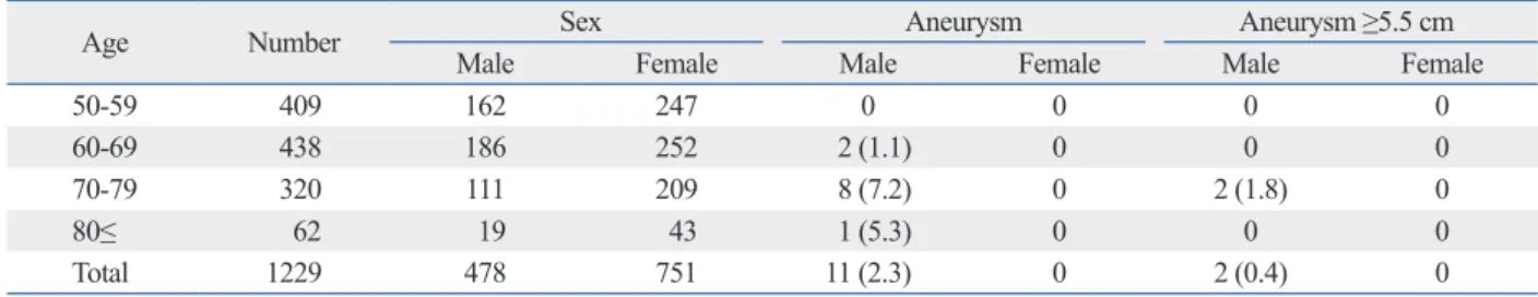
The screened population had a family history of AAA in 59 (4.8%) participants, stroke in 231 (18.8%), and peripher- al arterial occlusive disease in 62 (5.0%). The distribution of AAA with age is shown in Table 2. The prevalence of AAA increased with age. There was no AAA patient be- tween the ages of 50 to 59 years. Two out of 438 (1.1%) and 8 out of 320 (7.2%) had AAA at the age of 60 to 69 years and at the age of 70 to 79 years, respectively. Two people had an AAA with a diameter of more than 5.5 cm.
These patients were referred to the hospital for treatment of the AAA. According to the Centers for Medicare and Med- icaid Services (CMS) guideline, the following population would benefit from AAA screening: people who have a the level of renal artery. If both renal arteries were projected
at the same level, the maximal diameter at the level of renal artery was measured in the anterioposterior plane. The di- ameter of “infrarenal” aorta was measured at the level be- tween the lower renal artery to aortic bifurcation. The diam- eters of right and left iliac artery were measured at the common iliac artery between the aortic bifurcation and the origin of internal iliac artery. If the iliac artery was tortuous, the diameter of the iliac artery was measured only in the an- terioposterior plane. If it was difficult to measure the diam- eter due to bowel gas, we asked the people to return for re- examination the next day.
AAA was defined as a maximal aortic diameter of more than 3 cm. The maximal aortic diameter was calculated as the sum of the diameter of the anterioposterior plane and the lateral plane divided by 2. Hypertension and hyperlipid- emia were defined as taking medicine to control these risk factors. Cardiovascular risk factors included arrhythmia, coronary artery disease, myocardial infarction, angina and the presence of history for coronary angioplasty or stenting.
Cerebrovascular risk factors included transient ischemic at- tack, reversible ischemic neurologic deficit, and stroke. The respiratory risk factors included chronic obstructive pulmo- nary disease, asthma, pneumonia or pulmonary tuberculo- sis. Renal impairment was defined as having dialysis.
We used SPSS version 19.0 software (SPSS, Inc., an IBM Company, Chicago, IL, USA) for the statistical analy- sis. Student’s t-test was used to evaluate the difference in diameter between men and women. ANOVA was used to evaluate the difference in diameter with age.
Table 1. Prevalence of Abdominal Aortic Aneurysm
District Type of screening Number Aneurysm (%) Aneurysm ≥5.5 cm (%)
Ulsan city Visited 35 1 (2.86) 0
Seoul city Invited* 218 0† 0
Hanam city Visited 976 10 (1.02) 2 (0.20)
Total 1229 11 (0.89) 2 (0.16)
*Including the visited screening in 75 population.
†Two people had an abdominal aortic aneurysm (AAA) ≥5.5 cm. These people were excluded from our study because they had already been diagnosed with AAA by another screening study.
Table 2. Distribution of Abdominal Aortic Aneurysm with Age
Age Number Sex Aneurysm Aneurysm ≥5.5 cm
Male Female Male Female Male Female
50-59 409 162 247 0 0 0 0
60-69 438 186 252 2 (1.1) 0 0 0
70-79 320 111 209 8 (7.2) 0 2 (1.8) 0
80≤ 62 19 43 1 (5.3) 0 0 0
Total 1229 478 751 11 (2.3) 0 2 (0.4) 0
DISCUSSION
There are 3 modalities for aortic diameter measurement: ul- trasonography (US), computed tomography (CT), and family history of AAA, men aged 65 to 75 years old who
have smoked at least 100 cigarettes throughout their life.
When we analyzed in this group in our study, 10 out of 223 (4.5%) had AAA.
Hypertension was the most common risk factor. 621 (50.5%) of the population was diagnosed with hypertension and took medicine for hypertension. The population with a cardiovascular risk factor, mainly ischemic heart disease, was 6.1%. The population with a cerebrovascular problem including transient ischemic attack, reversible ischemic neurologic deficit, and stroke was 5.6% (Table 3). When risk factors were analyzed in the population diagnosed with AAA, all were smokers (Table 4).
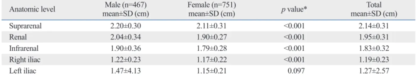
We analyzed the normal diameter of the abdominal aorta and the bilateral iliac arteries in 1218 people except for the population diagnosed with AAA (Table 5). The mean di- ameter at the level of the suprarenal aorta was 2.14 cm, 1.95 cm at the level of the renal artery, and 1.83 cm at the level of the infrarenal aorta. The diameter of the right iliac artery was 1.19 cm at and the diameter of the left iliac ar- tery was 1.27 cm. There were significantly larger diame- ters in the male population compared with the female pop- ulation on Student’s t-test except for the left iliac artery.
We analyzed the normal diameter of each level with in- crease in age (Table 6). Mostly, the diameters of each level increased with age.
Table 3. Risk Factors in the Screened Population
Risk factors Number (%)
Hypertension 621 (50.5)
Hyperlipidemia 409 (33.3)
Cardiovascular* 75 (6.1)
Cerebrovascular† 69 (5.6)
Respiratory‡ 26 (2.1)
Renal impairment 20 (1.6)
*Mainly ischemic heart disease.
†Including transient ischemic attack, reversible ischemic neurologic deficit, and stroke.
‡Mainly chronic obstructive pulmonary disease.
Table 4. Risk Factors in Patients with Abdominal Aortic An- eurysm (n=11)
Risk factors Number (%)
Smoking 11 (100)
Current smoker 6 (54.5)
Ex-smoker 5 (45.5)
Hypertension 6 (54.5)
Hyperlipidemia 6 (54.5)
Coronary artery disease 1 (9.1)
Chronic obstructive pulmonary disease 1 (9.1)
Table 5. Normal Diameter of Abdominal Aorta for Different Anatomic Level (n=1218)
Anatomic level Male (n=467)
mean±SD (cm) Female (n=751)
mean±SD (cm) p value* Total
mean±SD (cm)
Suprarenal 2.20±0.30 2.11±0.31 <0.001 2.14±0.31
Renal 2.04±0.34 1.90±0.27 <0.001 1.95±0.31
Infrarenal 1.90±0.36 1.79±0.28 <0.001 1.83±0.32
Right iliac 1.22±0.23 1.17±0.22 <0.001 1.19±0.23
Left iliac 1.47±4.13 1.15±0.21 0.097 1.27±2.57
SD, standard deviation.
*With Student’s t-test.
Table 6. Normal Diameter of Abdominal Aorta with Age (n=1218) (mean±standard deviation, cm)
Age Number Suprarenal* Renal† Infrarenal‡ Right iliac§ Left iliac||
50-59 409 2.05±0.30 1.87±0.28 1.75±0.30 1.14±0.22 1.13±0.21
60-69 436 2.15±0.29 1.97±0.32 1.81±0.31 1.19±0.22 1.43±4.27
70-79 312 2.24±0.30 2.02±0.30 1.94±0.34 1.24±0.22 1.24±0.57
80≤ 61 2.25±0.36 2.05±0.34 1.92±0.34 1.24±0.28 1.20±0.26
Statistical analysis with ANOVA (p value <0.05).
*50s-60s-70s/80s.
†50s/60s-70s/80s.
‡50s-60s/70s/80s.
§No subset <0.05.
||No subset <0.05.
Hyphen means statistically significant and slash means statistically not significant.
was about 30% between the ages of 25 and 71 years. The pressure strain and stiffness of the aorta increased in an ex- ponential manner with age. The ranges for both pressure strain and stiffness were much larger in the aneurysm group than in the control group, indicating possible involvement of pressure strain and stiffness in the pathogenesis of ab- dominal aortic aneurysm.
There are many reports that provide evidence supporting the value of abdominal aortic aneurysm screening. Ashton, et al.16 enrolled a population-based sample of men (n=67800) aged 65 to 74 years. They were randomly allocated to ei- ther receive an invitation for an abdominal ultrasound scan (invited group, n=33839) or not receive an invitation (con- trol group, n=33961). There were 65 (0.19%) aneurysm-re- lated deaths in the invited group, and 113 (0.33%) in the control group, with a 53% reduction of mortality in those who attended screening. The results provided reliable evi- dence of the benefit of screening for abdominal aortic aneu- rysm. Lindholt, et al.17 performed the screening to deter- mine whether screening Danish men aged 65 years or more for AAA reduced mortality. 4860 men were screened. The prevalence of abdominal aortic aneurysm was 4.0%. Deaths due to abdominal aortic aneurysms occurred in nine pa- tients in the screened group and 27 in the control group.
They concluded that screening for AAA in men aged 65 or more reduced mortality from AAA. Thompson, et al.18 in- vestigated the mortality benefit of screening men aged 65- 74 years for AAA in the longer term. The 10-year follow- up data showed that there was a 48% reduction in relative risk of mortality. A meta-analysis of the data showed that there is evidence of a significant reduction in mortality from AAA in men aged 65 to 79 years who undergo ultra- sound screening. However, there is insufficient evidence to demonstrate a benefit for women.19
AAA was not uncommon in Korean population. In this study, AAA was detected in 11 (0.89%) people among the 1229 person population. AAA ≥5.5 cm, which needed elec- tive repair was detected in 2 (0.16%). Ten people (4.5%) had AAA in the high risk group (223 people), which was suggested by CMS. Darwood, et al.20 reported the results of Gloucestershire Aneurysm Screening Program in men aged more than 65. 2412 (4.57%) had AAA after ultrasound screening. One hundred and forty eight men among 52690 had an AAA ≥5.4 cm in diameter and were referred for pos- sible treatment. According to the National Health Service abdominal aortic aneurysm screening programme in men
≥65, the prevalence of AAA was 1.7%.21 Norman, et al.22 magnetic resonance imaging. In our study, the measure-
ment of aortic diameter was made using US. The CT is less operator-dependent and more objective. In addition, CT- based measurements are not affected by gastrointestinal gas or other body features. Lederle, et al.12 analyzed the varia- tion in aortic diameters measured with both CT and ultraso- nography in 258 patients. They reported a difference of less than 0.2 cm in 44% and at least 0.5 cm in 33%. The US- based measurements were smaller than the CT-based mea- surements by an average of 0.27 cm. But Wanhainen, et al.13 reported that US-based measurements were larger by 2.8 mm than CT-based measurements. The difference and vari- ability of measurements between US and CT depends on the diameter of the aorta and how it is measured. There is no gold standard of measuring the aortic diameter. US is used as the most practical method for screening and the fol- low-up of small sized infrarenal AAA, while CT has be- come the preferred preoperative imaging technique for con- ventional open repair or endovascular repair. However, US have several advantages such as ease of use, low cost, and no radiation.
There are a few reports evaluating racial differences in the aortic diameter. In this study, the diameter of the infrare- nal aorta was 19.0 mm in males and 17.9 mm in females in a Korean population. In a similar study on an American population by Ouriel, et al.,5 the diameter was 23 mm in males and 19 mm in females. In another study, Sariosm- anoglu, et al.4 reported that the mean aortic diameters were 16 mm in males and 15 mm in females in a Turkish popula- tion. Differences in the infrarenal aortic diameter are due to different methods of measurement and different levels where the aorta was measured. There is a report evaluating the difference in aortic diameter between the races. Laugh- lin, et al.14 reported that the aortic diameter of people of Chinese, African, and Hispanic descent is smaller than the aortic diameter of Caucasians even after adjusting for dif- ferences in body size and other factors.
In this study, the diameter of the abdominal aorta in- creased with age increment. The infrarenal aortic diameter was measured at 17.5 mm for people in their 50s, 18.1 mm for people in their 60s, and 19.4 mm for people in their 70s.
In another study, aortic diameter showed significant corre- lation with age.4 Länne, et al.15 investigated the changes in the diameter of the distal abdominal aorta in 76 healthy Caucasian males aged 5 to 71 years old by means of an ul- trasound phase-locked echo-tracking system. The diameter of the abdominal aorta increased with age, and the increase
agement of patients with peripheral arterial disease (lower extremi- ty, renal, mesenteric, and abdominal aortic): a collaborative report from the American Association for Vascular Surgery/Society for Vascular Surgery, Society for Cardiovascular Angiography and In- terventions, Society for Vascular Medicine and Biology, Society of Interventional Radiology, and the ACC/AHA Task Force on Prac- tice Guidelines (Writing Committee to Develop Guidelines for the Management of Patients With Peripheral Arterial Disease): en- dorsed by the American Association of Cardiovascular and Pulmo- nary Rehabilitation; National Heart, Lung, and Blood Institute; So- ciety for Vascular Nursing; TransAtlantic Inter-Society Consensus;
and Vascular Disease Foundation. Circulation 2006;113:e463-654.
8. Wilmink AB, Quick CR. Epidemiology and potential for preven- tion of abdominal aortic aneurysm. Br J Surg 1998;85:155-62.
9. Lederle FA, Johnson GR, Wilson SE, Chute EP, Littooy FN, Ban- dyk D, et al. Prevalence and associations of abdominal aortic an- eurysm detected through screening. Aneurysm Detection and Management (ADAM) Veterans Affairs Cooperative Study Group. Ann Intern Med 1997;126:441-9.
10. Spark JI, Baker JL, Vowden P, Wilkinson D. Epidemiology of ab- dominal aortic aneurysms in the Asian community. Br J Surg 2001;88:382-4.
11. Yii MK. Epidemiology of abdominal aortic aneurysm in an Asian population. ANZ J Surg 2003;73:393-5.
12. Lederle FA, Wilson SE, Johnson GR, Reinke DB, Littooy FN, Acher CW, et al. Variability in measurement of abdominal aortic aneurysms. Abdominal Aortic Aneurysm Detection and Manage- ment Veterans Administration Cooperative Study Group. J Vasc Surg 1995;21:945-52.
13. Wanhainen A, Bergqvist D, Björck M. Measuring the abdominal aorta with ultrasonography and computed tomography - difference and variability. Eur J Vasc Endovasc Surg 2002;24:428-34.
14. Laughlin GA, Allison MA, Jensky NE, Aboyans V, Wong ND, Detrano R, et al. Abdominal aortic diameter and vascular athero- sclerosis: the Multi-Ethnic Study of Atherosclerosis. Eur J Vasc Endovasc Surg 2011;41:481-7.
15. Länne T, Sonesson B, Bergqvist D, Bengtsson H, Gustafsson D. Di- ameter and compliance in the male human abdominal aorta: influ- ence of age and aortic aneurysm. Eur J Vasc Surg 1992;6:178-84.
16. Ashton HA, Buxton MJ, Day NE, Kim LG, Marteau TM, Scott RA, et al. The Multicentre Aneurysm Screening Study (MASS) into the effect of abdominal aortic aneurysm screening on mortali- ty in men: a randomised controlled trial. Lancet 2002;360:1531-9.
17. Lindholt JS, Juul S, Fasting H, Henneberg EW. Screening for ab- dominal aortic aneurysms: single centre randomised controlled tri- al. BMJ 2005;330:750.
18. Thompson SG, Ashton HA, Gao L, Scott RA; Multicentre Aneu- rysm Screening Study Group. Screening men for abdominal aortic aneurysm: 10 year mortality and cost effectiveness results from the randomised Multicentre Aneurysm Screening Study. BMJ 2009;338:b2307.
19. Cosford PA, Leng GC. Screening for abdominal aortic aneurysm.
Cochrane Database Syst Rev 2007:CD002945.
20. Darwood R, Earnshaw JJ, Turton G, Shaw E, Whyman M, Poskitt K, et al. Twenty-year review of abdominal aortic aneurysm screening in men in the county of Gloucestershire, United King- dom. J Vasc Surg 2012;56:8-13.
21. Darwood RJ, Brooks MJ. The impact of decreasing abdominal aortic aneurysm prevalence on a local aneurysm screening pro- gramme. Eur J Vasc Endovasc Surg 2012;44:45-50.
reported the results of AAA screening in Western Australia.
The AAA prevalence was 7.2% for aortic diameter ≥3 cm and 0.5% for diameter ≥5.5 cm in 41000 men aged 65-83 years.22
There are several risk factors for AAA. Fleming, et al.23 reported the odds ratio for the risk factors of AAA. After adjustment for other risk factors, significant risk factors for an AAA 4.0 cm or greater include family history (1.94), coronary artery disease (1.52), hypercholesterolemia (1.44) and cerebrovascular disease (1.28). Family history of AAA is the strongest risk factor among other factors. Smoking is a significant risk factor for the development of AAA. Kent, et al.24 reported the effect of smoking history on the risk of AAA. Risk of AAA was higher for current smokers than for past smokers and increased with duration of smoking and quantity of cigarettes smoked per day as well as de- clined over time after quitting. Male gender was another strong risk factor. In the study of Cornuz, et al.,25 the odds ratio of male gender for AAA was 5.69. The development of AAA increased with age.24-26 The odds ratio for an age increase of 7 years was 1.7.9
In conclusion, the normal reference diameter of the infra- renal abdominal aorta in the Korean population is 19.0 mm in males and 17.9 mm in females. The diameter of the ab- dominal aorta increases with age.
REFERENCES
1. Wanhainen A. How to define an abdominal aortic aneurysm--in- fluence on epidemiology and clinical practice. Scand J Surg 2008;97:105-9.
2. Sterpetti AV, Schultz RD, Feldhaus RJ, Cheng SE, Peetz DJ Jr.
Factors influencing enlargement rate of small abdominal aortic aneurysms. J Surg Res 1987;43:211-9.
3. Johnston KW, Rutherford RB, Tilson MD, Shah DM, Hollier L, Stanley JC. Suggested standards for reporting on arterial aneu- rysms. Subcommittee on Reporting Standards for Arterial Aneu- rysms, Ad Hoc Committee on Reporting Standards, Society for Vascular Surgery and North American Chapter, International Soci- ety for Cardiovascular Surgery. J Vasc Surg 1991;13:452-8.
4. Sariosmanoglu N, Ugurlu B, Karacelik M, Tuzun E, Yorulmaz I, Manisali M, et al. A multicentre study of abdominal aorta diame- ters in a Turkish population. J Int Med Res 2002;30:1-8.
5. Ouriel K, Green RM, Donayre C, Shortell CK, Elliott J, DeWeese JA. An evaluation of new methods of expressing aortic aneurysm size: relationship to rupture. J Vasc Surg 1992;15:12-8.
6. al-Zahrani HA, Rawas M, Maimani A, Gasab M, Aba al Khail BA. Screening for abdominal aortic aneurysm in the Jeddah area, western Saudi Arabia. Cardiovasc Surg 1996;4:87-92.
7. Hirsch AT, Haskal ZJ, Hertzer NR, Bakal CW, Creager MA, Hal- perin JL, et al. ACC/AHA 2005 Practice Guidelines for the man-
aneurysm in a cohort of more than 3 million individuals. J Vasc Surg 2010;52:539-48.
25. Cornuz J, Sidoti Pinto C, Tevaearai H, Egger M. Risk factors for asymptomatic abdominal aortic aneurysm: systematic review and meta-analysis of population-based screening studies. Eur J Public Health 2004;14:343-9.
26. Wanhainen A, Bergqvist D, Boman K, Nilsson TK, Rutegård J, Björck M. Risk factors associated with abdominal aortic aneu- rysm: a population-based study with historical and current data. J Vasc Surg 2005;41:390-6.
22. Norman PE, Jamrozik K, Lawrence-Brown MM, Le MT, Spencer CA, Tuohy RJ, et al. Population based randomised controlled trial on impact of screening on mortality from abdominal aortic aneu- rysm. BMJ 2004;329:1259.
23. Fleming C, Whitlock EP, Beil TL, Lederle FA. Screening for ab- dominal aortic aneurysm: a best-evidence systematic review for the U.S. Preventive Services Task Force. Ann Intern Med 2005;
142:203-11.
24. Kent KC, Zwolak RM, Egorova NN, Riles TS, Manganaro A, Moskowitz AJ, et al. Analysis of risk factors for abdominal aortic