• 검색 결과가 없습니다.

Inhibitory Effects of Ginsenoside-Rb2 on Nicotinic Stimulation- Evoked Catecholamine Secretion

N/A
N/A
Protected

Academic year: 2022

Share "Inhibitory Effects of Ginsenoside-Rb2 on Nicotinic Stimulation- Evoked Catecholamine Secretion"

Copied!
9
0
0

로드 중.... (전체 텍스트 보기)

전체 글

(1)

431 http://dx.doi.org/10.4196/kjpp.2014.18.5.431

ABBREVIATIONS: Rb2, Ginsenoside-Rb2; ACh, acetylcholine; Bay- K-8644, methyl-1,4-dihydro-2,6-dimethyl-3-nitro-4-(2-trifluoro-methyl- phenyl)-pyridine-5-carboxylate; CA, catecholamines; DMPP, 1.1- dimethyl-4-phenyl piperazinium iodide; NO, nitric oxide.

Received August 12, 2014, Revised September 3, 2014, Accepted September 16, 2014

Corresponding to: Dong-Yoon Lim, Department of Pharmacology, College of Medicine, Chosun University, 375, Seosuk-dong, Dong-gu, Gwangju 501-759, Korea. (Tel) 82-62-230-6335, (Fax) 82-62-227- 4693, (E-mail) [email protected]

This paper was presented at the 24th Scientific Meeting of the International Society of Hypertension, Athens, Greece, June 13-16, 2014.

This is an Open Access article distributed under the terms of the Creative Commons Attribution Non-Commercial License (http://

creativecommons.org/licenses/by-nc/3.0) which permits unrestricted non-commercial use, distribution, and reproduction in any medium, provided the original work is properly cited.

Inhibitory Effects of Ginsenoside-Rb2 on Nicotinic Stimulation- Evoked Catecholamine Secretion

Hyo-Jeong Lim

1

, Hyun-Young Lee

2

, and Dong-Yoon Lim

3

1

Department of Internal Medicine (Division of Pulmonary and Critical Care Medicine), Veterans Health Service Medical Center, Seoul 134-791,

2

Department of Anesthesiology and Pain Medicine,

3

Department of Pharmacology, School of Medicine, Chosun University, Gwangju 501-759, Korea

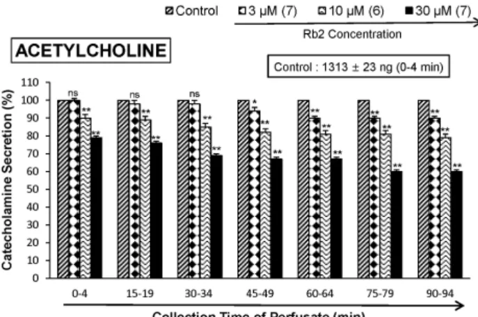
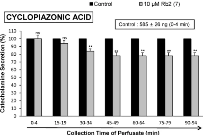
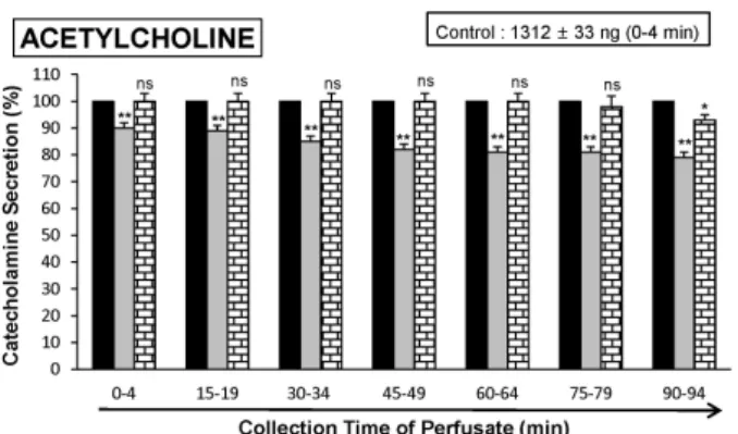
The aim of the present study was to investigate whether ginsenoside-Rb2 (Rb2) can affect the secre- tion of catecholamines (CA) in the perfused model of the rat adrenal medulla. Rb2 (3∼ 30 μ M), per- fused into an adrenal vein for 90 min, inhibited ACh (5.32 mM)-evoked CA secretory response in a dose- and time-dependent fashion. Rb2 (10 μ M) also time-dependently inhibited the CA secretion evoked by DMPP (100 μ M, a selective neuronal nicotinic receptor agonist) and high K

+

(56 mM, a direct membrane depolarizer). Rb2 itself did not affect basal CA secretion (data not shown). Also, in the presence of Rb2 (50 μ g/mL), the secretory responses of CA evoked by veratridine (a selective Na

+

channel activator (50 μ M), Bay-K-8644 (an L-type dihydropyridine Ca

2+

channel activator, 10 μ M), and cyclopiazonic acid (a cytoplasmic Ca
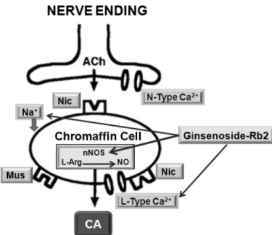
2+

-ATPase inhibitor, 10 μ M) were significantly reduced, respectively. Interestingly, in the simultaneous presence of Rb2 (10 μ M) and L-NAME (an inhibitor of NO synthase, 30 μ M), the inhibitory responses of Rb2 on ACh-evoked CA secretory response was considerably recovered to the extent of the corresponding control secretion compared with the inhibitory effect of Rb2-treatment alone. Practically, the level of NO released from adrenal medulla after the treatment of Rb2 (10 μ M) was greatly elevated compared to the corresponding basal released level.

Collectively, these results demonstrate that Rb2 inhibits the CA secretory responses evoked by nicotinic stimulation as well as by direct membrane-depolarization from the isolated perfused rat adrenal medulla. It seems that this inhibitory effect of Rb2 is mediated by inhibiting both the influx of Ca

2+

and Na

+

into the adrenomedullary chromaffin cells and also by suppressing the release of Ca

2+

from the cytoplasmic calcium store, at least partly through the increased NO production due to the activation of nitric oxide synthase, which is relevant to neuronal nicotinic receptor blockade.

Key Words: Adrenal Medulla, Catecholamine secretion, Ginsenoside-Rb2 (Rb2), Nitric oxide synthase (NOS), NO production

INTRODUCTION

Previously, Lim and his coworkers [1-3] have reported that all of total giseng saponin (TGS), panaxadiol and pan- axatriol cause the increased secretion of catecholamines (CA) from the isolated perfused rabbit adrenal glands in

a Ca

2+

-dependent fashion, which are mediated by the acti- vation of cholinergic (both nicotinic and muscarinic) re- ceptors and partly the direct action on the rabbit adrenome- dullary chromaffin cells. However, it has been reported that TGS shows the inhibitory effect on the CA release evoked by stimulation of nicotinic receptors in the perfused rat adrenal medulla [4], and also that several ginseng saponins (ginsenosides) from the ginseng root inhibit the CA secre- tion from bovine adrenal chromaffin cells stimulated by ACh [5-8].

In previous study, Tachikawa and his co-workers [6] have

shown that most of the ginsenosides (1∼100 μM) had a

tendency to reduce the ACh-evoked CA secretion. The order

of inhibitory potency (at the concentration of 10 μM) was

as follows: Rg

2

>Rf>Re>Rh1>Rb2, Rg1>Rb1>Rc>Rb3,

Rd, Ro, Rs

1

. The inhibition of ginsenoside-Rg2 at 10 μM

was 72%, but ginsenosides Rb3, Rd, Ro and Rs

1

did not

(2)

Fig. 1. Dose-dependent effects of ginsenoside-Rb2 (Rb2) on the secretory responses of catecholamines (CA) evoked by acetylcholine (ACh) from the perfused rat adrenal medulla. The CA secretion by a single injection of ACh (5.32×10

–3

M) in a volume of 0.05 mL was evoked at 15 min intervals during loading with 3, 10 and 30 μ M of Rb2 for 90 min as indicated by the arrow marks, res- pectively. The numbers in parentheses indicate the number of rat adrenal glands. Vertical bars on the columns represent the standard error of the mean (S.E.M.). Ordinate: the amounts of CA secreted from the adrenal gland (% of control). Abscissa: collection time of perfusate (min). Statistical difference was obtained by comparing the corresponding control with each concentration - treated group of Rb2. ACh-induced perfusate was collected for 4 minutes. *p<0.05, **p<0.01. ns, Not statistically significant.

show the inhibitory effect.

In the conscious rats, the releasing effect of NO of Korean Red Ginseng (KRG) may be partly contributed to the hypo- tensive effect [9]. It has also been shown that Rg3 relaxes the rat thoracic aorta as a consequence of NO production [10]. Han and his co-workers [11] have found evidence that NO levels in exhaled breath of human volunteers by KRG were increased along with decreased blood pressure and heart rate. In a series of studies, it has been found that ginsenosides reduce blood pressure via increases in pro- duction of endothelial nitric oxide [12] and that Rg3 is the most potent ginsenoside that activates eNOS in rat aorta [13]. Although some investigators have demonstrated that Rg3 induces eNOS activation in the vasculature of animal models [12,14], Hien and his co-workers [15] have found that Rg3 activates eNOS via eNOS phosphorylation in ECV 304 human endothelial cells and increases in eNOS expres- sion.

Several studies have shown that ginsenoside Rg1 can cause endothelial-dependent relaxation in the rat aorta [14]

and enhance endogenous NO production in human um- bilical vein endothelial cells [16], rat kidney [17] and in por- cine coronary arteries [18].

Despite of these many studies on various ginseng sap- onins, there are still a little known effects of ginsenoside- Rb2 on adrenal CA secretion. Therefore, in this study, we investigated whether ginsenoside-Rb2 affects nicotinic re- ceptor stimulation-evoked CA secretion from the perfused model of the isolated rat adrenal medulla and tried to es- tablish its mechanism of action.

METHODS Experimental procedure

Mature male Sprague-Dowley rats (purchased from DAMOOL SCIENCE, International Customer Service, Seoul, Korea), weighing 200 to 300 grams, were used in the experiment. The animals were housed individually in sepa- rate cages, and food (Cheil Animal Chow) and tap water were allowed ad libitum for at least a week to adapt to experimental circumstances. On the day of experiment, a rat was anesthetized with thiopental sodium (50 mg/kg) in- traperitoneally, and tied in supine position on fixing panel.

1. Isolation of adrenal glands

The adrenal gland was isolated by the modification of previous method [19]. The abdomen was opened by a mid- line incision, and the left adrenal gland and surrounding area were exposed by the placement of three-hook retrac- tors. The stomach, intestine and portion of the liver were not removed, but pushed over to the right side and covered by saline-soaked gauge pads and urine in bladder was re- moved in order to obtain enough working space for tying blood vessels and cannulations. A cannula, used for perfu- sion of the adrenal gland, was inserted into the distal end of the renal vein after all branches of adrenal vein (if any), vena cava and aorta were ligated. Heparin (400 IU/mL) was injected into vena cava to prevent blood coagulation before ligating vessels and cannulations. A small slit was made into the adrenal cortex just opposite entrance of adrenal vein. Perfusion of the gland was started, making sure that no leakage was present, and the perfusion fluid escaped only from the slit made in adrenal cortex. Then the adrenal

gland, along with ligated blood vessels and the cannula, was carefully removed from the animal and placed on a platform of a leucite chamber. The chamber was con- tinuously circulated with water heated at 37±1

o

C (Fig. 1).

Perfusion of adrenal gland

The adrenal glands were perfused by means of peristaltic pump (Isco, St. Lincoln, NE, USA) at a rate of 0.31 mL/

min. The perfusion was carried out with Krebs-bicarbonate solution of following composition (mM): NaCl, 118.4; KCl, 4.7; CaCl

2

, 2.5; MgCl

2

, 1.18; NaHCO

3

, 25; KH

2

PO

4

, 1.2; glu- cose, 11.7. The solution was constantly bubbled with 95 % O

2

+5% CO

2

and the final pH of the solution was maintained at 7.4∼7.5. The solution contained disodium EDTA (10 μg/

mL) and ascorbic acid (100 μg/mL) to prevent oxidation of catecholamines.

Drug administration

The perfusions of DMPP (10

–4

M) for 2 minutes and/or a single injection of ACh (5.32×10

–3

M) and KCl (5.6×10

–2

M) in a volume of 0.05 mL were made into perfusion stream via a three-way stopcock, respectively. McN-A-343 (10

–4

M), veratridine (10

–4

M), Bay-K-8644 (10

–5

M) and cyclopiazonic acid (10

–5

M) were also perfused for 4 min, respectively.

In the preliminary experiments, it was found that upon

administration of the above drugs, secretory responses to

ACh, KCl, McN-A-343, Bay-K-8644 and cyclopiazonic acid

returned to preinjection level in about 4 min, but the re-

sponses to DMPP in 8 min.

(3)

Collection of perfusate

As a rule, prior to stimulation with various secretagogues, the perfusate was collected for 4 min to determine the spon- taneous secretion of CA (background sample). Immediately after the collection of the background sample, collection of the perfusates was continued in another tube as soon as the perfusion medium containing the stimulatory agent reached the adrenal gland. Stimulated sample's perfusate was collected for 4 to 8 min. The amounts secreted in the background sample have been subtracted from that se- creted from the stimulated sample to obtain the net secre- tion value of CA, which is shown in all of the figures.

To study the effect of Rb2 on the spontaneous and evoked secretion, the adrenal gland was perfused with normal Krebs solution for 90 min, and then the perfusate was col- lected for a certain period (background sample). Then the medium was changed to the one containing the stimulating agent or along with Rb2, and the perfusates were collected for the same period as that for the background sample. The adrenal gland's perfusate was collected in chilled tubes.

Measurement of catecholamines

The content of CA (all of epinephrine, norepinephrine and dopamine) in perfusate was measured directly by the fluo- rometric method of Anton and Sayre [20] without the inter- mediate purification alumina for the reasons described ear- lier [19] using fluorospectrophotometer (Kontron Co., Mi- lano, Italy).

A volume of 0.2 mL of the perfusate was used for the reaction. The CA content in the perfusate of stimulated glands by secretagogues used in the present work was high enough to obtain readings several folds greater than the reading of control samples (unstimulated). The sample blanks were also lowest for perfusates of stimulated and non-stimulated samples. The CA content in the perfusate was expressed in terms of norepinephrine (base) equi- valents.

Measurement of NO release

NO release was measured using a NO-selective micro- electrode (ami700, Innovative Instruments Inc) and an am- plifier (inNo meter, Innovative Instruments Inc). Adreno- medullary NO production was quantified as the integrated signal detected by the microelectrode after perfusion of Rb2 into rat adrenal medulla, as previously described [21]. The electrode was calibrated by producing standardized concen- trations of NO in 0.5% (wt/vol) KI in 0.1 Mol/L H

2

SO

4

from NaNO

2

standards. NO release was quantitated as the cur- rent detected at the electrode after loading Rb2 into adrenal medulla. NO release was calculated as picomoles.

Statistical analysis

The statistical difference between the control and the pre- treated groups was determined by the Student's t and ANOVA tests. A p-value of less than 0.05 was considered to represent statistically significant changes unless specifi- cally noted in the text. Values given in the text refer to means and the standard errors of the mean (S.E.M.). The statistical analysis of the experimental results was made by computer program described by Tallarida and Murray [22].

Drugs and their sources

The following drugs were used: 1.1-dimethyl-4 -phenyl pi- perazinium iodide (DMPP), acetylcholine chloride, nor- epinephrine bitartrate, potassium chloride (KCl), N

ω

-nitro- L-arginine methyl ester hydrochloride (

L

-NAME), meth- yl-1,4-dihydro-2,6-dimethyl-3-nitro-4-(2-trifluoro-methyl- phenyl)-pyridine-5-carboxylate (BAY-K-8644), cyclopiazonic acid, veratridine and potassium nitrate (Sigma Chemical Co., U.S.A.). Ginsenoside-Rb2 was a gift from Dr. Geon- Wook Kang (Professor of Seoul National University, Seoul, Korea). Drugs were dissolved in distilled water (stock) and added to the normal Krebs solution as required except Bay-K-8644, which was dissolved in 99.5% ethanol and di- luted appropriately with Krebs-bicarbonate solution (final concentration of alcohol was less than 0.1%). Concentra- tions of all drugs were expressed in terms of molar base.

RESULTS

Influence of Ginsenoside-Rb2 on the CA secretion evo- ked by ACh, high K

+

, and DMPP from the perfused rat adrenal medulla

After the perfusion with oxygenated Krebs-bicarbonate solution for 1 hr, basal CA release from the isolated per- fused rat adrenal glands amounted to 20±2 ng for 2 min (n=12). Since Tachikawa and his co-workers [6] have re- ported that most of the ginsenosides (1∼100 μM) including Rb2 had a tendency to reduce the ACh-evoked CA secretion, it was attempted initially to examine the effects of Rb2 it- self on CA secretion from the perfused model of the rat adrenal glands. However, in the present study, Rb2 itself did not produce any effect on basal CA output from the per- fused rat adrenal glands (data not shown). Therefore, it was decided to investigate the effects of Rb2 on cholinergic nic- otinic receptor stimulation- as well as membrane depolari- zation-evoked CA secretory responses. Secretagogues were given at 15 to 20 min-intervals. Rb2 was present for 90 minutes after the establishment of the control release.

When ACh (5.32×10

–3

M) in a volume of 0.05 mL was injected into the perfusion stream, the amount of CA se- creted was 1313±23 ng for 4 min. However, the pretreat- ment with Rb2 in the range of 10∼30 μM for 90 min rela- tively concentration- and time-dependently inhibited ACh- stimulated CA secretion. As shown in Fig. 1, in the presence of Rb2, the CA releasing responses were inhibited to 60%

of the corresponding control release (100%). Also, it has been shown that depolarizing agent like high KCl stim- ulates markedly CA secretion (747±21 ng for 0∼4 min).

High K

+

(5.6×10

–2

M)-stimulated CA secretion after the pre-

treatment with 10 μM Rb2 was maximally inhibited to

80% of the control after 75 min period, although it was not

initially affected for the first period (0-4 min) as shown in

Fig. 2. DMPP (10

–4

M), which is a selective nicotinic re-

ceptor agonist in autonomic sympathetic ganglia, evoked a

sharp and rapid increase in CA secretion (1237±27 ng for

0∼8 min). However, as shown in Fig. 3, DMPP-stimulated

CA secretion after the pretreatment with Rb2 (10 μM) was

greatly reduced to 78% of the control release.

(4)

Fig. 2. Time-course effects of Rb2 on the high K

+

-evoked CA se- cretory responses from the perfused rat adrenal medulla. The CA secretion by a single injection of K

+

(5.6×10

–2

M) in a volume of 0.05 mL was evoked at 15 min intervals during loading with 10 μM of Rb2 for 90 min as indicated by the arrow marks. high K

+

-induced perfusate was collected for 4 minutes. Other legends are the same as in Fig. 1. **p<0.01. ns, Not statistically signi- ficant.

Fig. 3. Time-course effects of Rb2 on the CA secretory responses evoked by from the perfused rat adrenal medulla. The CA secretion by perfusion of DPPP (10

–4

M) for 2 min was induced at 20 min interval during loading with 10 μM of Rb2 for 90 min. DMPP- induced perfusate was collected for 8 minutes. Other legends are the same as in Fig. 1. **p<0.01. ns, Not statistically significant.

Fig. 4. Time-course effects of Rb2 on the CA secretion evoked by Bay-K-8644 from the perfused rat adrenal medulla. Bay-K-8644 (10

–5

M) was perfused into an adrenal vein for 4 min at 15 min intervals during loading with Rb2 (10 μM) for 90 min. Other legends are the same as in Fig. 1. **p<0.01. ns, Not statistically significant.

Fig. 5. Time-course effects of Rb2 on the CA secretion evoked by cyclopiazonic acid from the perfused rat adrenal medulla. Cyclo- piazonic acid (10

–5

M) was perfused into an adrenal vein for 4 min at 15 min intervals during loading with Rb2 (10 μM) for 90 min.

Other legends are the same as in Fig. 1. **p<0.01. ns, Not statistically significant.

Influence of Ginsenoside-Rb2 on the CA secretion evoked by Bay-K-8644, cyclopiazonic acid and veratri- dine from the perfused rat adrenal medulla Since Bay-K-8644 is known to be a calcium channel acti- vator, which enhances basal Ca

2+

uptake [23] and CA re- lease [24], it was of interest to determine the effects of Rb2 on Bay-K-8644-stimulated CA secretion from the isolated perfused rat adrenal glands. Bay-K-8644 (10

–5

M)-stimulat- ed CA secretion in the presence of Rb2 (10 μM) was re- duced to 73% of the control except for the early 15 min period as compared to the corresponding control release (640±33 ng for 0∼4 min) from 6 rat adrenal glands, as shown in Fig. 4.

Cyclopiazonic acid, a mycotoxin from Aspergillus and

Penicillium, has been described as a highly selective in- hibitor of Ca

2+

-ATPase in skeletal muscle sarcoplasmic re- ticulum [25,26]. The inhibitory action of Rb2 on cyclo- piazonic acid-evoked CA secretory response was observed as shown in Fig. 5. In the presence of Rb2 (10 μM) in 7 rat adrenal glands, cyclopiazonic acid (10

–5

M)-evoked CA secretion was also inhibited to 78% of the control response (585±26 ng for 0∼4 min), although it was not initially af- fected for the first and second periods (0∼19 min).

It has been known that veratridine-induced Na

+

influx mediated through voltage-dependent Na

+

channels incre- ased Ca

2+

influx via activation of voltage-dependent Ca

2+

channels and produced the exocytotic CA secretion in cul- tured bovine adrenal medullary cells [27]. As shown in Fig.

6, veratridine (5×10

–5

M) sharply produced CA secretion

(848±23 ng for 0∼4 min). In 8 rat adrenal glands, Rb2 (10

μM) also attenuated veratridine-induced CA secretion to

(5)

Fig. 6. Time-course effects of Rb2 on veratridine-evoked CA secretion from the perfused rat adrenal medulla. Veratridine (5×10

–5

M) was perfused into an adrenal vein for 4 min at 15 min intervals during loading with Rb2 (10 μM) for 90 min. Other legends are the same as in Fig. 1. **p<0.01.

Fig. 7. Effects of Rb2 plus L-NAME on the CA secretory responses evoked by acetylcholine from the perfused rat adrenal medulla. The CA secretion by a single injection of ACh (5.32×10

–3

M) in a volume of 0.05 mL was evoked at 15 min intervals during simultaneous loading with Rb2 (10 μM) plus L-NAME (30 μM) for 90 min.

Statistical difference was obtained by comparing the corresponding control (CONTROL) with Rb2-treated only group or group treated with Rb2+L-NAME. Other legends are the same as in Fig. 1. *p

< 0.05, **p<0.01. ns: Not statistically significant.

Fig. 8. Effects of Rb2 on nitric oxide (NO) production in the perfused rat adrenal medulla. Perfusate sample was taken for 8 min after loading the perfusion of Rb2 (10 μM) at a rate of 0.31 mL/min.

Ordinate: the amounts of NO released from the adrenal medulla (% of control). Abscissa: Treatment (before and after Rb2).

Statistical difference was made by comparing the control with Rb2-treated group. **p<0.01.

74% of the corresponding control release.

Influence of Ginsenoside-Rb2 plus L-NAME on Ach- Evoked CA secretion from the perfused rat adrenal medulla

It has also been found that, in this study, Rb2 inhibited the CA secretory response evoked by stimulation of chol- inergic receptors in the perfused rat adrenal glands. There- fore, to study the relationship between NO and Rb2- in- duced inhibitory effects on the CA release from the rat adre- nal glands, the effect of L-NAME on Rb2-induced inhibitory responses of CA secretion evoked by cholinergic receptor- stimulation as well as membrane depolarization was exa-

mined. In the present study, in the simultaneous presence of Rb2 (10 μM) and L-NAME (30 μM) for 90 min from 6 rat adrenal glands, ACh (5.32×10

–3

M)-evoked CA release was almostly recovered to 100∼93% of the corresponding control level compared to that of Rb2 (10 μM)-treated alone, as ishown in Fig. 7.

Influence of Ginsenoside-Rb2on the level of nitric oxide released from the perfused rat adrenal medulla As shown in Fig. 7, the inhibitory effects of Rb2 on ACh- evoked CA secretory responses were significantly recovered to the control level in the presence of L-NAME. Therefore, it was decided directly to determine the level of NO re- leased from rat adrenal medulla after the treatment of Rb2.

Moreover, it has been found that ginsenosides reduce blood pressure via increases in production of endothelial nitric oxide [12] and that ginsenoside Rg3 is the most potent gin- senoside that activates eNOS in rat aorta [13]. Although some investigators have demonstrated that Rg3 induces eNOS activation in the vasculature of animal models [13,14].

In 6 adrenal glands, the basal amount of NO released from medulla prior to administration of Rb2 was 29.6±2 picomoles. However, 8 min after loading with Rb2 (10 μM) it was greatly elevated to 40.6±2.5 picomoles, which was 137% of the basal release, as shown in Fig. 8.

DISCUSSION

The present experimental results showed that Rb2 in- hibited the CA secretory responses from the isolated per- fused rat adrenal gland evoked by stimulation of cholinergic nicotinic receptors as well as by direct membrane-depola- rization. It seems that this inhibitory effect of Rb2 is ex- erted by inhibiting influx of both Na

+

and Ca

2+

ions through their channels into the adrenomedullary chromaffin cells and also by suppression of Ca

2+

release from the cytoplas- mic calcium store at least partly via increased NO pro- duction due to activation of neuronal NO synthase, which is relevant to the blockade of neuronal nicotinic receptors.

 In the present study, in the simultaneous presence of Rb2

and L-NAME (an NO synthase inhibitor), the ACh-evoked

(6)

CA secretory responses was considerably recovered to the extent of the corresponding control secretion compared to those of Rb2-treatment alone. This result is well consistent with the report that, in a series of studies, ginsenosides reduce blood pressure via increases in production of endo- thelial nitric oxide [12] and that ginsenoside Rg3 is the most potent ginsenoside that activates eNOS in rat aorta [13]. Although several investigators have demonstrated that Rg3 induces eNOS activation in the vasculature of ani- mal models [12,14], Hien and his co-workers [15] have found that Rg3 activates eNOS via eNOS phosphorylation in ECV 304 human endothelial cells and increases in eNOS expression. Moreover, in this study, following treatment of Rb2 into adrenal medulla, NO production was greatly ele- vated as shown in Fig. 8. Taking into account these find- ings, in the present study, it is likely that Rb2 inhibits the CA secretory response evoked by various secretagogues through increasing NO production in adrenal chromaffin cells, since in the simultaneous presence of Rb2 and L- NAME, an inhibitor of NO synthase, ACh-evoked-CA secre- tion was nearly recovered to the corresponding control level compared to that of Rb2-treatment alone, and also Rb2 no- tably enhanced NO release from rat adrenal medulla.

In the support of this finding, it has been reported that the NOS inhibitor, L-NAME enhances K

+

-stimulated CA se- cretion in cultured bovine chromaffin cells [28] and that sodium nitroprusside (SNP) inhibits ACh-induced CA secre- tion in bovine chromaffin cells [29]. These studies suggest that NO may play an inhibitory role in the control of the CA secretion. Moreover, the presence of endothelial cells has been reported to inhibit the K

+

-induced or the nicotinic receptor agonist DMPP-induced CA secretion in cultured bovine chromaffin cells [28], suggesting that not merely nNOS but also eNOS may play roles in modulating adrenal CA secretion. In view of the results so far reported, the present studies strongly suggest that Rb2 possesses the ability partly to activate nNOS in the adrenomedullary chromaffin cells, resulting in enhancement of NO as well as inhibition of the CA release, in addition to the direct inhibitory effects on the CA secretion. In supporting this finding, among the ginsenosides of the protopanaxatriol and protopanaxadiol groups, ginsenoside Rg3 is the most potent vasodilator. Previously, it was shown that Rg3 in- hibited calcium-induced vascular contraction [13] as well as phenylephrine-induced vasocontraction as a consequence of NO production [10].

In contrast, it has been reported that L-NAME inhibits acetylcholine (ACh)-induced CA secretion in bovine chro- maffin cells [29], and that the NO donor SNP enhances nic- otine-induced CA secretion in cultured bovine chromaffin cells [30]. These findings suggest that NO may facilitate cholinergic agonist-induced CA secretion. On the other hand, a few in vivo studies have suggested that NO does not play a role in regulation of adrenal CA secretion [31,32].

Generally, the adrenal medulla has been employed as a model system to study numerous cellular functions involv- ing not only noradrenergic nerve cells but also neurons.

During neurogenic stimulation of the adrenal medulla, ACh is released from splanchnic nerve endings and activates cholinergic receptors on the chromaffin cell membrane [33].

This activation initiates a series of events known as stim- ulus-secretion coupling, culminating in the exocytotic re- lease of CA and other components of the secretory vesicles into the extracellular space. Usually, two mechanisms are involved in the secretion of adrenal medullary hormones.

Upon excitation of splanchnic nerves, ACh is released from the nerve terminals, and then activates nicotinic the CA secretion. Based on this fact, the present findings that Rb2 inhibited the CA secretory responses evoked by nicotinic receptor stimulation as well as by membrane depolarization in the adrenal medulla seem to support the fact that, ginse- nosides from Panax ginseng can decrease the blood pressure in both experimental animals and hypertensive patients [12,34-36]. These experimental results indicate that Rb2-in- duced inhibitory activity of the CA secretory response evoked by stimulation of nicotinic receptors might contrib- ute at least partly to its hypotensive mechanism. ACh, the physiological presynaptic transmitter at the adrenal me- dulla, which is released by depolarizing splanchnic nerve terminals and then activates nicotinic receptors, releases the CA, and induces dopamine β-hydroxylase by calcium- dependent secretory process [37,38]. In terms of this fact, the present results suggest that Rb2 may inhibit CA secre- tion evoked by nicotinic stimulation from the splanchnic nerve ending through the blockade of nicotinic receptors.

The CA release from the adrenal medulla in response to splanchnic nerve stimulation or nicotinic agonist is medi- ated by activation of nicotinic receptors located on the chro- maffin cells. The exocytotic CA release from the chromaffin cells appears to be essentially similar to that occurring in noradrenergic axons [39,40]. ACh-evoked CA secretion has shown to be caused through stimulation of both nicotinic and muscarinic receptors in guinea-pig adrenal gland [41]

as well as in the perfused rat adrenal glands [42]. In sup- port of this idea, it has been found that the ginseng sap- onins, ginsenoside Rg2, a panaxatriol [6] as well as ginseno- side Rg3, a panaxadiol [8] block the nicotinic ACh receptors or the receptor-operated Na

+

channels (but not voltage- sen- sitive Na

+

and Ca

2+

channels), inhibit Na

+

influx through the channels and consequently reduce both Ca

2+

influx and the CA secretion in bovine adrenal chromaffin cells.

In this study, Rb2 inhibited the CA secretory responses

evoked by ACh, high K

+

, and DMPP. It suggests that Rb2

can produce the similar effect as in adrenal medulla of the

normotensive rats [4] and also in bovine adrenal chromaffin

cells [6,8]. In previous study, ginsenoside-Rg3 inhibited

both ACh-induced Ca

2+

and Na

+

influxes in a concentration-

dependent manner similar to that observed with the ACh-

evoked CA secretion [8]. However, it had no or only a slight

effect on the CA secretion and Ca

2+

influx induced by high

K

+

concentration or veratridine, an activator of the volt-

age-sensitive Ca

2+

or Na

+

channels [5,6]. These results

strongly suggested that ginsenoside-Rg3 acts on the nic-

otinic ACh receptor-operated cation channels but not on the

voltage-sensitive Ca

2+

or Na

+

channels. Furthermore, the

ginsenoside-Rg3 inhibition was not overcome by increasing

the external ACh and Ca

2+

concentrations [8], indicating

that the inhibitory effect of ginsenoside-Rg3 is distinct from

that of the competitive antagonists of the nicotinic ACh re-

ceptors, such as trimethaphan [43,44], and that of blockers

of the L-type voltage-sensitive Ca

2+

channels, which are

competitive with external Ca

2+

concentrations, such as dil-

tiazem [45]. In fact, the mode of the ginsenoside-Rg3 antag-

onism was non-competitive with nicotine [8]. Anyway, these

results seem to be quite different from those of the present

study that Rb2 significantly inhibited the CA secretory re-

sponses evoked by cholinergic nicotinic stimulation as well

as an activator of the voltage-sensitive Ca

2+

or Na

+

channels

in the perfused model of rat adrenal medulla. The discrep-

ancy is due to the experimental employment of different

(7)

Fig. 9. Probable site of action of Rb2 at cholinergic nerve-chromaffin cell in the rat adrenal medulla.

accounting methodology and different components between the present and previous studies.

In the present study, Rb2 also time-dependently depres- sed the CA secretory response evoked by Bay-K-8644, which is known to activate L-type voltage-dependent Ca

2+

channels [23,46]. This result indicates that Rb2 may inhibit Ca

2+

influx through voltage-dependent Ca

2+

channels to the rat adrenomedullary cells. In support of this idea, in cul- tured bovine adrenal medullary cells, nicotinic (but not muscarinic) receptors mediate the Ca

2+

-dependent CA se- cretion [47,48]. It has also been known that the activation of nicotinic receptors stimulates the CA secretion by in- creasing Ca

2+

entry through receptor-linked and/or volt- age-dependent Ca

2+

channels in both perfused rat adrenal glands [42,49] and isolated bovine adrenal chromaffin cells [50-52]. Wada and his coworkers [53] have found that the adrenomedullary chromaffin cells have (i) nicotinic recep- tor-associated ionic channels, responsible for carbachol-in- duced Na

+

influx, (ii) voltage-dependent Na

+

channels, re- sponsible for veratridine-induced Na

+

influx and (iii) volt- age-dependent Ca

2+

channels (VDCC), suggesting that the influx of Na

+

caused either by carbachol or by veratridine leads to activate voltage-dependent Ca

2+

channels by alter- ing membrane potentials, whereas high K

+

directly acti- vates voltage-dependent Ca

2+

channels without increasing Na

+

influx. In the present study, the finding that the CA secretory responses evoked by Bay-k-8644 as well as by high K

+

were depressed in the presence of Rb2 indicates that this inhibitory effect of Rb2 is exerted by the direct inhibition of calcium influx through VDCC into the adrenal chromaffin cells. Furthermore, slight elevation in the ex- tracellular potassium concentration increases both the fre- quency of spontaneous action potentials and the CA secre- tion [54], suggesting that the influx of calcium that occurs during action potentials is directly linked to the rate of secretion. These findings that Rb2 inhibited the CA secre- tion evoked by Bay-K-8644 as well as by high K

+

suggest that Rb2 can inhibit directly the VDCC. In the bovine chro- maffin cells, stimulation of nicotinic, but not muscarinic ACh receptors is known to cause CA secretion by increasing Ca

2+

influx largely through VDCC [55,56]. Therefore, it seems that these inhibitory effects of Rb2 on the CA secre- tion evoked by ACh, DMPP, Bay-K-8644 and veratridine may be mediated by inhibiting Ca

2+

influx through volt-

age-dependent Ca

2+

channels due to activation of nicotinic receptor-associated ionic channels, responsible for carba- chol-induced Na

+

influx, as well as of voltage-dependent Na

+

channels, responsible for veratridine-induced Na

+

in- flux.

The present study has also shown that Rb2 inhibits the CA secretion evoked by cyclopiazonic acid. Cyclopiazonic acid is known to be a highly selective inhibitor of Ca

2+

- ATPase in skeletal muscle sarcoplasmic reticulum [25,26]

and a valuable pharmacological tool for investigating intra- cellular Ca

2+

mobilization and ionic currents regulated by intracellular Ca

2+

[57]. Therefore, it is felt that the in- hibitory effect of Rb2 on the CA secretion may also be asso- ciated with the mobilization of intracellular Ca

2+

from the cytoplasmic calcium store. This is consistent with the find- ings obtained in skinned smooth muscle fibers of the longi- tudinal layer of the guinea-pig ileum, where Ca

2+

-uptake was also inhibited by cylopiazonic acid [58]. Suzuki and his coworkers [57] have shown that cyclopiazonic acid easily penetrates into the cytoplasm through the plasma mem- brane and reduces Ca

2+

-ATPase activity in sarcoplasmic/

endoplasmic reticulum, resulting in an increase in the sub- sequent Ca

2+

release from those storage sites.

In conclusion, as shown in Fig. 9, the results of the pres- ent study have strongly suggested that Rb2 inhibits the CA secretion by stimulation of cholinergic nicotinic receptors as well as by direct membrane depolarization in the iso- lated perfused rat adrenal glands. It seems that this in- hibitory effect of Rb2 is exerted by blocking influx of Na

+

and Ca

2+

through ionic channels into the adrenomedullary chromaffin cells as well as by inhibiting the release of Ca

2+

from the cytoplasmic calcium store at least partly via the increased NO production due to the activation of nitric ox- ide synthase. Based on these results, the ingestion of Rb2 may be helpful to prevent or alleviate the cardiovascular diseases, through inhibition of CA secretion from adreno- medullary cells and consequent reduction of the CA level in the circulation.

ACKNOWLEDGEMENTS

This study was supported partly by the fund of Chosun University (2011-2012).

REFERENCES

1. Lim DY, Park KB, Kim KY, Lee KS, Moon JK, Kim YH.

Influence to total ginseng saponin on secretion of catecho- lamines in the isolated adrenal gland of rabbits. Korean Biochem J. 1987;20:230-238.

2. Lim DY, Park KB, Kim KH, Choi CH, Bae JW, Kim MW.

Studies on secretion of catecholamines evoked by panaxadiol in the isolated rabbit adrenal gland. Korean J Pharmacol.

1988;24:31-42.

3. Lim DY, Choi CH, Kim CD, Kim KH, Kim SB, Lee BJ, Chung MH. Influnce of Panaxatriol-type saponin on secretion of catecholamine from isolated perfused rabbit adrenal gland.

Arch Pharm Res. 1989;12:166-175.

4. Hong SP, Chi H, Cho SH, Lee YK, Woo SC, Kim IS, Oh SH, Yang WH, Lim DY. Influence of total Ginseng saponin on nicotinic stimulation-induced catecholamine secretion from the perfused rat adrenal gland. Korean J Hypert. 1999;5:1-10.

5. Kudo K, Akasaka Y, Miyate Y, Takahashi E, Tachikawa E,

Kashimoto T. Effects of red ginseng fractions on catecholamine

(8)

Soc WAKAN-YAKU. 1992;9:236-239.

6. Tachikawa E, Kudo K, Kashimoto T, Takahashi E. Ginseng saponins reduce acetylcholine-evoked Na

+

influx and catecho- lamine secretion in bovine adrenal chromaffin cells. J Phar- macol Exp Ther. 1995;273:629-636.

7. Kudo K, Tachikawa E, Kashimoto T, Takahashi E. Properties of ginseng saponin inhibition of catecholamine secretion in bovine adrenal chromaffin cells. Eur J Pharmacol. 1998;341:

139-144.

8. Tachikawa E, Kudo K, Nunokawa M, Kashimoto T, Takahashi E, Kitagawa S. Characterization of ginseng saponin ginseno- side-Rg(3) inhibition of catecholamine secretion in bovine adrenal chromaffin cells. Biochem Pharmacol. 2001;62:943-951.

9. Jeon BH, Kim CS, Kim HS, Park JB, Nam KY, Chang SJ.

Effect of Korean red ginseng on blood pressure and nitric oxide production. Acta Pharmacol Sin. 2000;21:1095-1100.

10. Kim ND, Kim EM, Kang KW, Cho MK, Choi SY, Kim SG.

Ginsenoside Rg3 inhibits phenylephrine-induced vascular contraction through induction of nitric oxide synthase. Br J Pharmacol. 2003;140:661-670.

11. Han K, Shin IC, Choi KJ, Yun YP, Hong JT, Oh KW. Korea red ginseng water extract increases nitric oxide concentrations in exhaled breath. Nitric Oxide. 2005;12:159-162.

12. Kim ND, Kang SY, Schini VB. Ginsenosides evoke endo- thelium-dependent vascular relaxation in rat aorta. Gen Phar- macol. 1994;25:1071-1077.

13. Kim ND, Kang SY, Park JH, Schini-Kerth VB. Ginsenoside Rg3 mediates endothelium-dependent relaxation in response to ginsenosides in rat aorta: role of K

+

channels. Eur J Phar- macol. 1999;367:41-49.

14. Kang SY, Schini-Kerth VB, Kim ND. Ginsenosides of the protopanaxatriol group cause endothelium-dependent relaxa- tion in the rat aorta. Life Sci. 1995;56:1577-1586.

15. Hien TT, Kim ND, Pokharel YR, Oh SJ, Lee MY, Kang KW.

Ginsenoside Rg3 increases nitric oxide production via increases in phosphorylation and expression of endothelial nitric oxide synthase: essential roles of estrogen receptor-dependent PI3- kinase and AMP-activated protein kinase. Toxicol Appl Phar- macol. 2010;246:171-183.

16. Leung KW, Cheng YK, Mak NK, Chan KK, Fan TP, Wong RN.

Signaling pathway of ginsenoside-Rg1 leading to nitric oxide production in endothelial cells. FEBS Lett. 2006;580:3211-3216.

17. Han SW, Kim H. Ginsenosides stimulate endogenous produc- tion of nitric oxide in rat kidney. Int J Biochem Cell Biol. 1996;

28:573-580.

18. Chai H, Zhou W, Lin P, Lumsden A, Yao Q, Chen C. Ginsen- osides block HIV protease inhibitor ritonavir-induced vascular dysfunction of porcine coronary arteries. Am J Physiol Heart Circ Physiol. 2005;288:H2965-2971.

19. Wakade AR. Studies on secretion of catecholamines evoked by acetylcholine or transmural stimulation of the rat adrenal gland. J Physiol. 1981;313:463-480.

20. Anton AH, Sayre DF. A study of the factors affecting the aluminum oxide-trihydroxyindole procedure for the analysis of catecholamines. J Pharmacol Exp Ther. 1962;138:360-375.

21. McVeigh GE, Hamilton P, Wilson M, Hanratty CG, Leahey WJ, Devine AB, Morgan DG, Dixon LJ, McGrath LT. Platelet nitric oxide and superoxide release during the development of nitrate tolerance: effect of supplemental ascorbate. Circulation. 2002;

106:208-213.

22. Tallarida RJ, Murray RB. Manual of pharmacologic calculation with computer programs. 2nd ed. New York: Speringer-Verlag;

1987. 132 p.

23. García AG, Sala F, Reig JA, Viniegra S, Frías J, Fontériz R, Gandía L. Dihydropyridine BAY-K-8644 activates chromaffin cell calcium channels. Nature. 1984;309:69-71.

24. Lim DY, Kim CD, Ahn GW. Influence of TMB-8 on secretion of catecholamines from the perfused rat adrenal glands. Arch Pharm Res. 1992;15:115-125.

25. Goeger DE, Riley RT. Interaction of cyclopiazonic acid with rat skeletal muscle sarcoplasmic reticulum vesicles. Effect on Ca

2+

3995-4003.

26. Seidler NW, Jona I, Vegh M, Martonosi A. Cyclopiazonic acid is a specific inhibitor of the Ca

2+

-ATPase of sarcoplasmic reticulum. J Biol Chem. 1989;264:17816-17823.

27. Wada Y, Satoh K, Taira N. Cardiovascular profile of Bay K 8644, a presumed calcium channel activator, in the dog.

Naunyn Schmiedebergs Arch Pharmacol. 1985;328:382-387.

28. Torres M, Ceballos G, Rubio R. Possible role of nitric oxide in catecholamine secretion by chromaffin cells in the presence and absence of cultured endothelial cells. J Neurochem. 1994;63:

988-996.

29. Uchiyama Y, Morita K, Kitayama S, Suemitsu T, Minami N, Miyasako T, Dohi T. Possible involvement of nitric oxide in acetylcholine-induced increase of intracellular Ca

2+

concen- tration and catecholamine release in bovine adrenal chromaffin cells. Jpn J Pharmacol. 1994;65:73-77.

30. O'Sullivan AJ, Burgoyne RD. Cyclic GMP regulates nicotine- induced secretion from cultured bovine adrenal chromaffin cells: effects of 8-bromo-cyclic GMP, atrial natriuretic peptide, and nitroprusside (nitric oxide). J Neurochem. 1990;54:1805- 1808.

31. Breslow MJ, Tobin JR, Bredt DS, Ferris CD, Snyder SH, Traystman RJ. Role of nitric oxide in adrenal medullary vasodilation during catecholamine secretion. Eur J Pharmacol.

1992;210:105-106.

32. Breslow MJ, Tobin JR, Bredt DS, Ferris CD, Snyder SH, Traystman RJ. Nitric oxide as a regulator of adrenal blood flow.

Am J Physiol. 1993;264:H464-469.

33. Viveros OH. Mechanism of secretion of catecholaminies from adrenal medulla. In: Blaschko H, Sayers G, Smith DA, eds.

Handbook of physiology, Endocrinology. Vol VI, Sect 7, The adrenal gland. Washington DC: American physiological society;

1975. 389-426 p.

34. Han KH, Choe SC, Kim HS, Sohn DW, Nam KY, Oh BH, Lee MM, Park YB, Choi YS, Seo JD, Lee YW. Effect of red ginseng on blood pressure in patients with essential hypertension and white coat hypertension. Am J Chin Med. 1998;26:199-209.

35. Jeon BH, Kim CS, Park KS, Lee JW, Park JB, Kim KJ, Kim SH, Chang SJ, Nam KY. Effect of Korea red ginseng on the blood pressure in conscious hypertensive rats. Gen Pharmacol.

2000;35:135-141.

36. Sung J, Han KH, Zo JH, Park HJ, Kim CH, Oh BH. Effects of red ginseng upon vascular endothelial function in patients with essential hypertension. Am J Chin Med. 2000;28:205-216.

37. Dixon WR, Garcia AG, Kirpekar SM. Release of catecholamines and dopamine beta-hydroxylase from the perfused adrenal gland of the cat. J Physiol. 1975;244:805-824.

38. Viveros OH, Arqueros L, Kirshner N. Release of catecholamines and dopamine beta-hydroxylase from the adrenal medulla. Life Sci. 1968;7:609-618.

39. Douglas WW. Stimulus-secretion coupling: the concept and clues from chromaffin and other cells. Br J Pharmacol. 1968;

34:451-474.

40. Sorimachi M, Yoshida K. Exocytotic release of catecholamines and dopamine-beta-hydroxylase from the perfused adrenal gland of the rabbit and cat. Br J Pharmacol. 1979;65:117-125.

41. Nakazato Y, Ohga A, Oleshansky M, Tomita U, Yamada Y.

Voltage-independent catecholamine release mediated by the activation of muscarinic receptors in guinea-pig adrenal glands.

Br J Pharmacol. 1988;93:101-109.

42. Lim DY, Hwang DH. Studies on secretion of catecholamines evoked by DMPP and McN-A-343 in the rat adrenal gland.

Korean J Pharmacol. 1991;27:53-67.

43. Rang HP, Colquhoun D, Rang HP. The action of ganglionic blocking drugs on the synaptic responses of rat submandibular ganglion cells. Br J Pharmacol. 1982;75:151-168.

44. Weaver WR, Wolf KM, Chiappinelli VA. Functional hetero- geneity of nicotinic receptors in the avian lateral spiriform nucleus detected with trimethaphan. Mol Pharmacol. 1994;46:

993-1001.

45. Wada A, Yanagihara N, Izumi F, Sakurai S, Kobayashi H.

(9)

Trifluoperazine inhibits 45Ca

2+

uptake and catecholamine secretion and synthesis in adrenal medullary cells. J Neuro- chem. 1983;40:481-486.

46. Schramm M, Thomas G, Towart R, Franckowiak G. Novel dihydropyridines with positive inotropic action through activation of Ca

2+

channels. Nature. 1983;303:535-537.

47. Fisher SK, Holz RW, Agranoff BW. Muscarinic receptors in chromaffin cell cultures mediate enhanced phospholipid labeling but not catecholamine secretion. J Neurochem. 1981;

37:491-497.

48. Yanagihara N, Isosaki M, Ohuchi T, Oka M. Muscarinic receptor-mediated increase in cyclic GMP level in isolated bovine adrenal medullary cells. FEBS Lett. 1979;105:296-298.

49. Wakade AR, Wakade TD. Contribution of nicotinic and muscarinic receptors in the secretion of catecholamines evoked by endogenous and exogenous acetylcholine. Neuroscience.

1983;10:973-978.

50. Kilpatrick DL, Slepetis R, Kirshner N. Ion channels and membrane potential in stimulus-secretion coupling in adrenal medulla cells. J Neurochem. 1981;36:1245-1255.

51. Kilpatrick DL, Slepetis RJ, Corcoran JJ, Kirshner N. Calcium uptake and catecholamine secretion by cultured bovine adrenal medulla cells. J Neurochem. 1982;38:427-435.

52. Knight DE, Kesteven NT. Evoked transient intracellular free Ca

2+

changes and secretion in isolated bovine adrenal medul-

lary cells. Proc R Soc Lond B Biol Sci. 1983;218:177-199.

53. Wada A, Takara H, Izumi F, Kobayashi H, Yanagihara N.

Influx of 22Na through acetylcholine receptor-associated Na channels: relationship between 22Na influx, 45Ca influx and secretion of catecholamines in cultured bovine adrenal medulla cells. Neuroscience. 1985;15:283-292.

54. Kidokoro Y, Ritchie AK. Chromaffin cell action potentials and their possible role in adrenaline secretion from rat adrenal medulla. J Physiol. 1980;307:199-216.

55. Burgoyne RD. Mechanisms of secretion from adrenal chro- maffin cells. Biochim Biophys Acta. 1984;779:201-216.

56. Oka M, Isosaki M, Yanagihara N. Isolated bovine adrenal medullary cells: studies on regulation of catecholamine synthesis and release. In: Usdin E, Kopin IJ, Brachas J, ed.

Catecholamines: basic and clinical frontiers. Oxford: Pergamon Press; 1979. 70-72 p.

57. Suzuki M, Muraki K, Imaizumi Y, Watanabe M. Cyclopiazonic acid, an inhibitor of the sarcoplasmic reticulum Ca

2+

-pump, reduces Ca

2+

-dependent K

+

currents in guinea-pig smooth muscle cells. Br J Pharmacol. 1992;107:134-140.

58. Uyama Y, Imaizumi Y, Watanabe M. Effects of cyclopiazonic acid, a novel Ca

2+

-ATPase inhibitor, on contractile responses in skinned ileal smooth muscle. Br J Pharmacol. 1992;106:

208-214.

수치

Fig. 1. Dose-dependent effects of ginsenoside-Rb2 (Rb2) on the  secretory responses of catecholamines (CA) evoked by acetylcholine  (ACh) from the perfused rat adrenal medulla
Fig. 4. Time-course effects of Rb2 on the CA secretion evoked by  Bay-K-8644 from the perfused rat adrenal medulla
Fig. 8. Effects of Rb2 on nitric oxide (NO) production in the perfused  rat adrenal medulla
Fig. 9. Probable site of action of Rb2 at cholinergic nerve-chromaffin  cell in the rat adrenal medulla.

참조

관련 문서