aAssistant Professor, cResearch Assistant, Department of Ortho- dontics, Faculty of Dentistry, Erciyes University, Kayseri, Turkey.
bProfessor and Chair, Department of Orthodontics, Faculty of Dentistry, Izmir Katip Celebi University, Izmir, Turkey and Department of Pediatric Dentistry and Orthodontics, College of Dentistry, King Saud University, Riyadh, Saudi Arabia.
dResearch Assistant, Department of Restorative Dentistry and Endodontics, Faculty of Dentistry, Erciyes University, Kayseri, Turkey.
Corresponding author: Tancan Uysal.
Erciyes Üniversitesi Dişhekimliği Fakültesi, Ortodonti A.D. Mel- ikgazi, Kampüs, Kayseri, 38039, Turkey.
+905055719338; e-mail, [email protected].
Received September 24, 2010; Last Revision December 17, 2010;
Accepted December 20, 2010.
DOI:10.4041/kjod.2011.41.2.121
Effects of a new desensitizing paste containing 8% arginine and calcium carbonate on the shear bond strength of
orthodontic brackets
Ahmet Yagci, DDS, PhD,
aTancan Uysal, DDS, PhD,
bHatice Akinci, DDS,
cBanu Uysal, DDS
dObjective: The purpose of this study was to evaluate shear bond strength (SBS) and failure site location of brackets bonded to enamel with or without desensitizer application. Methods: Sixty-six freshly extracted human premolar teeth were randomly divided into 3 groups of 22. Group 1 served as the control.
Desensitizer was applied to the remaining teeth at two time intervals (Group 2, bonded immediately after Pro-Relief
TM(Colgate-Palmolive Co., New York, NY, USA) application and Group 3, bonded 30 days after Pro-Relief
TMapplication with the teeth stored in artificial saliva during the 30 days). Orthodontic brackets were bonded with a light cure composite resin and cured with a halogen light. After bonding, the SBS of the brackets was tested using a universal testing device. Adhesive remnant index (ARI) scores were de- termined after the brackets failed. Data were analyzed with analysis of variance, Tukey’s HSD, and G tests.
Results: The SBS was significantly lower in Group 2 than in Groups 1 (p = 0.024) and 3 (p = 0.017).
Groups 1 and Group 3 did not differ (p = 0.991). ARI scores did not differ significantly among groups.
Conclusions: The Pro-Relief
TMdesensitizer agent applied immediately before bonding significantly reduces bond strength, but the SBS values still exceed the minimum 5.9 - 7.8 MPa required for adequate clinical performance. Immersing the teeth in artificial saliva for 30 days after applying the Pro-Relief
TMdesensitizer agent and before bonding increased the SBS to control levels. (Korean J Orthod 2011;41(2):121-126) Key words: Dentin desensitizing agents, Orthodontic brackets, Shear strength
INTRODUCTION
Dentinal hypersensitivity is a common condition and
is often a chief concern among patients. The pain asso-
ciated with dentinal hypersensitivity is caused by vari-
ous types of external stimuli and the intensity of the
sensitivity varies between patients. Dentinal hyper-
sensitivity is characterized by short, sharp pain arising
from exposed dentin in response to stimuli, typically
thermal, evaporative, tactile, osmotic, or chemical, in
the absence of any other dental defect or disease.
1The
most widely accepted mechanism of dentinal sensitivity
is the hydrodynamic theory, first described by Brann-
strom.
2According to this theory, the movement of flu-
ids within the dentinal tubules due to temperature or
physical osmotic changes stimulates pressure-sensitive nerve receptors, leading to transmission of the sti- muli.
3-5Dentinal hypersensitivity is sometimes observed in adolescence, but it is more typically found in the adult population.
6The prevalence of dentinal hypersensitivity is as high as 14.3% of all dental patients, between 3.8% and 57% of the adult dentate population, and up to 30% of adults at some time during their lifetime.
7The major portion of sufferers is in the age range of 20 to 49 years, with a peak incidence between 30 and 39 years.
1Buccal cervical regions of the permanent teeth are most commonly affected, and canine, pre- molar, and incisor teeth are more frequently affected than the molar teeth.
1Successful management of dentin hypersensitivity is often very challenging for dental professionals. Al- though some of the traditional methods provide some relief to patients, more effective, faster acting, and lon- ger lasting treatments for dentinal hypersensitivity are in demand. In 2002, Kleinberg
8reported the develop- ment of new anti-sensitivity technology based upon the role that saliva plays in naturally reducing dentinal hypersensitivity. This technology, called Pro-Argin, physically plugs and seals exposed dentinal tubules and effectively relieves hypersensitivity.
8In 2007, the Colgate-Palmolive Company introduced a new Pro- Argin technology for the treatment of hypersensitivity and in early 2009, Colgate
ⓇSensitive Pro-Relief
TM(Colgate-Palmolive Co., New York, NY, USA) in-of- fice desensitizing paste was introduced. This product contains 8% arginine and calcium carbonate, and mim- ics the natural process of plugging and sealing the pa- tient’s dentinal tubules.
3The need for orthodontic treatment in the adult pop- ulation is high, comprising 50% to 60% of young adults.
9Orthodontists may apply bonding brackets to hypersensitive teeth that have been treated with desensitizers.
7The effect of desensitizers on the bond strength of adhesives to dentin is well documented,
10,11and a consensus has been reached that these agents significantly affect bond strength.
7To our knowledge, however, there are few studies of the effects of de- sensitizer agents on the shear bond strength (SBS) of orthodontic brackets to human enamel.
7,12,13Colgate
ⓇSensitive Pro-Relief
TMin-office desensitizing paste is a new material and there is no literature investigating the effects of this desensitizing agent on the SBS of ortho- dontic brackets to human enamel.
The purpose of this in vitro study was to determine the effect of Pro-Relief
TMin-office desensitizer paste on the SBS and to determine the adhesive remnant in- dex (ARI) of metallic brackets bonded with ortho- dontic composite at two time intervals (bonded imme- diately after desensitizer paste application and bonded 30 days after desensitizer paste application).
MATERIAL AND METHODS
Sixty-six non-carious maxillary premolars, extracted for orthodontic indications, were used in this study.
Teeth with hypoplastic areas, cracks, and enamel struc- ture irregularities were excluded. The criteria for tooth selection included no pretreatment with a chemical agent such as alcohol, formalin, or hydrogen peroxide.
Immediately after extraction, the teeth were scraped of any residual tissue tags and washed under running tap water. The teeth were stored in distilled water, and the water was changed weekly to avoid bacterial growth.
The sample was randomly divided into three groups of 22 teeth each. Each tooth was mounted vertically in a self-cure acrylic block to expose the crown. The buccal surfaces were cleaned and polished with a rubber cup and slurry with pumice and water, followed by rinsing with a water spray and drying with compressed air.
Specimens were prepared for bracket bonding ac- cording to one of the following procedures.
Group 1 (Control group): A 37% phosphoric acid gel (3M Dental Products, St Paul, MN, USA) was used to acid-etch the premolars for 15 seconds. The teeth were rinsed with water for 20 seconds and dried with an oil-free source for 20 seconds. In all etched samples, the enamel appeared frosty white. Standard edgewise premolar stainless steel brackets (G&H Wire Company, Greenwood, IN, USA), with a base surface area of 10 mm
2(according to the manufacturer’s speci- fication), were bonded to the teeth using standard pro- tocols according to the manufacturer’s instructions.
Transbond XT primer (3M Unitek, Monrovia, CA,
USA) was applied to the etched surface in a thin film.
Transbond XT adhesive paste (3M Unitek, Monrovia, CA, USA) was applied to the bracket base, and the bracket was positioned on the tooth and pressed firmly into place. Excess resin was removed with an explorer before it was polymerized. Then, a light-emitting diode (Blue Swan Digital, Dentanet, Istanbul, Turkey) was used to cure the specimens for 20 seconds.
Group 2: Colgate
ⓇSensitive Pro-Relief
TMin-office desensitizing paste was applied to the surface for 15 seconds using a rubber cup with a slow speed hand- piece at 3000 rpm using moderate to light pressure.
14After polishing, the samples were rinsed in tap water and then bonding procedure was applied as in Group 1.
Group 3: This group was treated the same as Group 2, but the teeth were stored in artificial saliva for 30 days at room temperature after applying the desensitiz- ing paste and before bonding. The artificial saliva was changed every day.
Debonding procedure
After completion of the procedures, the embedded specimens were secured in a jig attached to the base plate of an Instron Universal Testing Machine (Instron Corp., Norwood, MA, USA). A chisel-edge plunger was mounted in the movable crosshead of the testing machine and positioned so that the leading edge was aimed at the enamel-adhesive interface. A crosshead speed of 0.5 mm/min was used, and the maximum load necessary to debond the bracket was recorded.
The force required to remove the brackets was meas- ured in Newtons (N), and the SBS (1 MPa = 1 N/
mm
2) was then calculated by dividing the force values by the bracket base area (10 mm
2).
ARI scores
After debonding, all teeth and brackets were exam- ined under 10X magnification. Any adhesive remaining after bracket removal was assessed using the ARI.
15,16The criteria were as follows: score 0 = no adhesive re- maining on the tooth; score 1 = less than half of the adhesive remaining on the tooth; score 2 = more than half of the adhesive remaining on the tooth; and score
3 = all adhesive remaining on the tooth with a distinct impression of the bracket mesh.
Statistical methods
All statistical analyses were performed with the Statistical Package for the Social Sciences software package (SPSS for Windows 13.0, SPSS, Chicago, IL, USA) and Applet “Frequency Matrix Applet” Version 3.1. The Shapiro-Wilks normality test and Levene’s variance homogeneity test were applied to the data.
The data were normally distributed, and there was ho- mogeneity of variance among the groups. Thus, the statistical evaluation of SBS values among test groups was performed using parametric tests.
Descriptive statistics, including the mean, standard deviation, and minimum and maximum values were calculated for the three groups of teeth tested. Compar- isons of means of SBS values were made using an analysis of variance (ANOVA). Post-hoc multiple com- parisons were done by Tukey’s HSD test. The G-test was used to determine significant differences in the ARI scores among the groups.
RESULTS
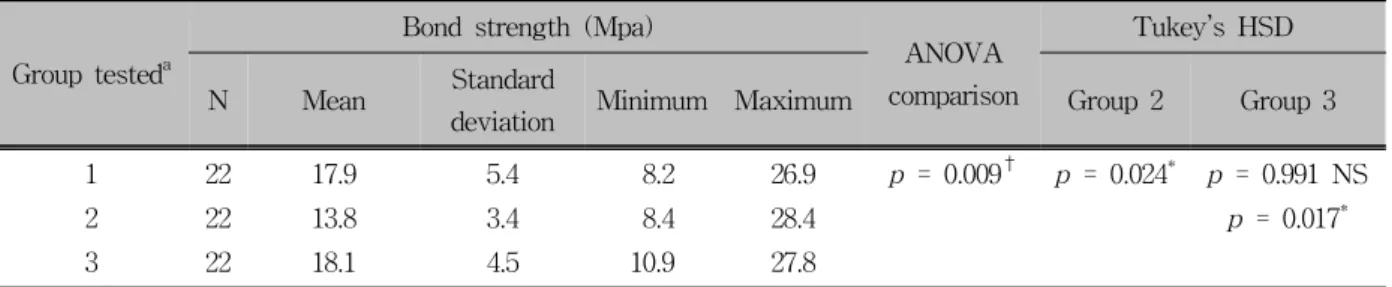
The descriptive statistics for the SBSs of the three groups tested are presented in Table 1. The results of the ANOVA indicated statistically significant differ- ences in the SBS among the three groups (p < 0.01).
Tukey’s HSD test showed that the SBS of Group 1 (control group, mean: 17.9 ± 5.4 MPa) and Group 3 (bonded 30 days after Pro-Relief
TMapplication, mean:
18.1 ± 4.5 MPa) were similar (p = 0.991), whereas the SBS of Group 2 (bonded immediately after Pro- Relief
TMapplication, mean: 13.8 ± 3.4 MPa) was sig- nificantly lower (Group 1 vs. Group 2: p = 0.024 and Group 2 vs. Group 3: p = 0.017). There were no sig- nificant differences between Groups 1 and 3 (p = 0.991).
The ARI scores for the different groups tested are
listed in Table 2. The results of the G test comparisons
indicated no significant differences among the three
groups.
Table 1. Descriptive statistics and the results of ANOVA comparing the SBS of the three groups tested
Group tested
aBond strength (Mpa)
ANOVA comparison
Tukey’s HSD
N Mean Standard
deviation Minimum Maximum Group 2 Group 3
1 22 17.9 5.4 8.2 26.9
p = 0.009† p = 0.024* p = 0.991 NS2 22 13.8 3.4 8.4 28.4
p = 0.017*3 22 18.1 4.5 10.9 27.8
a
Group 1, Control; Group 2, bonded immediately after desensitizer paste application; Group 3, bonded 30 days after desensitizer paste application. NS, Not significant; SBS, shear bond strength.
*p < 0.05; †p < 0.01.Table 2. Adhesive remnant index (ARI) scores (%)
Group tested
aN ARI score
bG-Test
0 1 2 3
1 22 2 (9.1%) 9 (40.9%) 6 (27.3%) 5 (22.7%)
p = 0.9071, NS2 22 2 (9.1%) 8 (36.4%) 1 (4.5%) 11 (50%)
3 22 2 (9.1%) 5 (22.7%) 6 (27.3%) 9 (40.9%)
a
Group 1, Control; Group 2, bonded immediately after desensitizer paste application; Group 3, bonded 30 days after desensitizer paste application.
bARI scores: Score 0, No adhesive remaining on the tooth; Score 1, less than half of the adhesive left on the tooth; Score 2, more than half of the adhesive left on the tooth; Score 3, all adhesive left on the tooth with a distinct impression of the bracket mesh. NS, Not significant.
DISCUSSION
Dentin hypersensitivity is an uncomfortable and un- pleasant condition that affects up to 57% of patients within a dental practice setting.
17A variety of products and methods are available for the treatment of dentin hypersensitivity. Treatment for dentinal hypersensitivity can involve occlusion of the dentinal tubules through the application of sedative agents, cavity varnishes, an- tiinflammatory agents, dentin bonding agents, or re- storative resin along with promotion of dentin remine- ralization.
18Several desensitizer agents have been used to provide desensitization of the natural teeth. In the present study, a new desensitizer paste was used prior to bonding and its effect on the SBS of orthodontic brackets was compared at two time intervals.
From a clinical perspective, orthodontists do not routinely desensitize teeth. Rather, general dentists do the desensitizing and orthodontists apply the brackets sometime later. Thus, the time span between desensi-
tizer application and bracket bonding should be consid- ered a possible factor in the effect of the desensitizer on bond strength. For this reason, the brackets in Group 2 were immediately bonded to enamel treated with desensitizer and the brackets in Group 3 were bonded 30 days after application of the desensitizer.
Group 2 had the lowest SBSs.
Power analysis using the G*Power Ver. 3.0.10.
(Franz Faul, Universität Kiel, Germany) software, based on a 1:1 ratio among groups, indicated that a sample size of 21 teeth would give more than 80%
power to detect significant differences with a 0.35 ef- fect size at a significance level of α = 0.05.
Several theories have been suggested to explain the
mechanism of tooth sensitivity, but the “hydrodynamic
theory” is widely accepted.
3-5According to this theory,
the aspiration of odontoblasts into the dentinal tubules,
as an immediate effect of physical stimuli applied to
exposed dentin, results in the outward flow of the tub-
ular contents (dentinal fluids) through capillary action.
Saliva provides calcium and phosphate, which over time occludes and blocks open dentinal tubules from external stimuli associated with dentinal hypersen- sitivity.
8,19The mechanism providing the clinical effec- tiveness of Pro-Relief
TMdesensitizer agent utilizes argi- nine, an amino acid; bicarbonate, a pH buffer; and cal- cium carbonate, a source of calcium. The technology is proposed to block dentinal hypersensitivity pain by occluding dentinal tubules with arginine, which is pos- itively charged at a physiologic pH of 6.5 to 7.5 to bind to the negatively charged dentin surface, and helps attract a calcium-rich layer from the saliva to in- filtrate and block the dentinal tubules.
19Türkkahraman and Adanir
7evaluated the effects of potassium nitrate and oxalate desensitizer agents on the SBS of orthodontic brackets and reported significantly lower SBS values in the groups receiving potassium nitrate and oxalate desensitizers. In the present study, SBS values were significantly lower in Group 2 (bonded immediately after Pro-Relief
TMapplication) than in the other groups. We found no statistically sig- nificant differences in the bond strength between Group 1 (control group) and Group 3 (teeth bonded 30 days after Pro-Relief
TMapplication).
Garcia-Godoy et al.
14investigated the effect of a de- sensitizing paste containing 8% arginine and calcium carbonate on the surface roughness of dental enamel.
In that study, the 3D non-contact profilometry images showed slight roughness after using the desensitizing paste but these changes were not statistically signifi- cant. Covering the surface with desensitizing agents and remnants may affect adhesive bonding. Malkoc et al.
12reported a remarkably decreased bond strength of orthodontic adhesives used to attach the bracket to the etched enamel surface after application of a desen- sitizer. Alterations in bond strength might be sig- nificant with regard to clinical operative procedures that involve composite resin bonding, such as bonding orthodontic brackets, porcelain veneers, composite ve- neers, or future composite restorations.
20Yip et al.
9demonstrated that application of argi- nine-calcium carbonate in office desensitizing paste to teeth exhibiting sensitivity following a dental prophy- laxis resulted in instant relief from discomfort and that the relief lasted for 28 days after a single application.
Schiff et al.
21applied this product immediately follow- ing scaling and 4 weeks later. In that study, the argi- nine-calcium carbonate paste group demonstrated stat- istically significant reductions in dentin hypersensitivity with respect to baseline adjusted mean air blast and mean tactile hypersensitivity scores, and no statistically significant differences were exhibited between paste groups at the post-scaling and 12-week examinations.
Reynolds
22determined the clinically acceptable min- imum bond strength values in direct orthodontic bond- ing systems to be 5.9 to 7.8 MPa. All bond strength values of composites used in this study were greater than this minimum requirement and fell within the clinically acceptable range. Clinical conditions may significantly differ, however, from an in vitro setting.
Moreover, the oral cavity is a complex environment with variations in temperature, stresses, humidity, acid- ity, and plaque.
23Because of the probable differences between in vivo and in vitro conditions, a direct com- parison cannot be made with the findings of the other studies.
Most orthodontic bonding studies have shown a mixed or cohesive-type failure.
15,16In those studies, af- ter bond strength testing, a part of the composite resin remained either on the enamel surface or the bracket base, causing cohesive failure rather than adhesive fail- ure between enamel and composite resin. Bond failure at the bracket-resin interface or within the resin is more desirable than at the resin-enamel interface be- cause enamel fractures and cracks have been reported during bracket debonding.
24For mechanically retained brackets, the most common failure site was the brack- et-resin interface, and, on average, more than 50% of resin remains on teeth after debonding.
25The ARI score comparisons in the present study indicated no significant differences among the three groups tested.
There are no published data about comprehensive
observations or intraoral applications concerning Pro-
Relief
TMin-office desensitizer paste and the effects on
the bond strength of brackets. Thus, the clinical sig-
nificance of this new desensitizer paste should be fur-
ther clarified in detail under in vivo conditions.
CONCLUSION
The use of a Pro-Relief
TMdesensitizer agent imme- diately before bonding significantly reduces the SBS, but the SBS still exceeds the minimum 5.9 to 7.8 MPa required to expect adequate clinical performance.
Immersion of teeth applied with Pro-Relief
TMde- sensitizer agent in artificial saliva for 30 days before bonding increased the SBS value to that of controls.
The use of desensitizer procedures with arginine and calcium carbonate immediately before bonding ortho- dontic brackets is not recommended.
REFERENCES
1. Addy M. Dentine hypersensitivity: new perspectives on an old problem. Int Dent J 2002;52:367-75.
2. Brannstrom M. Dentin sensitivity and aspiration of odon- toblasts. J Am Dent Assoc 1963;66:366-70.
3. Cummins D. Dentin hypersensitivity: from diagnosis to a breakthrough therapy for everyday sensitivity relief. J Clin Dent 2009;20:1-9.
4. Walters PA. Dentinal hypersensitivity: a review. J Contemp Dent Pract 2005;6:107-17.
5. Swift EJ Jr. Causes, prevention, and treatment of dentin hypersensitivity. Compend Contin Educ Dent 2004;25:95-106.
6. West NX. Dentine hypersensitivity. In: Lussi A editor. Dental erosion. Basel: Karger; 2006. p. 173-89.
7. Türkkahraman H, Adanir N. Effects of potassium nitrate and oxalate desensitizer agents on shear bond strengths of ortho- dontic brackets. Angle Orthod 2007;77:1096-100.
8. Kleinberg I. SensiStat. A new saliva-based composition for simple and effective treatment of dentinal sensitivity pain.
Dent Today 2002;21:42-7.
9. Yip CK. The need and demand of orthodontics among Chinese adults in Hong Kong (dissertation). Hong Kong: Univ of Hong Kong, 1993.
10. Sengun A, Koyuturk AE, Sener Y, Ozer F. Effect of desensi- tizers on the bond strength of a self-etching adhesive system to caries-affected dentin on the gingival wall. Oper Dent 2005;30:430-5.
11. Aranha AC, Siqueira Junior Ade S, Cavalcante LM, Pimenta LA, Marchi GM. Microtensile bond strengths of composite to
dentin treated with desensitizer products. J Adhes Dent 2006;
8:85-90.
12. Malkoc S, Demir A, Sengun A, Ozer F. The effect on shear bond strength of different antimicrobial agents after acid etching. Eur J Orthod 2005;27:484-8.
13. Holzmeier M, Ernst CP, Willershausen B, Hirschfelder U.
In-vitro shear bond strength of self-etching versus traditional adhesives for orthodontic luting. J Orofac Orthop 2006;67:
244-59.
14. Garcia-Godoy F, Garcia-Godoy A, Garcia-Godoy C. Effect of a desensitizing paste containing 8% arginine and calcium car- bonate on the surface roughness of dental materials and human dental enamel. Am J Dent 2009;22:21A-4A.
15. Artun J, Bergland S. Clinical trials with crystal growth con- ditioning as an alternative to acid-etch enamel pretreatment.
Am J Orthod 1984;85:333-40.
16. Oliver RG. The effect of different methods of bracket removal on the amount of residual adhesive. Am J Orthod Dentofacial Orthop 1988;93:196-200.
17. Addy M. Etiology and clinical implications of dentine hyper- sensitivity. Dent Clin North Am 1990;34:503-14.
18. Trowbridge HO, Silver DR. A review of current approaches to inoffice management of tooth hypersensitivity. Dent Clin North Am 1990;34:561-81.
19. Panagakos F, Schiff T, Guignon A. Dentin hypersensitivity: ef- fective treatment with an in-office desensitizing paste contain- ing 8% arginine and calcium carbonate. Am J Dent 2009;22:3A-7A.
20. Josey AL, Meyers IA, Romaniuk K, Symons AL. The effect of a vital bleaching technique on enamel surface morphology and the bonding of composite resin to enamel. J Oral Rehabil 1996;23:244-50.
21. Schiff T, Delgado E, Zhang YP, Cummins D, DeVizio W, Mateo LR. Clinical evaluation of the efficacy of an in-office desensitizing paste containing 8% arginine and calcium carbo- nate in providing instant and lasting relief of dentin hypersensitivity. Am J Dent 2009;22:8A-15A.
22. Reynolds IR. A review of direct orthodontic bonding. Br J Orthod 1975;2:171-8.
23. Zachrisson YO, Zachrisson BU, Büyükyilmaz T. Surface prep- aration for orthodontic bonding to porcelain. Am J Orthod Dentofacial Orthop 1996;109:420-30.
24. Bishara SE, Olsen ME, Von Wald L. Evaluation of debonding characteristics of a new collapsible ceramic bracket. Am J Orthod Dentofacial Orthop 1997;112:552-9.
25. Forsberg CM, Hagberg C. Shear bond strength of ceramic brackets with chemical or mechanical retention. Br J Orthod 1992;19:183-9.