Ⅰ. 서 론
구순구개열 환자에서 치조골 결손부 회복을 위해 고려되 는 이차골이식(secondary bone grafting)은 많은 장점을 가지는 술식으로 구순구개열의 치료에 광범위하게 이용되 고 있다1-3). 치조골 결손으로 인해 매복된 영구치를 정상적 으로 맹출시키기 위해서는 골결손부의 회복이 필요하며, 특
히 매복된 상악 견치의 맹출을 촉진시키기 위해서 이차골이 식이 시행된다3).
이차골이식은 사용되는 골 이식재의 종류에 따라 자가골 이식(autografts), 동종골이식(allografts), 이종골 이식 (xenografts)의 방법이 사용된다1,4). 이식된 골의 흡수를 줄 이고5,6), 성공적인 생착을 위해서는 자가골이식 및 동종골이 식이 추천되지만, 채취할 수 있는 골량의 한계 및 치배 손상 의 위험성, 부가적인 수술의 필요성, 수술시간의 지연 및 감 염율과 합병증의 증가위험과 같은 단점을 가진다4,7-13).
반면에 이종골 이식의 경우, 자가골 및 동종골 이식의 단 점을 피할 수 있으나 이식된 골의 생착 및 치아 맹출에 대한 영향에 대해 보고된 연구가 드물다. 이종골 이식재로는 주로
◆ 증 례
구순구개열 환자에서의 이종골 이식재를 통한 견치의 맹출
김지훈1*∙최병호2∙장채리1
연세대학교 원주기독병원1소아치과, 2구강악안면외과
CANINE ERUPTION THROUGH BIO-OSS
�GRAFT IN PATIENTS WITH CLEFT LIP & PALATE Ji-Hun Kim1*, Byung-Ho Choi
2, Cherry Chang
1
1
Department of Pediatric Dentistry,
2Department of Oral and Maxillofacial Surgery, Wonju College of Medicine, Yonsei University
Objective :
To report eruption of maxillary canine through Bio-Oss� graft in patients with secondary bone-grafted alveolar clefts.Methods :
Secondary alveolar bone grafts placed in the cleft alveolar defect have been shown to support dental eruption through the graft and may further affect the prevalence of impacted teeth. As the case may be, it could be difficult to do secondary alveolar bone graft with autologous bone. In particular, few reports have been shown the secondary bone graft with heterogenous bone(Bio-Oss�).In this report, the eruption of canine into bone-grafted alveolar clefts was recorded as panoramic, occlusal radiographs, in 3 patients grafted with Bio-Oss�.
Results :
Like autologous bone graft, the canine was erupted and developed into the cleft alveolar defect through Bio-Oss�graft.Conclusion :
In some cases that autologous bone graft is not available, we can consider heterogenous bone graft into the cleft alveolar defect for dental development and eruption of impacted teeth.Key words :
Cleft, Bone graft, Teeth eruptionAbstract
교신저자 : 김 지 훈
220-701 강원도 원주시 일산동 162 연세대학교 원주기독병원 소아치과
Tel: 033-741-0673 Fax: 033-741-1442 E-mail: [email protected]
원고접수일: 2010.11.20 / 원고최종수정일: 2010.11.30 / 원고채택일: 2010.12.15
Bio-Oss�(Geistlich-Pharma, Wolhusen, Switzerland) 가 사용되며, 소뼈에서 채취된 탄산인회석(carbonate ap- atite)을 포함한 무기물(anorganic bovine bone marix)로 서 상악동 이식재로 널리 사용되고 있다14-16). Bio-Oss�는 화학적 처리를 통해 이종골이 가질 수 있는 항원성이 제거 되며, 골 재생 단계와 비슷한 생리적 반응을 일으키는 기질 로 작용하여 골의 생착을 유도하는 것으로 알려져 있다17).
본 교실에서는 동물 실험을 통해 이종골 이식(Bio-Oss� xenograft)을 통한 치아 맹출에 대해 연구해 왔으며18), 기 존 문헌에서도 성공적인 치아맹출이 보고된 바 있다4). 따라 서 이번 연구에서는 이차골이식을 받은 구순구개열 환자들 중 자가골 및 동종골 이식이 어려워 이종골 이식을 시행하 고 성공적인 치아맹출이 관찰된 증례를 보고하고자 한다.
Ⅱ. 증 례
이차골이식은 매복된 치아의 정상적 맹출을 돕기 위해 매 복된 치아의 치근이 1/2 에서 2/3 정도 형성되었을 때 시행
되었고, 이차골이식의 시기는 매복된 치아의 예후에 영향을 줄 수 있음이 보고된 바 있다3).
1. 증례 1(10세 2개월, 여)
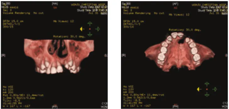
이차골 이식 당시 10세 2개월된 여아로, 구순구개열로 인 해 좌측 치조골의 골결손 및 상악 좌측 측절치 및 견치가 매 복되어 있었다(Fig. 1). 견치의 치근 형성은 2/3 정도로 이 차골이식 시기에 적절하였고, 전신마취하에 Bio-Oss�를 사 용하여 골결손부 이차골이식을 시행하였다(Fig. 1-3).
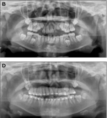
이식 한달 후, 수술부위의 치유가 완료되고 이식재의 생 착이 시작되었으나 아직 치아는 이동하지 않았다(Fig.
1B). 이식 7개월 후, 매복된 측절치 및 견치의 치아 이동이 관찰된다(Fig. 1C). 골이식 및 구순구개열 수술로 인한 반 흔 형성으로 총생이 관찰되었고 이로 인해 교정치료가 이루 어졌으며, 매복되었던 측절치 및 견치는 성공적으로 맹출이 완료되었다(Fig. 1D, 1E).
Fig. 1.
Panoramic view before & after secondary bone graft with Bio-Oss�in Case 1.A: Panoramic view before secondary bone graft, B: 1 month after secondary bone graft, C: 7 months after secondary bone graft, D: 20 months after secondary bone graft, E: 25 months after secondary bone graft.
A
C
E
B
D
2. 증례 2(9세 1개월, 남)
이차골 이식 당시 9세 1개월된 남아로, 양측성 구순구개 열로 인해 좌우측 치조골의 결손 및 상악 우측 측절치의 결 손, 상악 좌측 측절치의 왜소(peg lateralis), 상악 좌우측
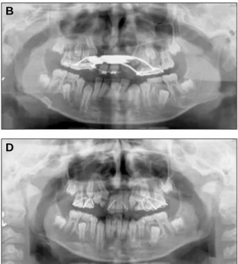
견치의 매복이 관찰된다(Fig. 4). 전산화 단층촬영을 통해 이식부위의 골결손 상태를 확인하였고, 구개확장 후 이차골 이식이 이루어졌다(Fig. 4B, 4C, 5). 3개월 후 매복된 좌 우측 견치의 맹출이 관찰되며, 8개월 후 정상적인 치아 맹 출 및 발달이 이루어지고 있다(Fig. 4D, 4E).
Fig. 2.
Computed tomography of alveolar bone defect in Case 1 patient.Fig. 3.
Clinical photograph of secondary bone grafting with Bio-Oss� particle in Case 1.Fig. 4.
Panoramic view before & after secondary bone graft with Bio-Oss� in Case 2.A: Panoramic view before secondary bone graft, B: Palatal expansion before secondary bone graft, C: 1 months after secondary bone graft, D: 3 months after secondary bone graft, E: 8 months after secondary bone graft.
A
C
E
B
D
Fig. 5.
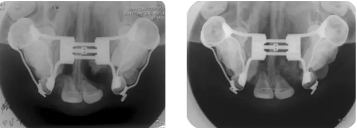
Computed tomography of alveolar bone defect in Case 2 patient.3. 증례 3(12세 1개월, 남)
이차골 이식 당시 12세 1개월된 남아로, 양측성 구순구개 열로 인해 좌우측 치조골의 결손 및 상악 우측 측절치의 결 손, 상악 좌측 측절치의 매복 및 왜소(peg lateralis), 상악 좌우측 견치의 매복이 관찰된다(Fig. 7). 구개확장 후 이차 골 이식이 이루어졌으며, 교합 방사선 사진을 통해 이차골 이식 전후의 상태를 확인할 수 있다(Fig. 6, 7A). 골이식 후 정상적으로 치아 이동 및 맹출이 이루어지고 있음이 술 후 16개월 파노라마에서 관찰된다(Fig. 7C).
Ⅲ. 고 찰
증례를 통해 골결손부위로의 이종골(Bio-Oss�)을 이용한 이차골이식 후 매복된 측절치 및 견치가 성공적으로 골내 이동 및 맹출하는 것이 관찰되었다(Fig. 1, 4, 7). 이는 동 물 실험을 통한 이전의 연구 결과와도 일치하며18), 기존 문 헌을 통해 보고된 바와도 부합한다4).
보고된 증례에서 골이식 직후 열개의 발생 및 이로 인한 창상 치유의 지연이 일부 발견되었으나, 적절한 소독 및 구 강위생 관리로 합병증 없이 치유되었다. 이식된 이종골의
Fig. 6.
Occlusal radiographs before & after secondary bone graft in Case 3 patient.Fig. 7.
Panoramic view before & after secondary bone graft with Bio-Oss� in Case 3.A: Panoramic view before secondary bone graft, B: 6 month after secondary bone graft, C: 16 months after secondary bone graft
A
C
B
성공적인 생착을 위해서는 구개열 부위의 긴밀한 봉합과 연 조직 관리가 필요하며 지속적인 술후 관리를 통해 감염 및 합병증의 위험을 줄일 수 있다.
이전 연구에 의하면 성공적인 이식재의 생착을 위해 자가 골, 특히 하악 정중부 골(chin bone)이 막내골(intramem- braneous)로서 연골내골(enchondral bone)에 비해 골흡 수율이 적은 최적의 골이식재로 추천된다1,19). 그러나 술후 합병증 및 치배손상의 위험성, 골채취량의 한계, 부가적인 수술로 인한 감염 및 합병증으로 인해 선택이 제한되기도
한다4,7-13). 동종골 역시 채취량에 한계가 있고, 높은 비용 및
술후 면역반응 및 골흡수로 제한적으로 선택된다4,20,21). 이차골이식이 필요한 구순구개열 환자의 경우, 어릴 때 부터 영양섭취를 위한 구개폐쇄술 및 일차골이식, 입술 및 비성형(lip revision & rhinoplasty) 등과 같은 잦은 수술 로 인해 수술에 대한 거부감이 크고, 공여부(donor site)의 골채취가 제한적인 경우가 많다2,7,22-24).
따라서, 이종골을 이용한 골이식을 통해 공여부에 대한 부가적인 수술 위험 및 합병증을 줄이고, 골채취 및 이식재 의 양에 대한 부담을 감소시킬 수 있다25). 그러나 임플란트 식립시 상악동 골이식에 자주 사용되는 이종골(Bio-Oss�) 의 치아맹출에 대한 영향에 대한 연구는 적을 뿐더러, 이에 대한 임상연구는 찾아보기 힘들다. 본 교실에서는 이종골의 치아맹출에 대한 기존 동물실험연구를 바탕으로, 자가골 및 동종골 이식이 어려운 환자에서 이종골을 이식하여 성공적 으로 매복된 치아의 맹출을 유도하였다.
그러나, 본 연구의 한계점으로 이식된 이종골(Bio-Oss�) 이 치근의 형성 및 치조골에 미치는 영향에 대한 조직학적 연구의 보완이 필요하다. Thaller 등26)에 따르면, 이식된 이 종골은 자가골에 비해 골유도 능력(osteoinductive capac- ity)이 부족하기 때문에 생착에 더 많은 시간이 요구되며, 따라서 치조골로 생착되기보다는 대부분의 이종골이 구강 내로 빠져나간다고 한다26). 하지만, 기존 연구에 따르면 이 식재로 인한 치아 맹출의 지연이나 방해는 관찰되지 않았으 며, 치관 및 치근의 형성에도 영향을 끼치지 않음이 보고된
바 있다4,18). 추후 이식된 이종골이 어떻게 흡수되고 생착되
는지에 대한 골대사의 기전 및 이에 대한 조직학적 연구가 좀 더 필요할 것으로 사료된다.
Ⅳ. 요 약
이종골의 치아맹출에 대한 기존 문헌 및 동물실험연구를 바탕으로, 자가골 및 동종골 이식이 어려운 구순구개열 환 자에서 이종골(Bio-Oss�)을 이식하여 성공적으로 매복된 치아의 맹출을 유도하였고, 치근의 형성 및 치아의 골내 이 동도 정상적으로 이루어졌다.
참고문헌
1. Freihofer, H. P., Borstlap, W. A., Kuijpers- Jagtman, A. M., Voorsmit, R. A., van Damme, P. A., Heidbuchel, K. L., Borstlap-Engels, V. M.
: Timing and transplant materials for closure of alveolar clefts. A clinical comparison of 296 cas- es. J Craniomaxillofac Surg 21(4): 143-148, 1993.
2. Kalaaji, A., Lilja, J., Friede, H., Elander, A.:
Bone grafting in the mixed and permanent denti- tion in cleft lip and palatepatients: long-term re- sults and the role of the surgeon's experience. J Craniomaxillofac Surg 24(1): 29-35, 1996.
3. Witsenburg, B., Remmelink, H. J.: Reconstruction of residual alveolo-palatal bone defects in cleft patients. A retrospective study. J Craniomaxillofac Surg 21(6): 239-244, 1993.
4. Merkx, M. A., Maltha, J. C., van’t Hoff, M., Kuijpers-Jagtman, A. M., Freihofer, H. P. : Tooth eruption through autogenous and xenogenous bone transplants: a histological and radiographic evaluation in beagle dogs. J Craniomaxillofac Surg 25(4): 212-219, 1997.
5. Koole, R., Bosker, H., van der Dussen, F. N. : Late secondary autogenous bone grafting in cleft patients comparing mandibular (ectomesenchy- mal) and iliac crest (mesenchymal) grafts. J Craniomaxillofac Surg 17 Suppl 1: 28-30, 1989.
6. Zins, J. E., Whitaker, L. A. 1983. “Membranous versus endochondral bone: implications for cran- iofacial reconstruction”. Plast Reconstr Surg 72(6): 778-785.
7. Borstlap, W. A., Heidbuchel, K. L., Freihofer, H.
P., Kuijpers-Jagtman, A. M. : Early secondary bone grafting of alveolar cleft defects. A comparison between chin and rib grafts. J Craniomaxillofac Surg 18(5): 201-205, 1990.
8. Cohen, M., Figueroa, A. A., Haviv, Y., Schafer, M. E., Aduss, H. : Iliac versus cranial bone for secondary grafting of residual alveolar clefts.
Plast Reconstr Surg 87(3): 423-427; discussion 428, 1991.
9. Hoppenreijs, T. J., Nijdam, E. S., Freihofer, H.
P. : The chin as a donor site in early secondary osteoplasty: a retrospective clinical and radiologi-
cal evaluation. J Craniomaxillofac Surg 20(3):
119-124, 1992.
10. Koole, R., Visser, W. J., Klein, W. R., Suiker, A.
M. : A comparative investigation on autologous mandibular and iliac crest bone grafts. An exper- imental study in sheep. J Craniomaxillofac Surg 19(4): 133-143, 1991.
11. Kortebein, M. J., Nelson, C. L., Sadove, A. M. : Retrospective analysis of 135 secondary alveolar cleft grafts using iliac or calvarial bone. J Oral Maxillofac Surg 49(5): 493-498, 1991.
12. Sugimoto, A., Ohno, K., Michi, K., Kanegae, H., Aigase, S., Tachikawa, T. : Effect of calcium phosphate ceramic particle insertion on tooth eruption. Oral Surg Oral Med Oral Pathol 76(2):
141-148, 1993.
13. Witsenburg, B., Peter, H., Freihofer, M. : Autogenous rib graft for reconstruction of alveolar bone defects in cleft patients. Long-term follow- up results. J Craniomaxillofac Surg 18(2): 55- 62, 1990.
14. Froum, S. J., Wallace, S. S., Cho, S. C., Elian, N., Tarnow, D. P. : Histomorphometric compari- son of a biphasic bone ceramic to anorganic bovine bone for sinus augmentation: 6- to 8- month postsurgical assessment of vital bone for- mation. A pilot study. Int J Periodontics Restorative Dent 28(3): 273-281, 2008.
15. Hislop, W. S., Finlay, P. M., Moos, K. F. : A preliminary study into the uses of anorganic bone in oral and maxillofacial surgery. Br J Oral Maxillofac Surg 31(3): 149-153, 1993.
16. Wetzel, A. C., Stich, H., Caffesse, R. G. : Bone apposition onto oral implants in the sinus area filled with different grafting materials. A histo- logical study in beagle dogs. Clin Oral Implants Res 6(3): 155-163, 1995.
17. Klinge, B., Alberius, P., Isaksson, S., Jonsson, J.
: Osseous response to implanted natural bone mineral and synthetic hydroxylapatite ceramic in the repair of experimental skull bone defects. J Oral Maxillofac Surg 50(3): 241-249, 1992.
18. 김지훈, 최병호, 장채리 : Bio-Oss�골이식이 치아맹
출에 미치는 영향에 관한 동물실험 연구. 대한구강악안 면외과학회지 36(6), 2010.
19. Smith, J. D., Abramson, M. : Membranous vs endochondrial bone autografts. Arch Otolaryngol 99(3): 203-205, 1974.
20. Froum, S. J., Wallace, S. S., Elian, N., Cho, S.
C., Tarnow, D. P. : Comparison of mineralized cancellous bone allograft (Puros) and anorganic bovine bone matrix (Bio-Oss) for sinus augmen- tation: histomorphometry at 26 to 32 weeks after grafting. Int J Periodontics Restorative Dent 26(6): 543-551, 2006.
21. Wang, H. L., Tsao, Y. P. : Histologic evaluation of socket augmentation with mineralized human allograft. Int J Periodontics Restorative Dent 28(3): 231-237, 2008.
22. Batra, P., Sharma, J., Duggal, R., Parkash, H. : Secondary bone grafting in cleft lip and palate with eruption of tooth into the graft: a case re- port. J Indian Soc Pedod Prev Dent 22(1): 8-12, 2004.
23. da Silva Filho, O. G., Teles, S. G., Ozawa, T. O., Filho, L. C.: Secondary bone graft and eruption of the permanent canine in patients with alveolar clefts: literature review and case report. Angle Orthod 70(2): 174-178, 2000.
24. Ozawa, T., Omura, S., Fukuyama, E., Matsui, Y., Torikai, K., Fujita, K. Factors influencing secondary alveolar bone grafting in cleft lip and palate patients: prospective analysis using CT image analyzer. Cleft Palate Craniofac J 44(3):
286-291, 2007.
25. Norton, M. R., Odell, E. W., Thompson, I. D., Cook, R. J.: Efficacy of bovine bone mineral for alveolar augmentation: a human histologic study. Clin Oral Implants Res 14(6): 775-783, 2003.
26. Thaller, S. R., Hoyt, J., Dart, A., Borjeson, K., Tesluk, H. : Repair of experimental calvarial de- fects with Bio-Oss particles and collagen sponges in a rabbit model. J Craniofac Surg 5(4): 242- 246, 1994.