| Abstract |
Purpose: This study identified the co-activation of quadriceps and hamstring muscles during hamstring strengthening exercises in healthy adults.
Methods: Twenty-one participants were required for the present study design to achieve 80% power, 0.8 effect size (η2), and an alpha level of 0.05. Thus, this study recruited 21 healthy adults. All participants performed Nordic exercises, bridge exercises, and one-leg deadlifts randomly. The activity of the rectus femoris, vastus medialis (VM), vastus lateralis (VL), biceps femoris (BF), and semitendinosus (SM) were measured. In addition, the ratios of VM/VL and hamstring/quadriceps (HQ) were measured during the three hamstring strengthening exercises using electromyography. One-way ANOVA was used to compare the co-activation of quadriceps and hamstring muscles in the three exercises.
Results: The activity of VM and VL during the performance of one-leg deadlifts was significantly higher than the other two exercises. The BF had significantly higher activity during the Nordic exercises compared to the other two exercises. In addition, the SM activation was significantly greater during Nordic exercises than one-leg deadlifts. Additionally, there was significant difference in HQ ratio among hamstring strengthening exercises. In specific, the one-leg deadlifts yielded a significantly lower HQ ratio.
Conclusion: This study revealed that one-leg deadlifts are effective in rehabilitation for anterior cruciate ligament injury. In
†Corresponding Author : Du-Jin Park ([email protected])
Original Article Open Access
뒤넙다리근 강화 운동 시 넙다리네갈래근과 뒤넙다리근의 동시 수축에 대한 근전도 분석
황영인⋅문상재1⋅박두진2†1)
호서대학교 물리치료학과, 1진주경상대학교병원 재활치료실, 2부산가톨릭대학교 물리치료학과
Electromyographic Analysis of Quadriceps and Hamstrings Co-activation during Hamstring Strengthening Exercises
Young-In Hwang, P.T., Ph.D⋅Sang-Jae Moon, P.T., B.S.1⋅Du-Jin Park, P.T., Ph.D2†
Department of Physical Therapy, College of Life and Health Science, Hoseo University
1Department of Physical Therapy, Jinju Gyeongsang National University Hospital
2Department of Physical Therapy, College of Health Sciences, Catholic University of Pusan
Received: September 16, 2019 / Revised: October 1, 2019 / Accepted: October 1, 2019
ⓒ 2019 Journal of Korea Proprioceptive Neuromuscular Facilitation Association
This is an Open Access article distributed under the terms of the Creative Commons Attribution Non-Commercial License (http://creativecommons.org/licenses/by-nc/3.0) which permits unrestricted non-commercial use, distribution, and reproduction in any medium, provided the original work is properly cited.
addition, Nordic exercises can be recommended to facilitate hamstring muscle activation.
Key Words: Hamstring strengthening exercise, Co-activation, Electromyography
Ⅰ. 서 론
앞무릎통증증후군(anterior knee pain syndrome) 또는 무릎넙다리통증증후군(patellofemoral pain syndrome, PFPS)은 주로 15∼30세의 젊은 청년들이 왕성한 신체 활동을 하면서 겪는 가장 대표적인 다리 질환이다 (Boling et al., 2019). 이는 많은 운동선수의 스포츠 활동 을 제한하는 원인이 되며(Blønd & Hansen, 1998), 장기 간 지속되게 되면 퇴행성 관절염까지도 초래할 수 있 다(Myer et al., 2010). PFPS의 원인으로는 엉덩정강근 막띠(iliotibial band), 넙다리네갈래근(quadriceps), 뒤넙 다리근(hamstring), 장딴지근(gastrocnemius) 등의 유연 성 감소, 다리 근육의 약증(weakness), 그리고 무릎뼈 의 부정렬 등이 있다(Waryasz & McDermott, 2008).
다양한 발생 원인 중 넙다리네갈래근에 대한 약증 은 대표적인 PFPS의 발생 인자이다. PFPS 환자의 경 우, 기능적인 활동인 계단 오르기와 내리기를 하는 동안 가쪽넓은근(vastus lateralis, VL)이 안쪽넓은근 (vastus medialis, VM)에 비해 선행적으로 활성화되며 (Cowan et al., 2001), 이로 인해 대부분의 환자는 안쪽 넓은근의 위축이 나타났다(Pattyn et al., 2011). 이 근육 의 약증은 무릎뼈의 가쪽 이동을 발생시키고, 무릎관 절에 가해지는 스트레스를 증가시켜 PFPS를 야기시 킨다(Tang et al., 2001). 이에 많은 연구자들은 PFPS 환자의 VM/VL 비율에 대해 많은 관심을 가졌으며 (Mostamand et al., 2011; Ryan & Rowe, 2006), PFPS를 개선시킬 수 있는 중재는 1에 가까운 VM/VL 비율을 보이는 운동을 적용하는 것이다(Kushion et al., 2012).
다른 원인으로는 뒤넙다리근의 약증을 들 수 있으며, Waryasz와 McDermott (2008)는 PFPS를 경험한 운동선 수의 약 73∼81%가 뒤넙다리근의 근력 감소를 보였음 을 보고하였다.
뒤넙다리근의 약증은 PFPS뿐만 아니라 앞쪽십자 인대(anterior cruciate ligament, ACL)의 손상도 초래할 수 있다. 약화된 뒤넙다리근은 불균형한 뒤넙다리근 과 넙다리네갈래근의 활동(hamstring to quadriceps, HQ) 비율을 초래하여 ACL 손상 위험을 높일 수 있다 (Dedinsky et al. 2017). 특히 무릎을 펴는 동작을 수행하 는 동안 뒤넙다리근의 역할은 정강뼈의 움직임을 조 절하여 무릎관절 앞쪽으로 발생하는 전단력(shear force)을 감소시켜 ACL 손상을 최소화할 수 있다(More et al., 1993). 최근 연구에서도 점진적 플라이오메트릭 운동을 하는 동안 뒤넙다리근의 증가된 활동 없이 나 타나는 넙다리네갈래근의 과도한 활동은 ACL 손상 위험을 증가시킨다고 보고하였다(Ford et al., 2014).
이처럼, 뒤넙다리근의 활동 감소와 약증은 불균형한 HQ 비율을 발생시키고, PFPS와 ACL 손상에 직접적인 원인이 될 수 있다.
Myer 등(2015)의 연구에 의하면, ACL 손상이 있는 대상자의 HQ 비율은 0.51이며, PFPS가 있는 대상자의 HQ 비율은 0.68이었다. 0.6이하의 HQ 비율은 이러한 질환에 대한 발생 가능성을 높일 수 있으며, 이를 예방 하기 위해서는 0.6이상의 HQ 비율을 보이는 한다리 스쿼트 및 변형된 한다리 스쿼트 같은 치료적 운동을 권장하고 있다(Dedinsky et al., 2017; Wright et al., 2009). 이와 같이 뒤넙다리근의 활동이 ACL 손상과 PFPS의 발생에 매우 중요한 요소임에도 불구하고, 대 부분의 선행 연구는 넙다리네갈래근 강화 운동을 규 명하는데 초점을 두고 있으며, 무릎관절의 안정성에 기여하는 뒤넙다리근의 활동을 촉진할 수 있는 뒤넙 다리근 강화 운동에 대한 연구는 부족한 실정이다.
대표적인 뒤넙다리근 강화 운동에는 노르딕 운동 (nordic exercise), 교각 운동(bridge exercise), 그리고 한 발서기 데드리프트(single deadlift)가 있다. 노르딕 운
동은 과거에는 러시안 뒤넙다리근 운동(russian hamstring exercise)으로 불렸으며, 특별한 기구없이 파 트너와 함께 쉽게 뒤넙다리근을 강화할 수 있다 (Ebben, 2009). 교각 운동은 역시 뒤넙다리근의 강화를 위해 많이 사용되며, 지면과 짐볼(gymball)에서 다양 하게 적용할 수 있다(Ekstrom et al., 2008). 그리고 데드 리프트는 뒤넙다리근과 엉덩이근을 훈련시키는데 사 용되며(Weingroff, 2014), 한발서기 데드리프트의 경우 에는 이들 근육에 대한 더 빠른 훈련 효과를 얻기 위해 주로 실시된다(Liebenson, 2015). 이와 같은 뒤넙다리 근 강화 운동을 수행하는 동안 넙다리네갈래근과 뒤 넙다리근의 활성도, VM/VL 및 HQ 비율을 통해 PFPS 와 ACL 손상이 있는 대상자에게 효과적인 운동을 제 시하고자 본 연구를 실시하였다.
Ⅱ. 연구 방법
1. 연구 대상
본 연구는 21명의 건강한 성인 대상자를 모집하여 진행하였다. 모든 대상자는 연구의 과정과 목적에 대해 설명을 듣고, 자발적으로 동의한 자에 한하여 선정하였으며, 본 연구에서 실시된 모든 실험 과정은 헬싱키 선언에 따라 진행되었다. 선천적 혹은 후천적 인 근골격계 질환이 있는 자, 운동 수행이 불가능한 자, 신경계 질환이 있는 자는 연구 대상자에서 제외하 였다.
2. 측정방법 및 도구
1) 근활성도 측정
본 연구에서는 넙다리네갈래근과 뒤넙다리근의 활 성도를 측정하기 위한 도구로 표면 근전도(Free EMG 300, BTS Bioengineering, Italy)를 사용하였으며, 피부 에서 발생하는 저항을 최소화시키기 위해 알코올 솜
을 이용하여 이물질을 닦아낸 후 Ag/AgCI 전극을 부착 하였다. 표면 전극은 우세측 다리에 부착하였으며, 우 세측 다리는 공을 가장 멀리 찬 쪽으로 선정하였다 (Letafatkar et al., 2019). 우세측 다리의 전극은 다음과 같은 근육 부위에 부착하였다.
넙다리곧은근(rectus femoris)은 무릎과 위앞엉덩 뼈가시(anterior superior iliac spine, ASIS) 사이 중앙 지점, 안쪽넓은근은 ASIS에서 앞쪽 무릎관절의 내측 인대를 연결한 선의 80% 지점, 가쪽넓은근은 ASIS에 서 무릎뼈(patella) 가쪽 부위를 잇는 선의 2/3 지점, 넙다리두갈래근(biceps femoris)은 정강뼈(tibia)의 가 쪽 위관절융기(lateral epicondyle)과 궁둥뼈 결절 (ishial tuberosity)을 연결한 선의 중간 위치, 반힘줄근 (semitendinosus)은 정강뼈의 안쪽 위관절융기와 궁둥 뼈 결정을 잇는 선의 중간 위치에 부착하였다(Hermens et al., 2000). 기준 전극(reference electrode)은 우세측 정강뼈 결절(tibial tuberosity)에 부착하여 실험을 진행 하였다(Chmielewski et al., 2005). 근전도 신호의 표본 추출률과 주파수 대역폭은 각각 1,000Hz와 10∼450Hz 로 설정하였다. 수집된 근활성도 신호는 프로그램을 이용하여 실효치 진폭(root mean square, RMS)을 분석 하였다.
2) 최대 수의적 등척성 수축(maximal voluntary isometric contraction, MVIC)
각 근육들의 활동을 표준화하기 위해 최대 수의적 등척성 수축을 실시하였다. 모든 근육의 최대 수의적 등척성 수축은 Hislop 등(2013)의 문헌을 토대로 다음 과 같이 진행되었다. 넙다리네갈래근에 대한 측정은 대상자가 검사대 가장자리에 걸터앉은 상태에서 발목 위 먼쪽부위에서 종아리의 앞쪽면을 잡고 저항을 주 어 실시하였다. 반힘줄근은 엎드려 눕은 자세에서 무 릎관절을 90°이하로 굽힘한 상태에서 발목을 잡고 무 릎을 폄하면서 가쪽으로 비스듬히 저항을 준다. 넙다 리두갈래근은 반힘줄근과 동일한 자세에서 무릎을 폄 하면서 안쪽으로 비스듬히 저항을 준다.
3) VM/VL 및 HQ 비율
안쪽넓은근과 가쪽넓은근의 동시 수축을 분석하기 위해 VM/VL 비율을 사용하였으며, 선행 연구의 정량 화 방식인 안쪽넓은근의 %MVIC값에 가쪽넓은근의
%MVIC값을 나누어 계산하였다(Abdelraouf et al., 2019). 넙다리네갈래근과 뒤넙다리근의 동시 수축을 분석하기 위해 HQ 비율을 사용하였으며, 넙다리두갈 래근의 %MVIC값에 안쪽넓은근의 %MVIC값을 나누 는 정량화 방식으로 계산하였다(Nishiwaki et al., 2006).
3. 실험 절차
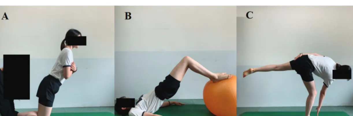
본 연구의 대상자는 뒤넙다리근 강화 운동인 노르 딕 운동, 교각 운동, 한다리 데드리프트를 숙지하기 위해서 실험 전 약 30분간 교육을 받았다(Fig. 1). 또한 신호음을 숙지하여 운동의 시작과 끝을 숙지하게 하 였으며, 모든 대상자는 우세측 다리를 이용하여 측정 하였다. 노르딕 운동은 대상자의 상반신을 수직으로 유지하여 바닥에 무릎을 꿇고 검사자가 대상자 발뒤 꿈치에 압력을 가해 발을 고정시킨 후, 대상자는 상체 를 일자로 유지하며 5초간 버틸 수 있는 최대의 각도까 지 앞으로 전진한다. 그리고 대상자는 3분의 휴식을 취하고 최대 각도의 50%범위에서 5초간 유지한다. 교 각 운동은 대상자의 발목을 짐볼 위에 올려놓고 무릎 을 편 상태에서 손바닥은 지면으로 향하게 한 뒤 골반
을 들어 허리를 중립 자세를 유지하도록 하였다. 그 후 무릎을 90°까지 굽힌 후 5초간 유지 하였다. 한다리 데드리프트는 대상자의 우세측 다리로 지지한 채, 반 대 다리를 바닥으로부터 충분히 떨어지도록 들고 동 시에 같은 편 손을 바닥으로 향해 뻗는다. 그리고 상체 를 구부려 독립적으로 5초간 자세를 유지한다. 각 운동 은 5초간 실시하였으며, 처음 1초와 마지막 1초를 제외 한 3초간 평균값을 자료분석에 사용하였다. 각 운동은 무작위 순서로 3회에 걸쳐 측정하였다. 근피로를 최 소화 하기 위해 운동에 따라 3분간의 휴식 시간과 동일한 운동 수행 시에는 1분간의 휴식시간을 제공하 였다.
4. 자료 분석
본 연구에서는 뒤넙다리근 강화 운동 사이에 넙다 리네갈래근과 뒤넙다리근의 활성도를 비교하기 위하 여 일원배치 분산분석(one-way ANOVA)를 사용하였 다. 각 운동 간 활성도 차이를 알아보기 위한 사후분석 방법으로는 Tukey의 다중비교분석을 실시하였다. 수 집된 자료는 SPSS 25.0 for Windows (SPSS Inc., USA) 프로그램을 이용하였으며, 통계적 유의수준은 0.05로 하였다.
Fig. 1. Hamstring strengthening exercises. A) Nordic exercise, B) Bridge exercise, C) One leg dead-lift.
Ⅲ. 연구 결과
1. 연구 대상자의 일반적인 특성
본 연구에 참가한 대상자는 21명의 건강한 성인으 로 평균연령은 22.48±1.21세, 평균키는 168.05±9.09
㎝, 평균몸무게는 60.71±10.5㎏, 평균체질량지수는 21.31±1.87㎏/㎡이었다. 그리고 참여 대상자는 남성 9 명과 여성 12명으로 구성되었다.
2. 운동 간 근활성도 비교
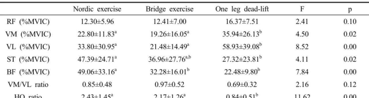
뒤넙다리근 강화 운동 시 각 근육의 활성도 및 동시 수축 비율은 다음과 같다(Table 1). 넙다리곧은근의
활성도는 모든 운동에 유의한 차이가 없었다(p>0.05).
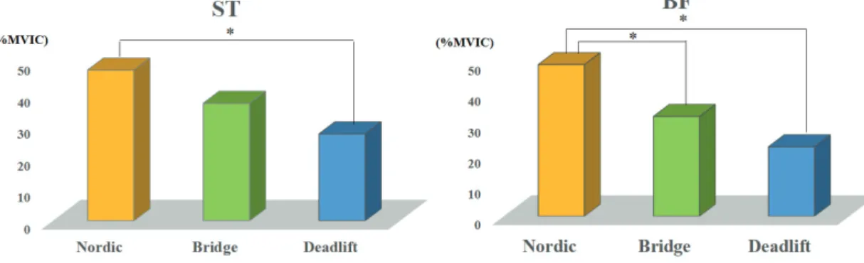
안쪽넓은근의 활성도는 노르딕 운동과 교각 운동이 한다리 데드리프트에 비해 유의하게 높게 나타났다 (Fig. 2)(p<0.05). 가쪽넓은근의 활성도는 노르딕 운동 과 교각 운동이 한다리 데드리프트에 비해 유의하게 높게 나타났다(Fig. 2)(p<0.05). 반힘줄근의 활성도는 노르딕 운동이 한다리 데드리프트에 비해 유의하게 높았다(Fig. 3)(p<0.05). 넙다리두갈래근의 활성도는 노르딕 운동이 교각 운동과 한다리 데드리프트에 비 해 유의하게 높게 나타났다(Fig. 3)(p<0.05). VM/VL 비율은 모든 운동에 유의한 차이는 없었다(p>0.05).
HQ 비율은 노르딕 운동과 교각 운동이 한다리 데드리 프트에 비해 유의하게 높게 나타났다(Fig. 4)(p<0.05).
Nordic exercise Bridge exercise One leg dead-lift F p
RF (%MVIC) 12.30±5.96 12.41±7.00 16.37±7.51 2.41 0.10
VM (%MVIC) 22.80±11.83a 19.26±16.05a 35.94±26.13b 4.50 0.02 VL (%MVIC) 33.80±30.95a 21.48±14.49a 58.93±39.08b 8.52 0.00 ST (%MVIC) 47.39±24.71a 36.96±27.76a,b 27.32±23.81b 4.11 0.02
BF (%MVIC) 49.06±33.16a 32.28±16.01b 22.48±9.80b 7.84 0.00
VM/VL ratio 0.85±0.48 0.97±0.52 0.69±0.32 2.16 0.12
HQ ratio 2.43±1.45a 2.17±1.26a 0.84±0.51b 11.62 0.00
RF: rectus femoris, VM: vastus medialis, VL: vastus lateralis, ST: semitendinosus, BF: biceps femoris,
HQ: hamstrings/quadriceps, the values with different superscripts (a,b) in the same column are significantly different (p<0.05) Table 1. Comparison of activation of quadriceps and hamstring muscles among hamstring strengthening exercises
(n=21)
Fig. 2. Activation of vastus medialis and lateralis among hamstring strengthening exercises.
Fig. 4. HQ ratio among hamstring strengthening exercises.
Ⅳ. 고 찰
본 연구에서 실시한 뒤넙다리근 강화 운동 중 한다 리 데드리프트는 두 운동에 비해 증가된 안쪽넓은근 과 가쪽넓은근의 활성도를 보였다. 한다리 데드리프 트는 스쿼트(squat) 운동에 비해 허벅지 뒤쪽 근육인 뒤넙다리근과 엉덩이근을 강화시킬 수 있는 운동이지 만(Weingroff, 2014), 실제 허벅지 앞쪽에 위치한 넙다 리근의 활동을 촉진시키는데도 사용된다. Begalle 등 (2012)은 한다리 데드리프트 시 넙다리네갈래근 (VM+VL)의 평균 활성도가 65.71%MVIC로 뒤넙다리 근(BF+SM)의 평균 활성도인 24.15%MVIC에 비해 높 게 나타났음을 보고하였다. 그리고 뒤넙다리근의 활 동만을 분석한 선행 연구에서는 한다리 데드리프트가
노르딕 운동과 교각 운동에 비해 감소된 넙다리두갈 래근과 반힘줄근의 활성도를 보였다(Tsaklis et al., 2015). 이러한 결과를 종합하여 볼때, 한다리 데드리프 트는 몸통을 90°로 숙여 체중을 한발로 지탱하면 무릎 관절의 폄을 유지해야 하기 때문에, 두 발로 실시하는 두 운동에 비해 안쪽넓은근과 가쪽넓은근의 활성도가 유의하게 높게 나타난 것으로 생각된다.
더하여 한다리 데드리프트는 두 운동에 비해 높은 안쪽넓은근과 가쪽넓은근의 활성도를 보였지만, 가쪽 넓은근의 활성도가 안쪽넓은근에 비해 상대적으로 높 게 나타나 두 운동에 비해 상대적으로 낮은 VM/VL 비율을 보였다. 가쪽넓은근의 증가된 활성도는 높은 무릎벌림 모멘트(high knee abduction moments)를 발생 시켜 무릎뼈의 가쪽 이동을 초래하고, 이는 PFPS의 주된 원인이 된다(Fagan & Delahunt, 2008). 이러한 손 상 기전으로 인해 PFPS가 있는 대상자는 0.54의 낮은 VM/VL 비율 보인다고 하였다(Powers, 2000). 본 연구 의 교각 운동은 0.97의 VM/VL 비율을 보였으며, PFPS 를 위한 효과적인 운동의 기준 비율인 1에 가까웠다 (Kushion et al., 2012; Souza & Gross, 1991). 이는 본 연구의 짐볼처럼 불안정한 지지면에서 실시한 운동이 관절수용기의 자극과 고유수용성감각에 대한 들신경 (afferent) 정보 입력을 촉진시켜 VM/VL의 비율을 증 가시키는데 효과적임을 보고한 선행 논문의 결과(Kim et al., 2018)를 뒷받침하는 것이다. 반면 한다리 데드리 프트는 PFPS가 있는 대상자에게 적용하기에는 한계 가 있음을 보여주는 결과이다.
Fig. 3. Activation of semitendinosus and biceps femoris among hamstring strengthening exercises.
노르딕 운동은 뒤넙다리근의 편심성 수축을 통해 뒤넙다리근 좌상(hamstring strain)을 예방하는데 효과 적이며(van der Horst et al., 2015), 10주간의 운동 수행 으로 뒤넙다리근의 최대 편심성 토크(torque)를 매우 큰 폭으로 증가시킬 수 있다(Mjølsnes et al., 2004).
Tsaklis 등(2015)도 노르딕 운동 시 넙다리두갈래근과 반힘줄근 모두 약 60%MVIC 이상의 활성도를 보였으 며, 교각 운동(약 40∼50%MVIC)과 데드리프트(약 25∼30%MVIC)에 비해 상대적으로 뒤넙다리근의 활 성도가 높게 나타났음을 보고하였다. 본 연구에서도 넙다리두갈래근과 반힘줄근의 활성도가 노르딕 운동 에서 가장 높게 나타나 선행 연구의 결과들을 지지하 였다. 그리고 이러한 결과는 가장 높은 HQ 비율의 결과로 이어진 것으로 생각된다. 교각 운동 역시 상대 적으로 넙다리네갈래근에 비해 높은 뒤넙다리근의 활 성도로 인해 HQ 비율이 높게 나타난 것으로 보인다.
ACL 손상이 있는 대상자를 위한 운동으로 흔히 스쿼트 운동이 추천된다. 본 연구와 동일한 공식이 적용된 선행 연구의 HQ 비율을 살펴보면, 다양한 스쿼 트 운동 중 0.3이 가장 높은 HQ 비율이었으며 (Nishiwaki et al., 2006), Lynn 등(2012)도 적용한 스쿼트 운동 중 0.47이 가장 높은 HQ 비율이었다. 이는 ACL 손상을 예방하기 위해 추천되는 0.6이상의 HQ비율에 비해 부족한 수준이다. 최근 연구에서는 ACL 손상이 있는 대상자의 평균 HQ 비율은 0.51로 나타났으며 (Myer et al., 2015), 이를 위한 스쿼트 운동 적용 시 무릎의 관절의 각도를 약 42∼72°정도로 실시해야 0.6 이상의 HQ 비율의 기준을 충족할 수 있다고 하였다 (Dedinsky et al., 2017).
이외에도 HQ 비율이 1에 근접할수록 높은 뒤넙다 리근의 활동을 보이며, 이는 무릎관절의 앞쪽으로 전 달되는 전단력을 감소시켜 무릎의 안정성을 높이는데 기여한다고 하였다(Cheung et al., 2012). 본 연구의 한 다리 데드리프트는 0.84의 HQ 비율을 보였으며, ACL 손상 예방을 위해 제시한 0.6∼1.0에 해당하는 HQ 비 율을 보였다(Cheung et al., 2012; Dedinsky et al., 2017;
Wright et al., 2009). 이는 한다리 데드리프트가 ACL
손상 예방을 위한 치료적 운동으로 활용될 수 있음을 보여주는 결과이다. 연구의 결과를 종합하여 볼 때, 적절한 HQ 비율을 보인 한다리 데드리프트는 ACL 손상 예방 운동으로 사용될 수 있으며, PFPS 대상자에 게는 1에 가까운 VM/VL 비율을 보인 교각 운동의 적용을 권장한다. 그리고 노르딕 운동은 넙다리두갈래 근과 반힘줄근의 활동을 촉진시키는데 효과적이었다.
본 연구의 HQ 비율은 전통적인 방법인 동심성 뒤 넙다리근/동심성 넙다리네갈래근(Hcon/Qcon) 비율 을 이용하여 각 운동의 HQ 비율을 산출하였으며, 이 러한 방법은 가장 많이 사용되는 방법이다(Coombs
& Garbutt, 2002). 하지만 움직임을 수행하는 동안 뒤 넙다리근의 편심성 수축과 넙다리네갈래근의 동심성 수축의 기능적인 역할을 고려한 동적 조절 비율 (dynamic control ration, DCR) 혹은 편심성 뒤넙다리근 /동심성 넙다리네갈래근(Hecc/Qcon) 비율도 사용된 다(Coombs & Garbutt, 2002; Croisier et al., 2008). 정상 적인 Hcon/Qcon 비율의 기준은 재활 과정이나 손상 예방에서 0.6의 기준으로 제시되어 있으나(Kannus, 1994), DCR 혹은 Hecc/Qcon 비율은 일반적으로 1.0으 로 제시되며(Coombs & Garbutt, 2002), 기능적인 움직 임에 따라 0.9∼1.3정도의 비율을 보인다(Chan et al., 1996). 그리고 선행 연구를 의하면, DCR 혹은 Hecc/Qcon는 Hcon/Qcon에 비해 일반적으로 높게 나타 난다(Cometti et al., 2001). 국내 연구 중 뒤넙다리근 강화 운동의 HQ 비율을 전통적인 관점에서 분석한 연구도 부족한 실정이기에 본 연구에서는 전통적인 동시 수축을 형태로 실험을 진행하였다. 하지만 3가지 운동 모두 기본적으로 편심성 수축을 통해 뒤넙다리 근의 활동을 촉진할 수 있는 운동이기 때문에 Hecc/
Qcon 비율에 대한 연구도 이루어졌으면 한다.
Ⅴ. 결 론
뒤넙다리근 강화 운동 중 한다리 데드리프트는 안 쪽넓은근과 가쪽넓은근의 활성화를 통해 적절한 HQ
비율을 보여 ACL 손상 예방을 위한 운동으로 사용될 수 있을 것이다. 그리고 뒤넙다리근을 강화하기 위한 방법으로는 노르딕 운동을 추천한다.
References
Abdelraouf OR, Abdel-Aziem AA, Ahmed AA, et al. Backward walking alters vastus medialis oblique/vastus lateralis muscle activity ratio in females with patellofemoral pain syndrome. Turkish Journal of Physical Medicine and Rehabilitation. 2019;65(2):169-176.
Begalle RL, Distefano LJ, Blackburn T, et al. Quadriceps and hamstrings coactivation during common therapeutic exercises. Journal of Athletic Training.
2012;47(4):396-405.
Blønd L, Hansen L. Patellofemoral pain syndrome in athletes:
a 5.7-year retrospective follow-up study of 250 athletes. Acta Orthopaedica Belgica. 1998;64(4):
393-400.
Boling M, Padua D, Marshall S, et al. Gender differences in the incidence and prevalence of patellofemoral pain syndrome. Scandinavian Journal of Medicine and Science in Sports. 2010;20(5):725-730.
Chan KM, Maffulli N, Korkia P, et al. Principles and practice of isokinetics in sports medicine and rehabilitation.
Hong Kong. Williams & Wilkins. 1996.
Cheung RT, Smith AW, Wong del P. H:Q ratios and bilateral leg strength in college field and court sports players.
Journal of Human Kinetics. 2012;33:63-71.
Chmielewski TL, Hurd WJ, Rudolph KS, et al. Perturbation training improves knee kinematics and reduces muscle co-contraction after complete unilateral anterior cruciate ligament rupture. Physical Therapy. 2005;
85(8):740-754.
Cometti G, Maffiuletti NA, Pousson M, et al. Isokinetic strength
and anaerobic power of elite, subelite and amateur French soccer players. International Journal of Sports Medicine. 2001;22(1):45-51.
Coombs R, Garbutt G. Developments in the use of the hamstring/quadriceps ratio for the assessment of muscle balance. Journal of Sports Science and Medicine. 2002;1(3):56-62.
Cowan SM, Bennell KL, Hodges PW, et al. Delayed onset of electromyographic activity of vastus medialis obliquus relative to vastus lateralis in subjects with patellofemoral pain syndrome. Archives of Physical Medicine and Rehabilitation. 2001;82(2):183-189.
Croisier JL, Ganteaume S, Binet J, et al. Strength imbalances and prevention of hamstring injury in professional soccer players: a prospective study. The American Journal of Sports Medicine. 2008;36(8):1469-1475.
Dedinsky R, Baker L, Imbus S, et al. Exercises that facilitate optimal hamstring and quadriceps co-activation to help decrease ACL injury risk in healthy females:
a systematic review of the literature. International Journal of Sports Physical Therapy. 2017;12(1):3-15.
Ebben WP. Hamstring activation during lower body resistance training exercises. International Journal of Sports Physiology and Performance. 2009;4(1):84–96.
Ekstrom RA, Osborn RW, Hauer PL. Surface electromyographic analysis of the low back muscles during rehabilitation exercises. The Journal of Orthopaedic and Sports Physical Therapy. 2008;38(12):736-745.
Fagan V, Delahunt E. Patellofemoral pain syndrome: a review on the associated neuromuscular deficits and current treatment options. British Journal of Sports Medicine.
2008;42(10):789-795.
Ford KR, Myer GD, Schmitt LC, et al. Preferential quadriceps activation in female athletes with incremental increases in landing intensity. Journal of Applied Biomechanics. 2011;27(3):215-222.
Hermens HJ, Freriks B, Disselhorst-Klug C, et al. Development
of recommendations for SEMG sensors and sensor placement procedures. Journal of Electromyography and Kinesiology. 2000;10(5):361-374.
Hislop H, Avers D, Brown M. Daniels and Worthingham’s muscle testing, 9th ed. St. Louis (MS). Saunders.
2013.
Kannus P. Isokinetic evaluation of muscular performance:
implications for muscle testing and rehabilitation.
International Journal of Sports Medicine. 1994;15 Suppl 1:S11-18.
Kim YH, Kim BJ, Park DJ. Isolated activation ratio of the quadriceps femoris muscle on different support surfaces during squat exercise. PNF and Movement.
2018;16(1):125-132.
Letafatkar A, Rajabi R, Minoonejad H, et al. Efficacy of perturbation-enhanced neuromuscular training on hamstring and quadriceps onset time, activation and knee flexion during a tuck-jump task. International Journal of Sports Physical Therapy. 2019;14(2):
214-227.
Liebenson C. Learning the single leg dead lift. Journal of Bodywork and Movement Therapies. 2015;19(4):
732-735.
Lynn SK, Noffal GJ. Lower extremity biomechanics during a regular and counterbalanced squat. Journal of Strength and Conditioning Research. 2012;26(9):
2417-2425.
Mjølsnes R, Arnason A, Østhagen T, et al. A 10-week randomized trial comparing eccentric vs. concentric hamstring strength training in well-trained soccer players. Scandinavian Journal of Medicine and Science in Sports. 2004;14(5):311-317.
More RC, Karras BT, Neiman R, et al. Hamstrings--an anterior cruciate ligament protagonist. An in vitro study. The American Journal of Sports Medicine. 1993;21(2):
231-237.
Mostamand J, Bader DL, Hudson Z. The effect of patellar
taping on EMG activity of vasti muscles during squatting in individuals with patellofemoral pain syndrome. Journal of Sports Sciences. 2011;29(2):
197-205.
Myer GD, Ford KR, Barber Foss KD, et al. The incidence and potential pathomechanics of patellofemoral pain in female athletes. Clinical Biomechanics. 2010;
25(7):700-707.
Myer GD, Ford KR, Di Stasi SL, et al. High knee abduction moments are common risk factors for patellofemoral pain (PFP) and anterior cruciate ligament (ACL) injury in girls: is PFP itself a predictor for subsequent ACL injury? British Journal of Sports Medicine. 2015;
49(2):118-122.
Nishiwaki GA, Urabe Y, Tanaka K. EMG analysis of lower extremity muscles in three different squat exercises.
Journal of the Japanese Physical Therapy Association.
2006;9(1):21-26.
Pattyn E, Verdonk P, Steyaert A, et al. Vastus medialis obliquus atrophy: does it exist in patellofemoral pain syndrome?
The American Journal of Sports Medicine. 2011;39(7):
1450-1455.
Piva SR, Goodnite EA, Childs JD. Strength around the hip and flexibility of soft tissues in individuals with and without patellofemoral pain syndrome. Journal of Orthopaedic and Sports Physical Therapy. 2005;
35(12):793-801.
Powers CM. Patellar kinematics, part I: the influence of vastus muscle activity in subjects with and without patellofemoral pain. Physical Therapy. 2000;80(10):
956-964.
Ryan CG, Rowe PJ. An electromyographical study to investigate the effects of patellar taping on the vastus medialis/vastus lateralis ratio in asymptomatic participants. Physiotherapy Theory and Practice.
2006;22(6):309-315.
Souza DR, Gross MT. Comparison of vastus medialis obliquus:
vastus lateralis muscle integrated electromyographic ratios between healthy subjects and patients with patellofemoral pain. Physical Therapy. 1991;71(4):
310-320.
Tang SF, Chen CK, Hsu R, et al. Vastus medialis obliquus and vastus lateralis activity in open and closed kinetic chain exercises in patients with patellofemoral pain syndrome: an electromyographic study. Archives of Physical Medicine and Rehabilitation. 2001;82(10):
1441-1445.
Tsaklis P, Malliaropoulos N, Mendiguchia J, et al. Muscle and intensity based hamstring exercise classification in elite female track and field athletes: implications for exercise selection during rehabilitation. Open Access Journal of Sports Medicine. 2015;6:209-217.
Van der Horst N, Smits DW, Petersen J, et al. The preventive effect of the nordic hamstring exercise on hamstring injuries in amateur soccer players: a randomized controlled trial. The American Journal of Sports Medicine. 2015;43(6):1316-1323.
Waryasz GR, McDermott AY. Patellofemoral pain syndrome (PFPS): a systematic review of anatomy and potential risk factors. Dynamic Medicine. 2008;7:9.
Weingroff C. Dead lifts. In: Liebenson C. The functional training handbook. Philadelphia. Williams and Wilkins. 2014.
Wright J, Ball N, Wood L. Fatigue, H/Q ratios and muscle coactivation in recreational football players.
Isokinetics and Exercise Science. 2009;17(3):161–
167.