Isolation of mesenchymal stem cells from Pap smear samples
Junghyun Park, PhD 1,* , Daryeon Son, BS 1,* , Wonjun Hong, BS 1 , Jihoon Jang, BS 1 , Geum Joon Cho, MD, PhD 2 , Gwonhwa Song, PhD 1,3 , In Yong Kim, PhD 1,3 , Seungkwon You, PhD 1,3
1
Department of Biotechnology, College of Life Sciences and Biotechnology,
2Department of Obstetrics and Gynecology, College of Medicine,
3Institute of Animal Molecular Biotechnology, Korea University, Seoul, Korea
Objective
Exploiting their ability to differentiate into mesenchymal lineages like cartilage, bone, fat, and muscle, and to elicit paracrine effects, mesenchymal stem cells (MSCs) are widely used in clinical settings to treat tissue injuries and autoimmune disorders. One of accessible sources of MSC is the samples used for Papanicolaou (Pap) test, which is a cervical screening method for detecting potentially pre-cancerous and cancerous alterations in the cervical cells and to diagnose genetic abnormalities in fetuses. This study aimed to identify and isolate the stem cells from Pap smear samples collected from pregnant women, and to trace the origin of these cells to maternal or fetal tissue, and characterize their stem cell properties.
Methods
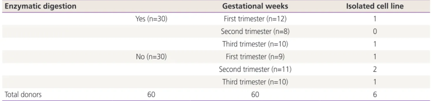
To investigate the possibility and efficiency of establishing MSC lines from the Pap smear samples, we were able to establish 6 cell lines from Pap smear samples from 60 pregnant women at different stages of gestation.
Results
The 3 cell lines randomly selected among the 6 established in this study, displayed high proliferation rates, several characteristics of MSCs, and the capacity to differentiate into adipocytes, osteocytes, and chondrocytes. Our study identified that the stem cell lines obtainable from Pap smear sampling were uterine cervical stromal cells (UCSCs) and had 10% efficiency of establishment.
Conclusion
Despite their low efficiency of establishment, human UCSCs from Pap smear samples can become a simple, safe, low- cost, and donor-specific source of MSCs for stem cell therapy and regenerative medicine.
Keywords: Papanicolaou test; Mesenchymal stem cells; Regenerative medicine
Received: 2020.03.18. Revised: 2020.05.21. Accepted: 2020.05.25.
Corresponding author: Geum Joon Cho, MD, PhD
Department of Obstetrics and Gynecology, College of Medicine, Korea University Guro Hospital, 148 Gurodong-ro, Guro-gu, Seoul 08308, Korea
E-mail: md_cho@hanmail.net https://orcid.org/0000-0001-6761-0944 Gwonhwa Song, PhD
Department of Biotechnology, College of Life Sciences and Biotechnology, 73 Goryeodae-ro, Seongbuk-gu, Seoul 02841, Korea E-mail: ghsong@korea.ac.kr
https://orcid.org/0000-0003-2817-5323
In Yong Kim, PhD
Department of Biotechnology, College of Life Sciences and Biotechnology, 73 Goryeodae-ro, Seongbuk-gu, Seoul 02841, Korea
E-mail: iykim@korea.ac.kr
https://orcid.org/0000-0001-7211-5710 Seungkwon You, PhD
Department of Biotechnology, College of Life Sciences and Biotechnology, 73 Goryeodae-ro, Seongbuk-gu, Seoul 02841, Korea E-mail: bioseung@korea.ac.kr
https://orcid.org/0000-0003-3006-0912
*These authors contributed equally to this work.
Articles published in Obstet Gynecol Sci are open-access, distributed under the terms of the Creative Commons Attribution Non-Commercial License (http://creativecommons.org/
licenses/by-nc/3.0/) which permits unrestricted non-commercial use, distribution, and reproduction in any medium, provided the original work is properly cited.
Copyright © 2020 Korean Society of Obstetrics and Gynecology https://doi.org/10.5468/ogs.20073
pISSN 2287-8572 · eISSN 2287-8580
Introduction
Adult mesenchymal stem cells (MSCs) are defined as undif- ferentiated multipotent cells that are capable of self-renewal, differentiating into several distinct tissue lineages, such as bone, adipose tissue, and cartilage, and in vitro expansion. To date, many types of MSCs, derived from bone marrow (BM), adipose tissue, and amniotic fluid, show tremendous poten- tial to be used for treating conditions ranging from organ failures to immunological diseases [1]. Although the thera- peutic mechanisms of MSCs have not been fully explored, accumulated data demonstrate that these cells hold promise for clinical applications [2]. Various approaches have been tried for MSC-mediated tissue regeneration. As these cells have the potential for multilineage differentiation, and can secrete soluble factors that enhance cell survival and func- tion, they can be used for patient-specific tissue regeneration without ethical restrictions, and are immune privileged. These special properties of MSCs have encouraged the researchers to find the best sources of these cells, preferably using non- invasive procedures. MSCs from various sources share many characteristics and generally meet the accepted criteria for MSCs, like multilineage differentiation potential, self-renewal capacity, expression of specific surface markers, and adher- ence to plastic surfaces. However, each of these cells also exhibit a set of unique and individualistic properties in their differentiation potentials, expression of specific surface mark- ers, cytokine production, gene expression profiles, and ability to establish cell lines in vitro. Moreover, the ease with which biopsies are obtained differs between sources [3].
The Papanicolaou (Pap) smear test is a cervical screening method to detect potentially pre-cancerous and cancerous abnormalities in the cervix. At 1942, Dr. Papanicolaou first discovered a deformed nuclear morphology in benign cells collected by scraping the cervix, indicating their cervical and uterine cancer potentials [4]. In addition, because Pap smear samples from pregnant women contain fetal trophoblastic cells, this test is also used for non-invasive prenatal diag- nosis since the 1950s [5]. Majority of the cells in Pap smear samples are epithelial cells, such as cervical cells from the mother and trophoblasts from the fetus. Because of the wide use and simplicity of the Pap smear test, the isolated human uterine cervical stromal cells (hUCSCs) have been recently proposed as a source of adult MSCs [6]. However, their reli- ability and accessibility for clinical applications are not clear
and their potential as a source of MSC should be further explored by comparing it with well-established MSCs. This study is designed to verify the hUCSCs harvested from Pap smear as a reproducible and expandable source of MSCs for their clinical application. The Pap smear test is non-invasive and has no consequent complications, and thus can be per- formed on a wide population of women. These advantages have greatly facilitated the establishment of MSCs in the cur- rent study. Supported by our previous experience in identify- ing and characterizing potential sources of adult stem cells [7], we isolated and expanded hUCSC lines from Pap smear samples, and identified, assessed, and compared their MSC properties in terms of self-renewal and multilineage differen- tiation potentials compared with MSCs from amniotic-fluid.
Importantly, although the Pap smear test was originally de- veloped to collect cervical epithelial cells for cancer screening in women, it has been reported that the samples from preg- nant women also contain fetus-derived cells [8]. We there- fore investigated whether the isolated cell population contain fetus-derived trophoblast stem cells, using human leukocyte antigen (HLA)-G, from a family of non-classical HLA class I molecules, typically expressed in embryos, embryonic stem cells, and trophoblasts [9].
Materials and methods
1. Isolation and culture of human uterine cervical stromal cells and amniotic fluid-derived mesenchymal stem cells
Pap smear samples were collected with informed consent, from 60 pregnant women at random, regardless of the length of their pregnancy. The collection procedure was ap- proved by the Institutional Review Board of Korea University (KUGH16060-001). These samples were washed twice with phosphate-buffered saline (PBS) containing 100 U penicillin/
streptomycin and centrifuged at 500 g for 10 minutes, and digested with 0.25% Trypsin/ethylenediaminetetraacetic acid (EDTA) (Hyclone, Waltham, MA, USA) for 30 minutes at 37°C. These were washed and centrifuged before the pel- let was seeded into a 100 mm plate and incubated for 72 hours in low-glucose Dulbecco’s modified Eagle’s medium (DMEM) (Invitrogen, Carlsbad, CA, USA) containing 10% fe- tal bovine serum (FBS), 100 U penicillin/streptomycin, and 1%
L-glutamine at 37°C and 5% CO
2. For expansion, adherent
cells obtained from Pap smear samples, and amniotic fluid- derived mesenchymal stem cells (AF-MSCs) were cultured in low-glucose DMEM containing 10% FBS, 100 U penicil- lin/streptomycin, 1% L-glutamine, 4 ng/mL basic fibroblast growth factor (R&D Systems, Minneapolis, MN, USA), and 50 μg/mL ascorbic acid (expansion medium). AF-MSCs were isolated and cultured in strict adherence to the guidelines issued by the Institutional Review Board of Korea University.
These cells were previously confirmed to possess character- istics of MSCs, based on their differentiation, proliferation, and immunological phenotypes [7].
2. Proliferation assay
To determine the growth rates of hUCSCs and AF-MSCs, cells were seeded in 12-well plates at a density of 3×10
4cells/well in expansion medium, cultured for 3 days, stained with 0.01% crystal violet solution, and de-stained with 10%
acetic acid. Finally, absorbance at 600 nm was spectrophoto- metrically determined.
3. Adipogenic differentiation
Adipogenic differentiation was induced using a previously described protocol [7]. Briefly, cells were seeded in 6-well plates at a density of 3×10
4cells/well, and cultured until they reached 100% confluency. Thereafter, cells were grown for 7 days in high-glucose DMEM (Invitrogen) supplemented with 1 mM dexamethasone (Sigma-Aldrich, St. Louis, MO, USA), 0.5 mM 3-isobutyl-1-methyl-xanthine (Sigma-Aldrich), 10 ng/mL recombinant human insulin (Sigma-Aldrich), 100 mM indomethacin (Sigma-Aldrich), and 10% FBS, to induce/
maintain differentiation. After this, the cells were fixed in 10% formalin (Sigma-Aldrich) and stained with 2% (w/v) Oil Red O (Sigma-Aldrich) for 20 minutes at room temperature to detect oil droplets in the cytoplasm.
4. Osteogenic differentiation
Osteogenic differentiation was induced according to a pre- viously described protocol [7]. Briefly, cells were seeded in 6-well plates at a density of 3×10
4cells/well, cultured in low- glucose DMEM (Gibco/Invitrogen) containing 10% FBS until they reached 100% confluency, and fed twice a week with osteogenic induction medium (high-glucose DMEM [Invitro- gen] supplemented with 100 nM dexamethasone, 10 mM β-glycerophosphate, 0.2 mM ascorbate, and 10% FBS). Os- teogenic differentiation was assessed by von Kossa staining.
5. Chondrogenic differentiation
To induce chondrogenic differentiation, cells were detached, transferred to a 15 mL polypropylene tube, pelleted via centrifugation at 200 g for 5 minutes, and cultured in high- glucose DMEM supplemented with 0.1 M dexamethasone, 50 µg/mL ascorbic acid (Sigma-Aldrich), 100 µg/mL sodium pyruvate (Sigma-Aldrich), 40 µg/mL proline (Sigma-Aldrich) 10 ng/mL transforming growth factor-1 (R&D Systems), 50 mg/mL ITS premix (Gibco/Invitrogen), 6.25 µg/mL insulin, 6.25 µg/mL transferrin (Sigma-Aldrich), 6.25 ng/mL seleni- ous acid (Sigma-Aldrich), 1.25 mg/mL bovine serum albumin (BSA; Sigma-Aldrich), and 5.35 mg/mL linoleic acid (Sigma- Aldrich). After 4 weeks of culture, cells were fixed in 4%
paraformaldehyde and stained with Alcian Blue (Sigma- Aldrich).
6. Reverse transcription polymerase chain reaction RNA was isolated and purified using TRIzol (Invitrogen) ac- cording to the manufacturer’s instructions. cDNA was syn- thesized using Reverse Transcriptase II (Invitrogen). To amplify target genes, 25 ng cDNA was mixed with forward and re- verse primers (Bioneer, Daejeon, Korea). The PCR conditions were as follows: 24–30 cycles of denaturation at 99°C for 30 seconds, annealing at 62°C for 30 seconds, and exten- sion at 72°C for 30 seconds, followed by a final amplifica- tion step at 72°C for 10 minutes. The levels of target genes amplified by 30 cycles of PCR were within the linear range.
Primer sequences are listed in Supplementary Table 1.
7. Fluorescence-activated cell sorting analysis
hUCSCs and AF-MSCs were trypsinized and transferred
to fluorescence-activated cell sorting (FACS) tubes (BD
Biosciences Clontech, Palo Alto, CA, USA) at a density of
1×10
6cells/tube. Cells were rinsed twice with cold Dulbec-
co’s PBS containing 1% BSA (pH 7.4), and incubated with a
primary antibody against CD29, CD31, CD34, CD44, CD45,
CD73, CD90, or CD120a (BD Biosciences) or against human
leukocyte antigen-G (HLA-G; Santa Cruz Biotechnology, Dal-
las, TX, USA) for 1 hour at 4°C. Thereafter, cells were washed
twice with PBS containing 1% BSA, resuspended in 100 μL
PBS containing 1% BSA and a fluorescein isothiocyanate-
labeled secondary antibody diluted 1:100, and incubated for
40 minutes at 4°C. Finally, cells were washed twice with PBS
containing 1% BSA and fixed in 4% paraformaldehyde for
FACS analysis. To identify nonspecific signals, control cells
were incubated with isotype-matched immunoglobulins.
8. Colony-forming unit assay
hUCSCs and AF-MSCs were seeded in 6-well plates at a density of 100 cells/well, cultured for 14 days, washed twice with PBS, and fixed in 10% formalin for 20 minutes at room temperature. To visualize the colonies, cells were stained with 0.01% crystal violet solution for 20 minutes at room temper- ature, washed with deionized water, and air-dried. Colonies were typically 5–8 mm in diameter, and were scored macro- scopically.
9. Karyotype analysis
Karyotyping was performed by the Cytogenomic Services Facility of Samkwang Medical Laboratories. hUCSCs were cultured in expansion medium as described above. Cell were treated with 0.05 μg/mL colcemid (Gibco/Invitrogen) for 1–2 hour to block the dividing cells at metaphase. Chromosomes were visualized by G-banding. At least 100 metaphase cells were analyzed, and a minimum of ten cells were karyotyped per line.
10. Immunofluorescence staining
To detect HLA-G, hUCSCs and AF-MSCs were incubated for 1 hour at 4°C with anti-HLA-G antibody (sc-21799; Santa Cruz Biotechnology) diluted in PBS containing 1% BSA, washed twice with PBS containing 1% BSA, and incubated for 40 minutes at 4°C with a fluorescein isothiocyanate- labeled secondary antibody diluted 1:100 in PBS containing 1% BSA. Nuclei were stained with DAPI diluted 1:1,000 in PBS containing 1% BSA.
11. Statistical analysis
All values are expressed as means±standard deviation. Data were compared using 1- or 2-way analysis of variances with post hoc Tukey’s test and paired 2-tailed Student’s t-tests.
Results
1. Isolation and characterization of the cell population from Papanicolaou smear samples We attempted to establish fetus-derived trophoblast cell lines by collecting the Pap smear sample from pregnant women.
The cell samples were collected by centrifugation and seed-
ed into cell culture dishes (Fig. 1A). Clusters of fibroblast- like cells with a homogenous morphology were observed in randomly selected areas after incubation for 72 hours (Fig. 1B). These adherent cells had a high proliferative poten- tial in serum-containing medium. Isolated cells were stably sub-cultured for at least 12 passages (>40 days) (Fig. 1C).
These cells became elongated and spindle-shaped upon re- peated passages (Fig. 1B). Expression of HLA-G was detected by immunofluorescence staining in AF-MSCs, but not in our cell lines (Fig. 1D). This observation was supported by the FACS analysis, in which HLA-G was detected in AF-MSCs, and negatively in the cell lines isolated by us (Fig. 1D). Fur- thermore, the G-banding karyotype analysis showed normal female chromosomes (Fig. 1E). These findings indicate that the cells from Pap smear samples were derived from the cervix, which is in agreement with Eiró et al. [6]; trophoblast stem cells derived from the fetus could not be established in the current study.
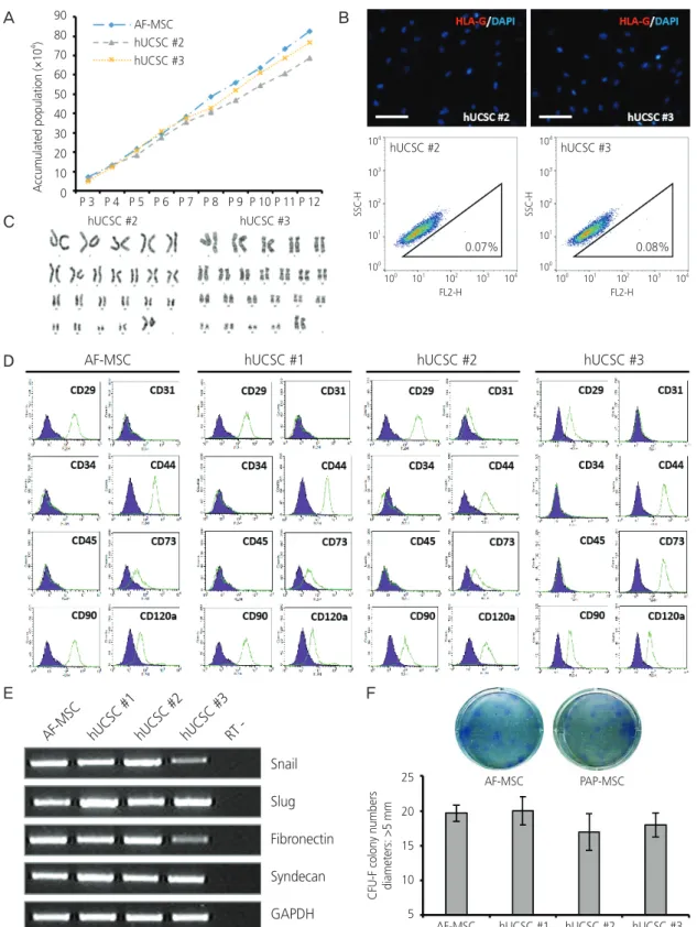
2. Characterization of human uterine cervical stromal cell lines
We focused on establishment of hUCSC lines from cells de- rived from Pap smears of pregnant women. Of the 60 sam- ples, only 6 hUCSC lines, including the one (Fig. 1, hUCSC
#1) assessed earlier, were established. Similar to hUCSC #1, the two other hUCSC lines (#2, #3) showed high prolifera- tion in serum-containing medium and could be stably sub- cultured for at least 12 passages (>40 days) (Fig. 2A). HLA- G expression profiles of hUCSC #2 and #3 were similar to those of hUCSC #1 as observed in the immunofluorescence and FACS analysis (Fig. 2B). Karyotype of hUCSC #2 and
#3 using G-banding showed normal female chromosomes
(Fig. 2C). The immunophenotype of hUCSCs was analyzed
by flow cytometry and it showed that AF-MSCs and hUCSC
lines were negative for CD31, CD34, and CD45, indicating
that they are not endothelial or hematopoietic in origin, but
were positive for the MSC-specific markers CD29, CD44,
CD73, CD90, and CD120a (Fig. 2D). In addition, both these
MSCs abundantly expressed mesenchymal markers (Snail
and Slug) and markers of extracellular matrix (fibronectin and
syndecan) (Fig. 2E). The clonogenic and proliferation capaci-
ties of AF-MSCs and hUCSCs were compared by the colony-
forming unit (CFU)-F assay. Colonies with a diameter larger
than 5 mm were counted. The number of colonies formed
did not significantly differ between hUCSCs and AF-MSCs or
between the hUCSC lines (Fig. 2F).
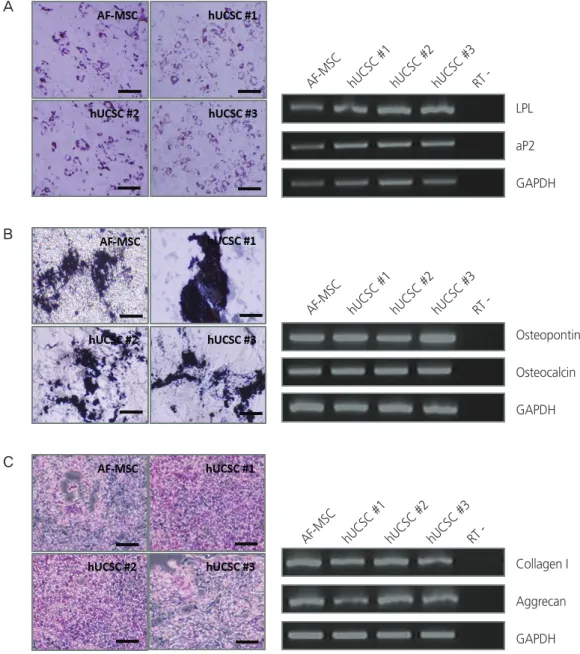
3. Adipogenic, osteogenic, and chondrogenic differentiation of human uterine cervical stromal cells in vitro
For successful and efficient tissue regeneration using MSCs, their multilineage differentiation capacity must be preserved upon in vitro expansion. To investigate the adipogenic and osteogenic differentiation potentials of hUCSCs, cells at pas- sage 4–6 were seeded at a density of 3×10
3cells/cm
2and
cultured in 10% FBS. At confluency, adipogenic and osteo- genic differentiation were induced. Expected morphological changes and formation of neutral lipid droplets, as detected by Oil Red O, were observed at 1.5 weeks after induction of adipogenic differentiation (Fig. 3A). Reverse transcription polymerase chain reaction (RT-PCR) analysis demonstrated that adipocyte markers, LPL and aP2, were highly expressed by these cells upon induction of adipogenic differentiation (Fig. 3A). On osteogenic differentiation, von Kossa staining exhibited dark brown or black labeling, indicating mineraliza-
Isolated cell (hUCSC #1)
0.04%
SSC-H
104
103 102
101
100
100 101 102 103 104 FL2-H
Fig. 1. Isolation of fibroblast-like cells from Papanicolaou (Pap) smear samples. (A) Method used to isolate cells from Pap smear samples.
(B) Morphology of isolated cell line at passage 0 and 5 (scale bar=500 µm). (C) Accumulated population comparison with isolated cell line and amniotic fluid-derived mesenchymal stem cell (AF-MSC) upon passaging. (D) Immunofluorescence staining and fluorescence-activated cell sorting (FACS) analysis of human leukocyte antigen (HLA)-G in AF-MSCs and isolated cells (scale bar=200 μm). (E) Karyotype of iso- lated cell line determined by G-banding.
Pap smear sample
Centrifugation and washing
Pellet
seeding 72 h culture
Accumulated population (×10
4) 90 80 70 60 50 40 30 20 10 0
AF-MSC Isolated cell
P 3 P 4 P 5 P 6 P 7 P 8 P 9 P 10 P 11 P 12
1 2 3 4 5
6 7 8 9 10 11 12
13 14 15 16 17 18
19 20 21 22 X Y
A
C
E
B
D
AF-MSC
99.91%
SSC-H
104
103 102
101
100
100 101 102 103 104 FL2-H
Fig. 2. Characterization of human uterine cervical stromal cell (hUCSC) lines. (A) Accumulated population of hUCSCs and amniotic fluid- derived mesenchymal stem cell (AF-MSC) upon passaging. (B) Immunofluorescence staining of human leukocyte antigen (HLA)-G in hUC- SC line #2 and #3 (scale bar=200 μm). Fluorescence-activated cell sorting (FACS) analysis of HLA-G expression in hUCSC line #2 and #3.
The percentages of HLA-G-positive cells are shown. (C) Karyotype of hUCSC line #2 and #3 determined by G-banding. (D) FACS analysis of the immunophenotypes of hUCSC lines (green). The isotype control is shown in purple. (E) mRNA expression of human MSC markers in hUCSC lines. (F) Colony-forming unit (CFU) assay investigating the self-renewal capacity of hUCSC lines.
Accumulated population (×10
4) 90 80 70 60 50 40 30 20 10 0
AF-MSC hUCSC #2 hUCSC #3
hUCSC #2
hUCSC #2
0.07% 0.08%
hUCSC #3
hUCSC #3
P 3 P 4 P 5 P 6 P 7 P 8 P 9 P 10 P 11 P 12
A
C
B
AF-MSC
AF-MSC
hUCSC #1
hUCSC #1
hUCSC #2
hUCSC #2
hUCSC #3
hUCSC #3 RT -
CFU-F colony numbers diameters: >5 mm 25 20 15 10 5
PAP-MSC AF-MSC
AF-MSC hUCSC #1 hUCSC #2 hUCSC #3 Snail
Slug Fibronectin Syndecan GAPDH D
E F
SSC-H SSC-H
104 103 102 101 100
104 103 102 101 100 100 101 102 103 104
FL2-H
100 101 102 103 104 FL2-H