Korean J Radiol 3(2), June 2002 133
Radiologic Findings of Multiple Myeloma with Gastric Involvement:
A Case Report
We report a case of multiple myeloma with gastric involvement occurring in a patient who underwent an upper gastrointestinal series (UGIS), CT and MRI.
UGIS depicted a luminal protruding mass, while contrast-enhanced CT demon- strated marked thickening of the gastric wall, with subtle contrast enhancement.
At T1- and T2-weighted MR imaging, the mass showed iso- and intermediate sig- nal intensity, respectively. After the administration of contrast material, subtle homogeneous enhancement was apparent.
n multiple myeloma, involvement of the gastrointestinal tract (gastric myeloma) is rare. Although more than 100 cases of gastric myeloma have been reported in the literature, only the UGIS and CT findings have, to our knowledge, been described (1 3). In this article, we report the findings in a case of gastric myeloma in which the patient underwent not only UGIS and CT, but also MR imaging studies.
CASE REPORT
A 61-year-old man presented with epigastric pain and weight loss. Two years previ- ously, a mass involving the left thigh had been diagnosed after wide excision as multi- ple myeloma and the patient had undergone chemotherapy. After 6 months, the disap- pearance of serumal monoclonal protein and a free light chain in the urine indicated complete remission. UGIS demonstrated a luminal protruding mass in the anterior wall of the gastric cardia and along the greater curvature of the body (Fig. 1A), while con- trast-enhanced CT revealed marked thickening of the gastric wall, with subtle contrast enhancement (Fig. 1B). There was no evidence of perigastric infiltration or lym- phadenopathy. MR imaging depicted a well-defined, homogeneous mass with promi- nent gastric wall thickening, but no necrotic portion. As demonstrated by T1-weighted imaging sequences, the signal intensity of the mass was less than that of the liver, though T2-weighting showed it as slightly higher, and homogeneous (Fig.1C). After the administration of contrast material, the observed homogeneous enhancement was similar to that of hepatic parenchyma (Fig. 1D).
Fiberoptic gastroscopy depicted a large, protruding, luminal mass and multiple biop- sies were performed. Histopathologic examination revealed dense and monotonous in- filtration by both mature and immature plasma cells (Fig. 1E), which at immunohisto- chemistry stained positive for IgG light chains.
Hyo-Sung Kwak, MD Gong-Yong Jin, MD Jeong-Min Lee, MD
Index terms : Stomach, neoplasms Stomach, radiography Stomach, magnetic resonance
imaging Stomach, CT
Korean J Radiol 2002;3:133-135 Received January 13, 2002; accepted after revision April 10, 2002.
All authors: Department of Diagnostic Radiology, Chonbuk National University Hospital
Address reprint requests to : Jeong-Min Lee, M.D., Department of Diagnostic Radiology, Chonbuk National University Hospital, 634-18 Keumam- dong, Chonju-shi, Chonbuk 561-712, South Korea.
Telephone: (8263) 250-1172 Fax: (8263) 272-0481 e-mail: [email protected]
I
DISCUSSION
The extraskeletal spread of multiple myeloma occurs more frequently than is currently recognized, but clinical
involvement of the gut is rarely reported. Where it does occur, the small bowel is most commonly involved, fol- lowed by the stomach, colon, and esophagus. Patients with gastric myeloma have presented with nonspecific gastroin- testinal symptoms such as epigastric pain, weight loss, and Kwak et al.
134 Korean J Radiol 3(2), June 2002
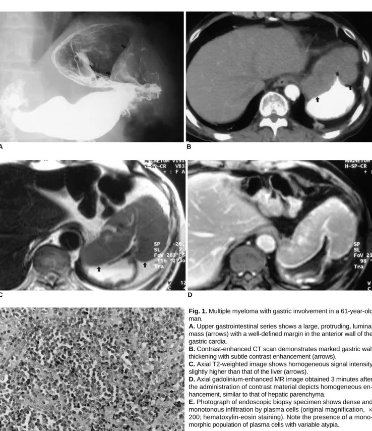
A B
C D
Fig. 1. Multiple myeloma with gastric involvement in a 61-year-old man.
A. Upper gastrointestinal series shows a large, protruding, luminal mass (arrows) with a well-defined margin in the anterior wall of the gastric cardia.
B. Contrast-enhanced CT scan demonstrates marked gastric wall thickening with subtle contrast enhancement (arrows).
C. Axial T2-weighted image shows homogeneous signal intensity slightly higher than that of the liver (arrows).
D. Axial gadolinium-enhanced MR image obtained 3 minutes after the administration of contrast material depicts homogeneous en- hancement, similar to that of hepatic parenchyma.
E. Photograph of endoscopic biopsy specimen shows dense and monotonous infiltration by plasma cells (original magnification, 200; hematoxylin-eosin staining). Note the presence of a mono- morphic population of plasma cells with variable atypia.
E
upper gastrointestinal bleeding (1 4). Pathologically, gastric myeloma is thought to originate from lymphoid follicles in the submucosa, or from plasma cells in the submucosa or mucosal lamina propria (5). Early gastric myeloma is con- fined to the mucosa or submucosa of the gastric wall. The most advanced form of gastric myeloma infiltrates the deep muscular layer or serosa, and to avoid misdiagnosis, deep endoscopic biopsy may be required (3).
Gastric myeloma can manifest in various forms: nodular, infiltrative, ulcerative, and polypoid (5), among which the nodular type is most common. In our case the infiltrative form occurred, manifesting as diffuse thickening of the an- terior wall of the gastric cardia and along the greater cur- vature of the body.
The radiologic findings of gastric myeloma have been re- ported in the literature (2, 3). UGIS has indicated the pres- ence of infiltrative and polypoid mass lesions, though to differentiate on the basis of those findings between gastric myeloma and a gastric stromal tumor, lymphoma, neu- roendocrine tumor, or adenocarcinoma is difficult. Yoon et al. (3) describe the CT findings in two patients with gastric myeloma: in both, homogeneous gastric wall thickening was apparent, and contrast enhancement was poor and similar in degree to that of paraspinal muscle. T1-weighted MR imaging sequences indicated that the signal intensity of the mass was less than that of the liver, while T2 weighting showed it as slightly higher. The administration of contrast material revealed no necrotic portion, and the homoge- neous enhancement observed was similar to that of hepatic
parenchyma. Although these findings simulate those of lymphoma, neuroendocrine tumor and gastric stromal tu- mor (6), differentiation is based on the fact that in lym- phoma the nodes are usually bulkier, while a neuroen- docrine or stromal tumor usually has a necrotic portion.
In conclusion, gastric myeloma is an uncommon entity that may mimic other malignant gastric tumors and tends to present as a homogeneous solid mass without a necrotic portion. Correct diagnosis depends upon a correctly inter- preted biopsy combined with the immunohistochemical study of immunoglobin.
References
1. Kinoshita Y, Watanabe M, Takahashi H, et al. A case of gastric plasmacytoma: genetic analysis and immunofixation elec- trophoresis. Am J Gastroenterol 1991;86:349-353
2. Spagnoli I, Gattoni F, Mazzoni R, Uslenghi C. Primary gastroin- testinal plasmacytoma: report of three cases. Diagn Imaging 1983;52:23-27
3. Yoon SE, Ha HK, Lee YS, et al. Upper gastrointestinal series and CT findings of primary gastric plasmacytoma: report of two cases. AJR 1999;173:1266-1268
4. Pimental RR, Van Stolk R. Gastric plasmacytoma: a rare cause of massive gastrointestinal bleeding. Am J Gastroenterol 1994;89:642-643
5. Remingio PA, Klaum A. Extramedullary plasmacytoma of the stomach. Cancer 1971;27:562-568
6. Marcos HB, Semelka RC. Stomach diseases: MR evaluation us- ing combined T2-weighted single-shot echo train spin-echo and gadolinium-enhanced spoiled gradient-echo sequences. J Magn Reson Imaging 1999;10:950-960
Multiple Myeloma with Gastric Involvement
Korean J Radiol 3(2), June 2002 135