Kor. J. Pharmacogn.
45(2) : 147∼ 153 (2014)
147
머위 분획물의 In Vitro 라디칼 소거능 및 신경세포의 산화적 스트레스 보호 효과
왕 천1·이아영1·최지명1·이동구2·김현영3·이상현2*·조은주1*
1부산대학교 식품영양학과 및 김치연구소, 2중앙대학교 식물시스템과학과, 3경남과학기술대학교 식품과학부
In Vitro Radical Scavenging Effect and Neuroprotective Activity from Oxidative Stress of Petasites japonicus
Qian Wang1, Ah Young Lee1, Ji Myung Choi1, Dong Gu Lee2, Hyun Young Kim3, Sanghyun Lee2* and Eun Ju Cho1*
1Department of Food Science and Nutrition and Kimchi Research Institute, Pusan National University, Busan 609-735, Korea
2Department of Integrative Plant Science, Chung-Ang University, Anseong 456-756, Korea
3Department of Food Science, Gyeongnam National University of Science and Technology, Jinju 660-758, Korea
Abstract − This study was focused on the evaluation of radical scavenging effect and the protective activity against oxidative stress of the extract and fractions from Petasites japonicus. P. japonicus was extracted with methanol and then fractionated into 4 fractions [n-butanol, ethyl acetate (EtOAc), methylene chloride, and n-hexane]. The extract and fractions showed strong 1,1- diphenyl-2-picrylhydrazyl radical scavenging activity. Among all the fractions, particularly, the EtOAc fraction showed the strongest effect with the IC50 value of 0.02µg/ml. In addition, the fractions also showed strong hydroxyl radical scavenging activity and nitric oxide scavenging activity as well. Furthermore, cell viability generated by the P. japonicus extract and 4 frac- tions were examined under C6 glial cellular model. The C6 glial cells showed high generation of reactive oxygen species (ROS) and decrease in cell viability by the treatment generator of hydrogen peroxide. However, the production of ROS formation was decreased by the treatment of the fractions of P. japonicus and also founded that the EtOAc fraction led to significant increase in the cell viability at concentration 100µg/ml. Results from this work indicated that P. japonicus showed protective effects against oxidative stress and its EtOAc fraction may be served as a useful natural antioxidant.
Key words − Petasites japonicus, C6 glial cells, Reactive oxygen species, Hydrogen peroxide, Oxidative stress
인간을 비롯한 모든 생물체들은 생체 내 생화학 반응 및 환경적 인자에 의해 superoxide radical(O2-), hydroxyl radical (·OH), peroxyl radical, hydrogen peroxide(H2O2)등과 같은 활성산소종(reactive oxygen species, ROS)이 생성되며,이들 은 산화력이 매우 강하기 때문에 인체 내에서 제거되지 못 하면 산화적 스트레스(oxidative stress)를 유발하게 된다.1-2) 생체 내 이들의 생성이 증가된 상태인 산화적 스트레스에 의해 세포 내 구성성분인 단백질 및 지질성분 등이 손상되
고과량의 활성산소나 지속적인 활성산소 생성으로 항산화
방어계의 균형이 깨지게 되면 조직을 손상시켜 암, 당뇨병, 동맥경화, 심장질환 및 알츠하이머와 같은 다양한 만성 질
환을 유발한다.3-5) 특히 뇌 신경세포에 있어 활성 산소에 의 한 산화적 손상은 신경퇴행성 질환의 유발기전과 밀접한 관 련이 있는 것으로 제시되었다.6)이로 인하여 생체 내 유리 라디칼의 생성을 억제하는 것이 질병예방을 위한 중요한 과 제로 대두되고 있으며 이에 관련된 연구도 다양하게 수행 되고 있다.7)
머위(Petasites japonicus S. et. Z. Max.)는 한국, 일본, 중 국 등 야산의 습지에서 자생하는 국화과(Compositae) 머위 속(Senecio)에 속하는 다년생 근경식물로, 머위의 떫은 맛과 정유성분의 향기 및 독특한 질감은 사람들의 기호에 적합 하여 쌈과 생채 및 나물로 이용하거나 말려서 탕의 재료로 사용 한다.8) 또한 머위는 민간에서 진해, 거담, 건위의 효과 가 있다고 알려져 있고 진정, 소종, 이뇨, 풍한 등의 치료에 오랫동안 사용되어 왔다.9) 그러나 머위의 생리활성에 관한
*교신저자(E-mail):[email protected], [email protected] (Tel): +82-31-670-4688, +82-51-510-2837
과학적 연구는 아직 미흡한 실정이다.
머위의 주요 성분으로는 머위 잎으로부터 분리된fukinolic acid10) 및 petaslignolide A11) 등이 있으며, 뿌리와 줄기에서 분리된 sesquiterpenoid인 eremopetasidioner과 phenolic compound인 petasiphenone 및 eremophilenolide 등이 분리 되었다는 보고가 있다.12-14) 이전 연구에서 머위 에탄올 추 출물이 알코올 투여로 손상된 간 조직을 보호하는 항산화 효과를 나타낼 뿐만 아니라 머위를 첨가한 식이를 먹인 mice 에서 체내 산화적 방어 능력을 높이는 것을 확인할 수 있었
다.15-16) 또한, 머위 추출물이 위암 및 간암 세포의 생육을
억제시켰으며 각질 형성세포에서 염증성 사이토카인 interleukin-1α와 prostaglandin E2의 생성 억제 효과 등이 보 고 되어 있다.17-18) 그러나 free radical에 의해 유도 되는 산 화적 스트레스 상태에서 머위의 신경 세포 보호 효과에 대 한 연구는 행해져 있지 않다. 따라서 본 연구에서는 머위의 추출물과 분획물을 이용하여 free radical에 의한 산화적 스 트레스 개선효과를 in vitro 라디칼 소거능과 C6 glial cell을 이용한 신경 세포 보호 효과를 검토해 보았다.
재료 및 방법
머위의 추출 및 분획물 조제 − 실험재료인 머위(P.
japonicus S. et. Z. Max.)는 국립수목원(2009년 울릉도 채 집)으로 제공 받았으며, 중앙대학교 안영희교수님으로부터 감정을 받은 후 사용하였다. 머위를 음건한 후 methanol (MeOH)로 추출하여 추출물을 조제하였다. MeOH 추출물 은 원형 플라스크에 놓고 MeOH을 채워서 환류 냉각장치 가 부착된 추출장치를 이용하여 3회에 걸쳐 추출하였다. 추 출 후 얻은 머위 MeOH 추출물은 극성별 유기용매인 n- butanol(BuOH), ethyl acetate(EtOAc), methylene chloride (MC), n-hexane(Hx)로 다시 표준 분획물을 조제하였다.
1,1-Diphenyl-2-Picrylhydrazyl(DPPH) 소거능 측정 − Ethanol에 녹인 각 농도 별 시료(5, 10, 50, 100 µg/ml) 100µl와 60 µM DPPH 용액 100 µL를 96-well plate에 혼합 하여 실온에서 30분간 방치시킨 후, 540 nm에서 흡광도를 측정하였다. 시료를 첨가하지 않은 대조군과 비교하여 free radical 소거효과를 IC50(DPPH radical 생성을 50%까지 억 제하는데 필요로 하는 시료 농도)로 나타내었다.19)
Hydroxyl Radical(·OH) 소거능 측정 − Fenton 반응에 따 라 10 mM FeSO4·7H2O-EDTA에 10 mM의 2-deoxyribose solution과 농도 별 시료 용액을 혼합한 다음 10 mM의 H2O2 를 첨가하여 37oC에서 4시간 동안 배양하였다. 이 혼합액에 2.8% trichloroacetic acid(TCA)와 1.0% thiobarbituric acid (TBA) solution을 각각 첨가하여 20분간 끓여 식힌 후 490 nm에서 흡광도를 측정하였다.20)
Nitric Oxide(NO) 소거 효과 측정 − NO 소거능은 Marcocci
등의 방법으로 측정하였다. 5 mM sodium nitroprusside(SNP) solution에 50 mM phosphate buffer(pH 7.4)에 녹인 각 농도 별 시료를 넣고 25oC에서 150분간 반응시켰다. 이 반응 혼 합액은 96 well plate에 주입하여 0.1%의 naphthyl ethy- lenediamine dihydrochlorid와 1%의 sulfanilamide를 함유한 5%의 phosphoric acid를 동량 섞은 griess reagent를 넣어 실 온에서 30분간 반응시킨 후 540 nm에서 흡광도를 측정하여 NO 저해활성을 확인하였다. 저해활성은 시료용액의 첨가구 와 무첨가구의 흡광도 감소율로 나타내었다.21)
세포 종류 및 시약 − C6 glial cell은 한국 세포주 은행 (KCLB, Seoul, Korea)에서 구입하고 배양을 위한 Dulbecco's modified eagle medium(DMEM)배지, fetal bovine serum(FBS)과 100 units/ml penicillin streptomycin, trypsin EDTA 용액은 Welgene(Daegu, Korea)에서 구입하여 사용하였다. 산화적 스트레스를 유도시키기 위해 사용한 H2O2는 Junsei(Junsei Chemical Co., Tokyo, Japan)회사에서 구입하여 사용하였다. 세포 생존율을 측정하기 위해 사용한 3-(4,5-dimethylthiazol-2-yl)-2,5-diphenyl tetrazolium bromide (MTT)는 BIO BASIC(BIO BASIC CANADA INC.)에서, dimethyl sulfoxide(DMSO)는 BIO PURE(Ontario, Canada) 에서 구입하여 사용하였다.
세포 배양 − C6 glial cell은 100 units/ml의 penicillin streptomycin과 10%의 FBS가 함유된 DMEM을 이용하여 37oC, 5% CO2 incubator에서 배양하였다. 배양된 세포는 1~2일에 한 번 배양액을 바꾸어 주면서 세포분화가 최대에 도달하였을 때 phosphate buffered salin(PBS)로 세포를 세 척한 후 0.05% trypsin과 0.02% EDTA 혼합액으로 부착된 세포를 분리하여 원심 분리해서 집적된 세포를 피펫으로 세 포가 골고루 분산되도록 잘 혼합하여 계대 배양하면서 실 험에 사용하였다.
Reactive Oxygen Species(ROS) 측정 − 세포가 con- fluence 상태가 되면 black 96 well plate에 5×10⁴ cells/ml로 seeding하여 2시간 동안 37oC에서 배양한 후 세포가 잘 부 착되면 산화적 스트레스를 유발하기 위하여 H2O2(250µM) 와 시료를 농도 별로(10, 50, 100 µg/ml) 처리하여 24시간 배양한 뒤 80 µM의 DCF-DA용액을 각 well에 주입하여 37oC에서 30분 동안 재배양한 후 FLUOstar OPTIMA(BMG lavtech, Ortenberg, Germany)에서 excitation-480 nm, emission-535 nm로 측정하였다.22)
Cell Viability 측정 − 세포가 confluence 상태가 되면 24 well plate에 well당 1×10⁵ cells/ml로 seeding하여 2시간 37oC에서 배양한 후 세포가 잘 부착되면 산화적 스트레스 를 유발하기 위하여 H2O2(500µM)를 첨가하여 6시간 배양 하였다. 산화적 스트레스 유발한 후, 머위 시료를 농도 별 로(5, 10, 50, 100 µg/ml) 처리하여 18시간 배양한 뒤 5 mg/
ml의 MTT solution을 각 well에 주입하였다. 37oC에서 4시
간 동안 재배양한 후 생성된 formazan 결정을 DMSO에 녹 여 540 nm에서 흡광도를 측정하였다.23)
통계분석 − 대조군과 각 시료들로부터 얻은 실험 결과들 은 평균 ± 표준편차로 나타내었고, SAS 4.2를 이용하여 각 실험 결과로부터 ANOVA(analysis of variance)를 구한 후 Duncan's multiple test(P<0.05)를 이용하여 각 군의 평균 간 의 유의성을 검정하였다.
결과 및 고찰
DPPH 소거 효과 − DPPH radical 소거활성 측정은 분자 내 radical을 가지고 있는 보라색의 화합물로 ascorbic acid, polyhydroxy 방향족 화합물 등에 의해 전자나 수소를 받아 불가역적으로 안정한 분자를 형성하여 환원되어짐에 따라 보라색에서 탈색되어 가는 원리를 이용한 것이다.24) 비교적 간단하면서도 대량으로 측정 가능하고 반복할 수 있기 때 문에 다양한 천연소재로부터 항산화 물질을 검색하는데 널 리 이용되고 있다. 머위 추출물과 분획물의 DPPH 소거효
과를 IC50로 나타낸 결과, Table I에서 보는 바와 같이 EtOAc 분획물,BuOH 분획물, MeOH 추출물, MC 분획물, Hx 분획물 순으로 각 0.02, 1.47, 6.09, 16.51, 22.78 µg/ml 의 IC50 값을 보여 EtOAc 분획물이 가장 우수한 DPPH 소 거능을 가지는 것을 확인할 수 있었다.
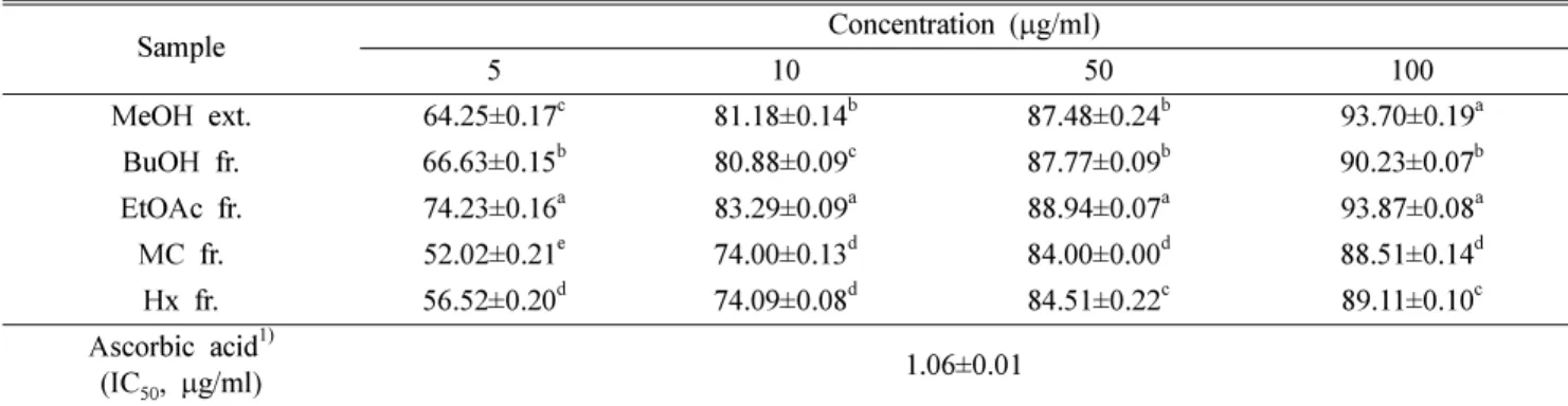
·OH 소거효과 − Free radical중에서도 가장 강한 독성을 나타내는 것으로 알려진 ·OH radical은 반응성이 매우 크고 반응속도가 빠르며 지질의 산화 및 DNA에 손상을 주고 돌 연변이를 유발함으로써 다양한 질환에 관여하는 것으로 알 려져 있다.25) Table II에 나타난 ·OH radical 소거능을 살펴 본 결과, 처리 농도에 따라 소거 효과가 유의적으로 높아지 는 것을 볼 수 있었고 MeOH 추출물과 4종 분획물 모두 10µg/ml에서 70% 이상의 ·OH radical 소거능을 확인할 수 있었다. 특히 EtOAc 분획물의 100 µg/ml 농도에서 93.87%
의 가장 높은 소거능을 나타내었고 MeOH, BuOH 분획물 또한 각 93.70%, 90.23%의 높은 소거능을 보였다. 따라서 머위 추출물과 분획물은 ·OH radical에 대한 독성 제거 효 과가 높은 것으로 확인되었다.
NO 소거효과 − NO는 혈액응고 및 혈압조절 기능, 암세 포에 대한 면역기능을 가지고 있지만,26) O2-와 반응하여 더 반응성이 크고 독성이 강한 산화제 peroxynitrite(ONOO-)를 생성한다. ONOO-는 in vitro 및 in vivo에서 가장 강력한 산 화제로 단백질, 아미노산, DNA 등과 반응하여 세포 및 조 직 손상을 야기할 뿐만 아니라 노화, 암, 관절염, 동맥경화, 피부 염증, 뇌막염, 알츠하이머병, 파킨슨병과 같은 퇴행성 질환에 중요한 요인으로 작용하는 것으로 알려져 있다.27-29) NO는 생리적 pH상태(pH 7.4)의 SNP용액에서 자연스럽게 생성 되며, 산소와 반응한 아질산염의 생성량을 griess reagent로 확인할 수 있다.30) 따라서 본 실험에서 SNP를 사 용하여 아질산염의 양으로 NO 소거활성을 산출하였다. 측 정한 결과(Table III), 농도 의존적으로 증가하는 경향을 보 였으며, 특히 EtOAc 분획물은 100 µg/ml에서 50.77%의 가 장 높은 NO 소거 효과를 나타내었다.
Table I. IC50 values of the MeOH extract and each fraction from P. japonicas against DPPH radical
Sample IC50 (µg/ml)1) MeOH ext. 6.09±0.04c
BuOH fr. 1.47±0.09d
EtOAc fr. 0.02±0.02e
MC fr. 16.51±0.23b
Hx fr. 22.78±0.54a
Ascorbic acid2) 2.43±0.01 Values are mean±SD.
a~eMeans with the different letters among same concentrations are significantly different (P<0.05) by Duncan's multiple range test.
1)IC50 is concentration in µg/ml required to inhibit DPPH radical formation by 50%.
2)Ascorbic acid was used as positive control.
Table II. Hydroxyl radical scavenging activity of the MeOH extract and each fraction from P. japonicus
Sample Concentration (µg/ml)
5 10 50 100
MeOH ext. 64.25±0.17c 81.18±0.14b 87.48±0.24b 93.70±0.19a BuOH fr. 66.63±0.15b 80.88±0.09c 87.77±0.09b 90.23±0.07b EtOAc fr. 74.23±0.16a 83.29±0.09a 88.94±0.07a 93.87±0.08a MC fr. 52.02±0.21e 74.00±0.13d 84.00±0.00d 88.51±0.14d Hx fr. 56.52±0.20d 74.09±0.08d 84.51±0.22c 89.11±0.10c Ascorbic acid1)
(IC50, µg/ml) 1.06±0.01
Values are mean±SD.
a~eMeans with the different letters among same concentrations are significantly different (P<0.05) by Duncan's multiple range test.
1)Ascorbic acid was used as positive control.
ROS 생성 억제 효과 − DCF-DA는 세포 내 과산화수소 를 측정하는 대표적 물질이고 세포막을 자유롭게 통과하여 세포 내 esterase에 의해 비형광성 DCFH로 탈아세틸화 되 며, DCFH는 활성산소종인 과산화수소에 의해 산화되어 강 한 형광을 나타내는 DCF가 되는 원리31)를 이용하여 머위 MeOH 추출물과 BuOH, EtOAc, MC, Hx 분획물의 세포 내 활성산소종 억제 효과를 알아보았다. C6 glial cell을 이용한
H2O2의 산화적 스트레스 실험 모델은 많은 연구자들에 의 해 확립되었다.32-33) 시료의 효과를 검증하기 위하여 H2O2를 시료와 동시에 처리하여 ROS의 생성능을 측정한 결과 ROS 의 생성량이 증가할 뿐만 아니라, 산화적 스트레스와 관련 된 인자들의 발현이 증가 되었다고 보고되었다.34)따라서 본 연구에서는 이러한 연구들을 바탕으로 시료와 H2O2를 동시 에 처리하여 머위 추출물 및 분획물의 효능을 검토하였다.
Table III. NO scavenging activity of the MeOH extract and each fraction from P. japonicus
Sample Concentration (µg/ml)
5 10 50 100
MeOH ext. 1.65±1.28c 13.43±0.51c 22.31±1.01c 43.80±0.64b BuOH fr. 9.30±0.51a 36.57±1.45a 38.02±0.00a 42.56±0.64b EtOAc fr. 4.05±0.47b 16.02±0.63b 32.63±0.47b 50.77±0.63a
MC fr. -1) - 8.03±0.67d 24.08±2.13c
Hx fr. - - - 5.98±1.14d
Ascorbic acid2)
(IC50, µg/ml) 0.18±0.03
Values are mean±SD.
a~dMeans with the different letters among same concentrations are significantly different (P<0.05) by Duncan's multiple range test.
1)No effect.
2)Ascorbic acid was used as positive control.
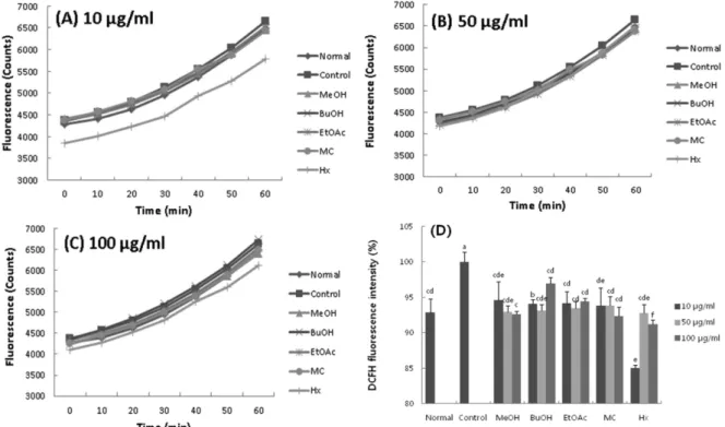
Fig. 1. Effect of fractions from P. japonicus on level of ROS in C6 glial cells treated with H2O2. (A: Time course of change in intensity of ROS fluorescence with fractions from P. japonicus at the concentration of 10µg/ml; B: Time course of change in inten- sity of ROS fluorescence with fractions from P. japonicus at the concentration of 50µg/ml; C: Time course of change in intensity of ROS fluorescence with fractions from P. japonicus at the concentration of 100µg/ml; D: The production of ROS treated with frac- tions from P. japonicus).
Values are mean ± SD.
a~fMeans with the different letters are significantly different (P<0.05) by Duncan's multiple range test.
결과를 살펴 보면(Fig. 1), 시간이 지남에 따라 모든 군에서 ROS 생성량이 증가하는 것을 보였으며, 이를 통해 산화적 스트레스가 유발되었음을 알 수 있었다. 60분 기준으로 control군을 100%로 하였을 때 머위 MeOH 추출물과 BuOH, EtOAc, MC, Hx 분획물을 농도 별로(10, 50, 100µg/ml) 처리한 결과, control군에 비해 ROS 생성량이 감 소한 것을 확인할 수 있었다. 특히, EtOAc 분획물은 normal 군(92.88%)과 비슷한 ROS 생성량을 보여 H2O2에 의해 유 도되는 세포 사멸을 억제하는 요인으로 사료되며 Hx 분획 물 또한 ROS 생성량을 억제함으로써 세포 독성을 보호한 것을 확인 할 수 있었다. 따라서 머위 추출물 및 분획물은 H2O2에 의해 유도되는 ROS의 생성을 효과적으로 감소시킴 으로써 ROS에 대한 신경세포 보호 효과가 있는 것을 알 수 있었다.
H2O2처리로 유도된 산화적 스트레스 개선 효과 − H2O2 는 세포 내에서 유전자 발현 변화를 조절하기도 하고, 세포 의 행동, 운동, 모양 등을 조절하는 신호전달 물질로 작용 하며, 생체의 정상적인 영위에서 10-9~10-7 M로 항상 생성 되고 있다. 하지만 구리(Cu2+)나 철(Fe2+)이온 존재 하에 ·OH 로 전환되어 산화적 스트레스를 유발함에 따라 세포 손상
을 일으켜 퇴행성 신경질환, 암 등의 각종 질병을 일으키게
된다.35-37) 본 연구에서는 H2O2가 유발하는 산화적 스트레스
로부터 C6 glial cell을 보호하는 머위 추출물과 분획물의 효 과를 확인하기 위해 MTT assay를 이용하여 아무것도 처리 하지 않은 normal군과 H2O2 처리한 control군 그리고 H2O2 와 머위 추출물/분획물을 농도별로 처리한 실험 군을 비교 하였다. 그 결과, normal군의 세포 생존율을 100%로 보았 을 때, H2O2를 처리한 control군에서는 72.24%로 나타내어 신경세포가 H2O2에 의해 손상을 받은 것을 확인할 수 있었 다. 반면 머위 추출물과 분획물을 처리하였을 때 세포생존 율이 증가하는 것을 볼 수 있었으며 BuOH 분획물과 EtOAc 분획물이 10 µg/ml의 낮은 농도에서도 80% 이상의 높은 세 포 생존율이 나타난 것을 볼 수 있었다. EtOAc 분획물을 처 리한 군은 농도(5, 10, 50, 100 µg/ml)에 따라서 세포 생존 율이 유의적으로 증가됨을 알 수 있었고 특히 50 µg/ml, 100µg/ml에서는 각 86.32%, 91.23%의 높은 세포 생존율을 나타내어 인체 내에서 발생하는 활성산소의 생성을 억제하 거나, 활성산소를 제거함으로써 신경세포를 보호하는 것으 로 사료된다.
이상의 결과를 종합하여 볼 때 머위는 free radical 소거
Fig. 2. Effect of fractions from P. japonicus on viability of C6 glial cells treated with H2O2. Values are mean±SD.
a~eMeans with the different letters are significantly different (P<0.05) by Duncan's multiple range test.
능과 산화적 스트레스 개선 효과가 있음을 알 수 있었고 그 중 EtOAc 분획물이 radical 소거능과 H2O2로 유발된 산화 적 스트레스 상태에서 세포생존율이 가장 높은 활성을 나 타내는 것을 확인할 수 있었다. 따라서 머위의 EtOAc 분획 물은 우수한 항산화제로서의 가능성을 나타낼 것으로 생각 된다.
결 론
본 연구는 머위 MeOH 추출물과 BuOH, EtOAc, MC, Hx 4종 분획물의 free radical 소거능과 C6 glial cell에서 H2O2에 의한 산화적 스트레스 개선 효과를 알아보았다. 그 결과 머위 추출물과 분획물은 우수한 free radical 소거능을 나타내었고, 특히 EtOAc 분획물은 DPPH 소거능에서 0.02µg/ml의 IC50 값을 보여 가장 우수한 radical 소거능이 있음을 확인할 수 있었다. ·OH radical 소거 효과를 알아본 결과, 처리농도에 따라 농도 의존적으로 ·OH radical 소거 능이 증가됨을 볼 수 있었으며 5 µg/ml의 낮은 농도에서도 70% 이상의 소거능을 나타내었고 100 µg/ml 농도에서는 93.87%의 가장 높은 소거능을 나타내었다. NO 소거능에서 는 EtOAc 분획물이 100 µg/ml에서 50.77%의 가장 높은 NO 소거 효과를 나타내었다. 또한 머위 추출물과 분획물이 H2O2로부터 유도된 산화적 스트레스 상태에서 세포 생존율 을 증가시켰으며, ROS 생성을 억제 시키는 것을 살펴볼 수 있었다. 이상의 결과에서 머위의 EtOAc 분획물은 우수한 항산화능과 산화적 스트레스 개선을 통한 신경세포 보호 효 과가 있음을 알 수 있었다.
사 사
이 논문은 2013년도 정부(교육과학기술부)의 재원으로 한 국연구재단의 기초연구사업 지원을 받아 수행된 것임 (2013R1A1A2011228).
인용문헌
1. Valko, M., Leibfritz, D., Moncol, J., Cronin, M. T. D., Mazur, M. and Telser, J. (2007) Free radicals and antioxidants in nor- mal physiological functions and human disease. Int. J. Bio- chem. Cell B. 39: 44-84.
2. Halliwell, B. and Gutteridge, J. M. C. (1999) Free Radicals in Biology and Medicine, 3rd ed., Oxford University Press, New York.
3. Ames, B. N. (1983) Dietary carcinogens and anticarcinogens.
oxygen radicals and degenerative diseases. Science 221:
1256-1264.
4. Cha, B. C., Lee, H. W. and Choi, M. Y. (1998) Antioxidative
and antimicrobial effects of nut species. Korean J. Phar- macogn. 29: 28-34.
5. Shin, C. H. (2001) Studies on the antioxidative character in the ethyl acetate extractions of Rumex crispus. Korean J.Bio- technol. Bioeng. 16: 592-602.
6. Barnham, K. J., Masters, C. L. and Bush, A. I. (2004) Neu- rodegenerative diseases and oxidative stress. Nat. Rev. Drug Discov. 3: 205-214.
7. Kim, S. D., Do, J. H. and Oh, H. I. (1981) Antioxidant activ- ity of Panax ginseng browning products. J. Korean Agric.
Chem. Soc. 24: 161-166.
8. Choi, O. B. (2002) Anti-allergic effects of Petasites japoni- cum. J. Korean Soc. Food Sci. Nutr. 15: 382-385.
9. Cho, B. S., Lee, J. J., Ha, J. O. and Lee, M. Y. (2006) Phys- icochemical composition of Petasites japonicus S. et Z. Max.
Korean J. Food Preserv. 13: 661-667.
10. Hasa, Y. and Tazaki, H. (2004) Biosynthesis of fukinolic acid isolated from Petasites japonicus. Biosci. Biotechnol. Bio- chem. 68: 2212-2214.
11. Min, B. S., Cui, H. S., Lee, H. K., Sok, D. E. and Kim, M.
R. (2005) A new furofuran lignan with antioxidant and anti- seizure activities from the leaves of Petasites japonicus. Arch.
Pharm. Res. 28: 1023-1026.
12. Yaoita, Y. and Kikuchi, M. (1994) Eremopetasidione a nor- sesquiterpenoid from the rhizomes of Petasites japonicus.
Phytochemistry 37: 1765-1766.
13. Yaoita, Y. and Kikuchi, M. (1994) Petasiphenone, a phenolic compound from rhizomes of Petasites japonicus. Phytochem- istry 37: 1773-1774.
14. Yaoita, Y. and Kikuchi, M. (1994) Structures of six new ere- mophilenolides from the rhizomes of Petasites japonicus Maxim. Chem. Pharm. Bull. 42: 1944-1947.
15. Cho, B. S., Lee, J. J. and Lee, M. Y. (2007) Effects of ethanol extracts from Petasites japonicus S. et Z. Max. on hepatic antioxidative systems in alcohol treated rats. J. Korean Soc.
Food Sci. Nutr. 36: 298-304.
16. Oh, S. H., Yang, Y. H., Kwon, O. Y. and Kim, M. R. (2006) Effects of diet with added butterbur (Petasites japonicus Maxim) of the plasma lipid profiles and antioxidant index of mice. J. East Asian. Soc. Dietary Life 16: 399-407.
17. Kim, J. H., Na, Y., Sim, G. S., Lee, B. C. and Pyo, H. B.
(2006) Antioxidative and anti-inflammatory effects of Peta- sites japonicus. J. Soc. Cosmet. Scientists Korea 32: 263-267.
18. Seo, H. S., Chung, B. H. and Cho, Y. G. (2008) The anti- oxidant and anticancer effects of butterbur (Petasites japoni- cus) extract. Korean J. Plant Res. 21: 265-269.
19. Hatano, T., Edamatsu, R., Hiramatsu, M., Mori, A., Fujita, Y., Yasuhara, T., Yoshida, T. and Okuda, T. (1989) Effects of the interaction of tannins with co-existing substances. VI: Effects of tannins and related polyphenols on superoxide anion rad- ical, and on 1,1-diphenyl-2-picrylhydrazyl radical. Chem.
Pharm. Bull. 37: 2016-2021.
20. Chung, S. K., Osawa, T. and Kawakishi, S. (1997) Hydroxyl radical-scavenging effects of spices and scavengers from brown mustard (Brassica nigra). Biosci. Biotechnol. Bio- chem. 61: 118-123.
21. Marcocci, L., Maguire, J. J., Droxylefaix, M. T. and Packer, L. (1994) The nitric oxide-scavenging properties of Ginkgo biloba extract EGb761. Biochem. Biophys. Res. Commun.
201: 748-755.
22. Byun, Y. J., Kim, S. K., Kim, Y. M., Chae, G. T., Jeong, S. W.
and Lee, S. B. (2009) Hydrogen peroxide induces autophagic cell death in C6 glioma cells via BNIP3-mediated suppres- sion of the mTOR pathway. Neurosci. Lett. 461: 131-135.
23. Mosmann, T. (1983) Rapid colormetric assay for cellular growth and survival: application to proliferation and cyto- toxicity assays. J. Immunol. Meth. 65: 55-63.
24. Wang, K. J., Zhang, Y. J. and Yang, C. R. (2005) Antioxidant phenolic compounds from rhizomes of Polygonum palea- ceum. J. Ethnopharmacol. 96: 483-487.
25. Halliwell, B. and Aruoma, O. I. (1991) DNA damage by oxy- gen-derived species. Its mechanism and measurement in mammalian systems. FEBS Lett. 281: 9-19.
26. Nakagawa, T. and Yokozawa, T. (2002) Direct scavenging of nitric oxide and superoxide by green tea. Food Chem. Tox- icol. 40: 1745-1750.
27. Lee, S. G., Kim, H. J., Lee, S. P. and Lee, I. S. (2009) Anti- oxidant and anticancer activities of defatted soybean grits fer- mented by Bacillus subtilis NUC1. J. Kor. Soc. Food. Sci.
Nutr. 38: 657-662.
28. Althaus, J. S., Oien, T. T., Fici, G. J., Scherch, H. M., Sethy, V. H. and VonVoigtlander, P. F. (1994) Structure activity rela- tionships of peroxynitrite scavengers an approach to nitric oxide neurotoxicity. Res. Commun. Chem. Patho. Pharmacol.
83: 243-254.
29. Lee, K. I. and Kim, S. M. (2009) Antioxidative and anti- microbial activities of Eriobotrya japonica Lindl. leaf extracts. J. Kor. Soc. Food Sci. Nutr. 38: 267-273.
30. Green, L. C., Wagner, D. A., Glogowski, J., Skipper, P. L., Wishnok, J. S. and Tannenbaum, S. R. (1982) Analysis of nitrate, nitrite and nitrate in biological fluids. Anal. Biochem.
126: 131-138.
31. Cathcart, R., Schwiers, E. and Ames, B. N. (1983) Detection of picomole levels of hydroperoxides using a fluorescent dichlorofluorescein assay. Anal. Biochem. 134: 111-116.
32. Quincozes-Santos, A., Bobermin, L. D., Latini, A., Wajner, M., Souza, D. O., Goncalves, C. A. and Gottfried, C. (2013) Resveratrol protects C6 astrocyte cell line against hydrogen peroxide-induced oxidative stress through heme oxygenase 1.
PLoS One 8: e64372
33. Mahesh, R. and Kim, S. J. (2009) The protective effects of insulin on hydrogen peroxide-induced oxidative stress in C6 glial cells. Biomol. Ther. 17: 395-402.
34. Chen, T. J., Jeng, J. Y., Lin, C. W., Wu, C. Y. and Chen, Y.
C. (2006) Quercetin inhibition of ROS-dependent and inde- pendent apoptosis in rat glioma C6 cells. Toxicol. 223: 113- 126.
35. Oda, A., Tamaoka, A. and Araki, W. (2010) Oxidative stress up-regulates presenilin 1 in lipid rafts in neuronal cells. J.
Neurosci. Res. 88: 1137-1145.
36. Pavlica, S. and Gebhardt, R. (2010) Protective effects of fla- vonoids and two metabolites against oxidative stress in neu- ronal PC12 cells. Life Sci. 86: 79-86.
37. Zaidi, A., Fernanders, D., Bean, J. L. and Michaelis, M. L.
(2009) Effects of paraquat-induced oxidative stress on the neuronal plasma membrane Ca2+-ATPase. Free Radic. Biol.
Med. 47: 1507-1514.
(2014. 3. 27 접수; 2014. 5. 2 심사; 2014. 6. 12 게재확정)