Curcuma longae Radix, Phellinus linteus 및 Scutellariae Radix 혼합추출물의 산화성 신경세포손상 보호효과
김주연*·권기연*·이홍규*·김승환**·유재국***·배기환****·성연희*
†*충북대학교 수의과대학, **경희대학교 체육대학, ***한국신약, ****충남대학교 약학대학
Protective Effect of an Ethanol Extract Mixture of Curcuma longae Radix, Phellinus linteus , and Scutellariae Radix on Oxidative Neuronal Damage
Joo Youn Kim * , Kiyeon Kweon * , Hong Kyu Lee * , Seung Hwan Kim ** , Jae Kuk Yoo *** , Ki Hwan Bae **** and Yeon Hee Seong *†
*
College of Veterinary Medicine, Chungbuk National University, Cheongju 361-763, Korea.
**
College of Physical Education, Kyunghee University, Youngin 446-701, Korea.
***
Han Kook Shin Yak, Nonsan 320-854, Korea.
****
College of Pharmacy, Chungnam National University, Taejon 305-764, Korea.
ABSTRACT : Previous work demonstrated that an ethanol extract (HS0608) of a mixture of three medicinal plants of Cur- cuma longae radix , Phellinus linteus , and Scutellariae radix markedly inhibits A β (25-35)-induced neurotoxicity. The present study was performed to further verify the neuroprotective effect of HS0608 on oxidative and ischemic cerebral injury using cultured rat cortical neurons and rats. Exposure of cultured cortical neurons to 100 µ M hydrogen peroxide (H
2O
2) induced neuronal apoptotic death. At 10-100 ㎍ / ㎖ , HS0608 inhibited neuronal death, elevation of intracellular calcium concentra- tion ([Ca
2+]
i), and generation of reactive oxygen species (ROS) induced by H
2O
2in primary cultures of rat cortical neurons.
In vivo, HS0608 prevented cerebral ischemic injury induced by 2-h middle cerebral artery occlusion (MCAO) and 24-h rep- erfusion. The ischemic infarct and edema were significantly reduced in rats that received HS0608 (200 ㎎ / ㎏ ). These results suggest that the anti-oxidative properties of HS0608 may be responsible for its neuroprotective effect against focal cerebral ischemic injury and that HS0608 may have a therapeutic role in neurodegenerative diseases such as stroke.
Key Words : Curcuma longae Radix , Phellinus linteus , Scutellariae Radix, Neuroprotection, H
2O
2, Cultured Cortical Neurons, Cerebral Ischemic Injury
INTRODUCTION
Pharmacological activities of Curcuma longae radix, the root of Curcuma longa , possessing anti-inflammatory (Guo et al ., 2008), anti-oxidant (Adaramoye et al. , 2002) and neuroprotective effects (Rajakrishnan et al ., 1999) have been extensively studied.
Phytochemical analyses have shown that the main constituents of Curcuma longae radix are curcumins, curcuminoids, zingiberine, phelandreen, and essential oils (Kapoor, 1990; Srinivasan, 1953).
Phellinus linteus has been used for its anti-cancer, anti-diabetes and anti-oxidant activities (Ajith and Janardhanan, 2002; Sliva et al ., 2008). It was defined that Phellinus linteus and its active component, hispolon, shows anti-inflammation and analgesic
effects via inhibition of nitric oxide and prostaglandin E
2production and antioxidative activity (Chang et al ., 2009; Kim et al ., 2007). Scutellariae radix from Scutellaria baicalensis Gergi (Labiatae) has been demonstrated to possess antipyretic, antibacterial, and anti-inflammatory properties (Bensky et al ., 1992; Huang, 1999). Several flavonoids such as baicalin, baicalein and wogonin have been isolated as active components of Scutellariae radix (Huang, 1999). Scutellariae radix and its flavonoids have also shown neuroprotection against ischemic brain damage, 6-hydroxydopamine-induced Parkinsonism, and A β (25-35)-induced amnesia (Mu et al ., 2009; Wang et al ., 2004;
Zhang et al ., 2006).
Ischemic stroke results from a transient or permanent reduction
†
Corresponding author: (Phone) +82-43-261-2968 (E-mail) [email protected]
Received 2010 October 21 / 1st Revised 2010 December 11 / 2nd Revised 2011 January 31 / Accepted 2011 February 14
in cerebral blood flow caused by the occlusion of a cerebral artery via an embolus or local thrombosis (Dirnagl et al ., 1999). Loss of blood flow results in depletion of metabolic substrates such as oxygen and glucose. Oxidative stress is believed to exacerbate the damage caused by cerebral ischemia (Chan, 2001). Since three medicinal plants of Curcuma longae radix , Phellinus linteus , and Scutellariae radix exhibit antioxidant and anti-inflammatory activities, we hypothesized that the combination of these three plants might protect neurons against neurodegenerative diseases such as Alzheimer disease and stroke. In a previous report, we demonstrated a significant neuroprotective effect of an ethanolic extract of a mixture of Curcuma longae radix , Phellinus linteus , and Scutellariae radix, which was named as HS0608, against A β (25-35)-induced neuronal cell damage and memory impairment (Kim et al ., 2009b). In the current study, we investigated the neuroprotective effect of HS0608 against ischemia-induced brain infarction following middle cerebral artery occlusion (MCAO) and reperfusion in rats, as well as the effect of HS0608 on hydrogen peroxide (H
2O
2)-induced neuronal damage in cultured rat cortical neurons, to elucidate the neuroprotective mechanism of HS0608.
MATERIALS AND METHODS
1. Plant materials and extraction
The dried Curcuma longae radix and Scutellariae radix were purchased at Kyungdong Folk Medicine market, Seoul, Korea, and fruiting bodies of cultured Phellinus linteus were supplied from Han Kook Shin Yak, Nonsan, Chungnam. These plants were identified by one of the authors, Dr. KiHwan Bae, Chungnam National University. One hundred g per plant from the three plants were mixed, refluxed in 3 L ethanol at room temperature for 3 h, filtered, and lyophilized to yield an ethanol extract (HS0608, 64 g), which was then stored at − 20 ℃ until required.
2. Experimental animals
Pregnant Sprague-Dawley (SD) rats and male SD rats (Daehan BioLink Co. Ltd., Chungbuk, Korea) were housed in an environmentally controlled room at 22 ± 2 ℃ , with a relative humidity of 55 ± 5%, a 12-h light/dark cycle, and food and water ad libitum . The procedures involving experimental animals complied with the animal care guidelines of the National Institutes of Health and the animal ethics committee of Chungbuk National University.
3. Measurements of H
2O
2-induced neuronal death and intracellular biochemical changes in primary cultures of rat cerebral cortical neurons
Primary cortical neuron cultures were prepared using embryonic day 15 to 16 SD rat fetuses, as previously described (Cho et al ., 2009). Experiments of H
2O
2-induced neurotoxicity were performed on neurons after 5-6 days in culture as previously described (Cho et al ., 2009; Kim et al ., 2009a).
Cultured neurons were treated with 100 µ M H
2O
2in a HEPES buffer at 37 ℃ for 15 min, followed by incubation for 12 h in H
2O
2- and serum-free DMEM medium to produce neuronal death and reactive oxygen species (ROS) generation. HS0608 was dissolved in dimethylsulfoxide (DMSO) at concentration of 100 ㎎ / ㎖ and further diluted in experimental buffers. The final concentration of DMSO was ≤ 0.1%, which did not affect cell viability. For each experiment, HS0608 was applied 15 min prior to treatment with 100 µ M H
2O
2, and then again during the H
2O
2exposure and post-exposure period.
A 3-(4,5-dimethylthiazol-2-yl)-2,5-diphenyl-tetrazolium bromide (MTT; Sigma) assay and Hoechst 33342 (Molecular Probes, Eugene, OR, USA) staining were performed to measure neuronal death as previously described (Cho et al ., 2009). Changes in [Ca
2+]
iimmediately after H
2O
2treatment were measured with Fluo-4 AM (Molecular Probes), a calcium-sensitive fluorescent dye, using a laser scanning confocal microscope (TCS SP2 AOBS; Leica, Heidelberg GmbH, Germany) with a 488-nm excitation argon laser and 515- ㎚ longpass emission filters (Cho et al ., 2009). The microfluorescence of 2',7'-dichlorofluorescein, the fluorescent product of 2',7'-dichlorodihydrofluorescein diacetate (H
2DCF-DA; Molecular Probes), and a laser scanning confocal microscope (TCS SP2 AOBS; Leica, Heidelberg GmbH, Germany) with 488- ㎚ excitation and 510- ㎚ emission filters were used to monitor the generation of ROS in neurons treated with 100 µ M H
2O
2(Cho et al ., 2009).
4. MCAO/reperfusion-induced focal cerebral ischemia in rats and evaluation of ischemic damage
Before surgery, male SD rats weighing 280-300 g were fasted
overnight with free access to water. Focal cerebral ischemia was
produced by MCAO for 2 h, followed by reperfusion for 24 h, as
previously described (Ban et al ., 2008). After MCAO/reperfusion,
rats were sacrificed by decapitation under anesthesia (diethyl
ether), and brains were quickly removed. The infarct and edema
volumes of brain tissue were measured using TTC staining, as
previously described (Ban et al ., 2008). HS0608 (100 and 200
㎎ / ㎏ ) was administered at three different time points (0.5 h before and 1 h after occlusion, as well as 1 h after reperfusion).
5. Statistical analysis
Data are expressed as mean ± SEM and statistical significance was assessed by one-way analysis of variance (ANOVA) and Tukey’s tests. P < 0.05 was considered significant.
RESULTS
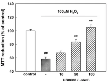
1. HS0608 inhibits H
2O
2-induced neuronal cell death The concentration of 100 µ M of H
2O
2was used for determining H
2O
2-induced neuronal cell damage in the present experiments based on our previous result (Park et al ., 2006).
When cortical neurons were exposed to 100 µ M H
2O
2, absorbance in the MTT assay was 58.4 ± 2.8% of that of the untreated controls (Fig. 1), indicating that H
2O
2caused neuronal cell death. Pretreatment of cortical neurons with 50 and 100 ㎍ / ㎖ HS0608 reduced the neuronal death induced by 100
µ M H
2O
2(absorbance, 83.5 ± 3.3% and 105.0 ± 3.7% of control, respectively; Fig. 1).
An additional experiment was performed with Hoechst 33342 staining to detect condensed or fragmented DNA, which is indicative of H
2O
2-induced neuronal apoptotic death. Treatment of neurons with 100 µ M H
2O
2produced apoptosis of 36.4 ±
2.7% of the total population of cultured cortical neurons,
compared with that of 10.6 ± 0.7% in control cultures. The addition of HS0608 (50 and 100 ㎍ / ㎖ ) significantly decreased the H
2O
2-induced apoptotic cell death, showing 28.6 ± 2.4 and 21.1 ± 1.7%, respectively (Fig. 2).
2. HS0608 inhibits H
2O
2-induced [Ca
2+]
ielevation Increases in [Ca
2+]
ihave been associated with H
2O
2-induced
Fig. 1.
Inhibitory effect of HS0608 on H
2O
2-induced neuronal cell death in cultured cortical neurons. Neuronal cell death was measured using the MTT assay. The MTT absorbance from untreated cells was normalized to 100%. Results are expressed as mean ± S.E.M. of data obtained from 5 independent experiments.
##P< 0.01 vs control; **
P< 0.01 vs 100
µM H
2O
2.
Fig. 2.
Inhibitory effect of HS0608 on H
2O
2-induced apoptosis of cultured cortical neurons. Apoptotic cells measured by Hoechst 33342 staining were counted in 5 to 6 fields per well. The values represent the apoptotic cells as a percentage of the total number of cells expressed as mean ± S.E.M. of data obtained from 4 independent experiments.
##P< 0.01 vs control, **
P< 0.01 vs 100
µ
M H
2O
2.
Fig. 3.
Inhibitory effect of HS0608 on H
2O
2-induced [Ca
2+]
ielevation in cultured cortical neurons. [Ca
2+]
iwas monitored using Fluo-4 AM dye and a confocal laser scanning microscope. All images were processed to analyze changes in [Ca
2+]
iat the single cell level. Results are expressed as the relative fluorescence intensity (RFI).
Each trace shows a single cell that is representative of at
least 3 independent experiments.
cell death. In our cell cultures, [Ca
2+]
ishowed initial rapid increase in response to treatment with 100 µ M H
2O
2with subsequently gradual and fluctuant increase for about 10 min (Fig. 3). In contrast, pretreatment with HS0608 (10 and 100
㎍ / ㎖ ) showed significant inhibitions of the increase of [Ca
2+]
iinduced by 100 µ M H
2O
2. HS0608 did not affect basal [Ca
2+]
i. 3. HS0608 inhibits H
2O
2-induced ROS generation
In H
2DCF-DA-loaded cerebral cortical neurons, 100 µ M H
2O
2increased the fluorescence intensity, indicating that ROS were generated. In neurons treated with 100 µ M H
2O
2, the relative fluorescence increased approximately 3-fold to 93.3 ± 5.7 compared with the value in control neurons (30.7 ± 2.0; Fig. 4).
The H
2O
2-induced increase in ROS generation was significantly inhibited by HS0608 (10, 50 and 100 ㎍ / ㎖ ).
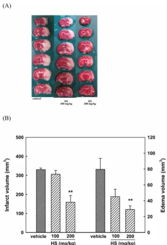
4. HS0608 inhibits MCAO/reperfusion-induced brain infarction
After MCAO/reperfusion, a large ipsilateral cerebral infarction was observed in the rat brain. TTC-stained coronal sections, in which normal brain tissue stains deep red, were used to determine the volume of a cerebral infarction; infarct tissues doese not stain (Fig. 5A). In coronal sections, the infarct size was significantly reduced by HS0608 (200 ㎎ / ㎏ ) compared to that of vehicle-treated controls (79.6 ± 13.8 mm
3for 200 ㎎ / ㎏ and 330.2 ± 8.4 mm
3for vehicle) (Fig. 5B). The edema volume increased by MCAO/reperfusion also was significantly reduced
by HS0608 treatment (29.0 ± 4.5 mm
3for 200 ㎎ / ㎏ ), compared to that of vehicle-treated controls (79.6 ± 13.8 mm
3) (Fig. 5B).
Animal body temperatures were monitored for 6 h after cerebral reperfusion commenced, and no significant differences were observed between the HS0608 and control groups (data not shown). Thus, the observed neuroprotective effect of HS0608 could not be attributed to hypothermic effects.
DISCUSSION
In vitro H
2O
2toxicity has been used for studying the neuropathology of oxidative stress in CNS disorders as a well- established model. H
2O
2-induced neurotoxicity in cultured neurons has been demonstrated to involve sustained elevation of [Ca
2+]
i, NMDA receptor modulation induced by glutamate
Fig. 4.
Inhibitory effect of HS0608 on H
2O
2-induced ROS generation in cultured cortical neurons. ROS was moni- tored using H
2DCF-DA dye and a confocal laser scanning microscope. Results are expressed as mean ± S.E.M. of RFI obtained from 4 independent experi- ments.
##P< 0.01 vs control; **
P< 0.01 vs 100
µM H
2O
2.
Fig. 5.
Protective effect of HS0608 on MCAO/reperfusion-induced
infarct and edema formation in rats. (A) A representative
rat brain stained with 2% TTC 24 h after reperfusion. (B)
The results show infarct and edema volumes caused in
the ipsilateral hemisphere and are expressed as mean ±
S.E.M. of data obtained from 6 rats. **
P< 0.01 vs vehicle.
release and ROS generation (Duffy and MacVicar, 1996; Mailly et al ., 1999). The present study also demonstrated that H
2O
2increased the levels of [Ca
2+]
iand ROS generation, resulting in apoptosis of cultured cortical neurons. H
2O
2exposure causes neuronal cells to exhibit increased permeability to Na
+ions, resulting in membrane depolarization and subsequent a large influx of Ca
2+ions via voltage-dependent Ca
2+channels (VDCC) (Wang and Joseph, 2000). H
2O
2has been demonstrated to inhibit the uptake of glutamate and enhance the release of glutamate, resulting in NMDA receptor overstimulation and a further increase in [Ca
2+]
i(Mailly et al ., 1999; Volterra et al ., 1994). In the present study, H
2O
2elicited a significant increase in [Ca
2+]
i, which was blocked by HS0608. Although these results suggest that HS0608 might prevent H
2O
2-induced Ca
2+entry through VDCC- and/or NMDA-receptor-coupled channels to inhibit ROS generation and then neuronal death, its underlying mechanism remains unclear. Curcuma longae radix, Phellinus linteus , and Scutellariae radix, the constituents of HS0608, have been reported to possess antioxidant principles, curcumin, hispolon, and baicalein, baicalin and wogonin (Chang et al ., 2009; Guo et al ., 2008; Su et al ., 2008; Zhang et al ., 2006), repectively, suggesting that inhibition of H
2O
2-induced neuronal death by HS0608 might be due to their ROS scavenging activity. Further study to elucidate the precise mechanism should be performed.
The rat model of MCAO followed by reperfusion, mimics many features of stroke in humans. In particular, the middle cerebral artery, which is the specific occlusion site in this model, is the most commonly affected vessel in both embolic and thrombotic stroke in humans (Longa et al ., 1989). Infarction and edema are the two main pathophysiological changes observed in the cerebral ischemia (Durukan and Tatlisumak, 2007). Cellular swelling causes brain edema, which is the earliest response to ischemic injury. The severity of the neurological deficit is known to correlate with the size of lesion (Tominaga and Ohnishi, 1989) and significant impairment in neurological function was observed after reperfusion. In addition to the in vitro neuroprotective effect of HS0608, we have shown that HS0608 functions in vivo as a potent neuroprotectant in transient brain ischemia, effectively decreasing infarct volume and edema in the rat brain. Ion pumps cannot function during ischemia and thus ATP levels become progressively depleted. This results in elevation of [Ca
2+]
i, which further accelerates ATP depletion. The rise in [Ca
2+]
iduring ischemia and reperfusion leads to mitochondrial Ca
2+accumu- lation, particularly when oxygen is reintroduced during reper- fusion. Reintroduction of oxygen allows generation of ATP;
however, damage to the electron transport chain results in increased mitochondrial generation of ROS (Murphy and Steenbergen, 2008). Evidence obtained over the past two decades shows that ROS are involved in brain lesions, including those due to cerebral ischemia-reperfusion. Many reports have demonstrated that antioxidants such as glutathione, superoxide dismutase, tocopherol and
L-NAME, as well as Ca
2+channel antagonists such as amlodipine and azelnidipine, provide protection against MCAO-induced focal ischemia in rats (Dawson et al ., 1994; Hurtado et al ., 2003; Lukic-Panin et al ., 2007). The neuroprotective effect that HS0608 provides against ischemic brain injury might be attributable to antioxidant activity or inhibition of Ca
2+influx.
In terms of neuroprotective activities of the three constituent plants of HS0608 and their active principles, Curcuma longae radix and curcumins have been shown to protect neurons against cerebral ischemia and A β -induced cognitive deficits and have anti-depressant activity (Frautschy et al ., 2001; Shukla et al ., 2008; Wang et al ., 2005; Yu et al ., 2002). Recent study has demonstrated that Phellinus linteus reduces infarction of ischemic rats (Suzuki et al ., 2009). Neuroprotective effects of Scutellariae radix and its active components, wogonin, baicalein and baicalin, have been widely studied (Heo et al ., 2009; Mu et al ., 2009; Wang et al ., 2004; Zhang et al ., 2006). It, thus, is presumed that the preparation of HS0608 might reveal synergistic effect of these three plants in protection of ischemia- induced neuronal damage.
In conclusion, we have demonstrated that HS0608 protects neurons from MCAO/reperfusion-induced ischemic brain damage in vivo and from H
2O
2-induced neurotoxicity in vitro . These results suggest that HS0608 represents promising a agent for the treatment and prevention of neurodegeneration in stoke. Further studies should determine the specific components in three plants of HS0608 that are responsible for preventing the ischemic neuronal death.
ACKNOWLEDGEMENTS
This work was supported by the research grant of the Chungbuk National University in 2010.
LITERATURE CITED
Adaramoye OA, Adesanoye OA, Olusola A and Akinloye O.
(2002). Antioxidant activity of turmeric extracts ( Curcuma
longa L.) and its effect on iron/ascorbate induced lipid peroxidation. Biokemistri. 12:127-135.
Ajith TA and Janardhanan KK . (2002). Antioxidant and antihepatotoxic activities of Phellinus rimosus (Berk) Pilat.
Journal of Ethnopharmacology. 81:387-391.
Ban JY, Cho SO, Choi SH, Ju HS, Kim JY, Bae K, Song KS and Seong YH . (2008). Neuroprotective effect of Smilacis chinae rhizome on NMDA-induced neurotoxicity in vitro and focal cerebral ischemia in vivo. Journal of Pharmacological Sciences. 106:68-77.
Bensky D, Gamble, A., Kaptchuk, T. (1992). Chinese herbal medicine materia medica. Eastland Press. Seattle, WA. USA.
p.107-109.
Chan PH. (2001). Reactive oxygen radicals in signaling and damage in the ischemic brain. Journal of Cerebral Blood Flow and Metabolism. 21:2-14.
Chang HY, Sheu MJ, Yang CH, Lu TC, Chang YS, Peng WH, Huang SS and Huang GJ. (2009). Analgesic effects and the mechanisms of anti-inflammation of hispolon in mice.
Evidence-Based Complementary and Alternative Medicine.
eCAM 2009:doi:10. 1093/ecam/nep027.
Cho SO, Ban JY, Kim JY, Jeong HY, Li S, Song KS, Bae K and Seong YH. (2009). Aralia cordata protects against amyloid
β
protein (25-35)-induced neurotoxicity in cultured neurons and has antidementia activities in mice. Journal of Pharmacological Sciences. 111:22-33.
Dawson DA, Graham DI, McCulloch J and Macrae IM.
(1994). Anti-ischemic efficacy of a nitric oxide synthase inhibitor and an N-methyl-D-aspartate receptor antagonist in models of transient and permanent focal cerebral ischaemia.
British Journal of Pharmacology. 113:247-253.
Dirnagl U, Iadecola C and Moskowitz MA. (1999). Pathobiology of ischaemic stroke : an integrated view. Trends in Neurosciences.
22:391-397.
Duffy S and MacVicar BA. (1996). In vitro ischemia promotes calcium influx and intracellular calcium release in hippocampal astrocytes. Journal of Neuroscience. 16:71-81.
Durunkan A and Tatlisumak. (2007). Acute ischemic stroke:
overview of major experimental rodent models, pathophysiology, and therapy of focal cerebral ischemia. Pharmacology Biochemistry and Behavior. 87:179-197.
Frautschy SA, Hu W, Kim P, Miller SA, Chu T, Harris-White ME and Cole GM. (2001). Phenolic anti-inflammatory antioxidant reversal of A beta-induced cognitive deficits and neuropathology. Neurobiology of Aging. 22:993-1005.
Guo LY, Cai XF, Lee JJ, Kang SS, Shin EM, Zhou HY, Jung JW and Kim YS. (2008). Comparison of suppressive effects of demethoxycurcumin and bisdemethoxycurcumin on expressions of inflammatory mediators in vitro and in vivo. Archives Pharmacal Research. 31:490-496.
Heo H, Shin Y, Cho W, Choi Y, Kim H and Kwon YK. (2009).
Memory improvement in ibotenic acid induced model rats by extracts of Scutellaria baicalensis . Journal of Ethnopharmacology.
122:20-27.
Huang KC. (1999). The pharmacology of chinese herbs. CRC press. Boca Raton, FL. USA. p.385
Hurtado O, De Cristobal J, Sanchez V, Lizasoain I, Cardenas
A, Pereira MP, Colado MI, Leza JC, Lorenzo P and Moro MA. (2003). Inhibition of glutamate release by delaying ATP fall accounts for neuroprotective effects of antioxidants in experimental stroke. FASEB Journal. 17:2082-2084.
Kapoor LD. (1990). Handbook of ayurvedic medicinal plants.
CRC Press. Boca Raton, FL. USA. p. 149-150.
Kim HG, Yoon DH, Lee WH, Han SK, Shrestha B, Kim CH, Lim MH, Chang W, Lim S, Choi S, Song WO, Sung JM, Hwang KC and Kim TW. (2007). Phellinus linteus inhibits inflammatory mediators by suppressing redox-based NF-kappaB and MAPKs activation in lipopolysaccharide-induced RAW 264.7 macrophage. Journal of Ethnopharmacology. 114:307-315.
Kim JY, Ju HS, Ban JY, Song KS, Bae K and Seong YH.
(2009a). Protective effect of Vitis amurensis stems and leaves extract on hydrogen peroxide-induced oxidative neuronal cell damage in cultured neurons. Korean Journal of Medicinal Crop Science. 17:444-450
Kim JY, Jeong HY, Ban JY, Yoo JK, Bae K and Seong YH.
(2009b). Ethanol extract of three plants of Curcuma longae radix, Phellinus linteus , and Scutellariae radix inhibits amyloid
β