• 검색 결과가 없습니다.

Green tea polyphenol (-)-epigallocatechin-3-gallate prevents ultraviolet-induced apoptosis in PC12 cells

N/A
N/A
Protected

Academic year: 2021

Share "Green tea polyphenol (-)-epigallocatechin-3-gallate prevents ultraviolet-induced apoptosis in PC12 cells"

Copied!
11
0
0

로드 중.... (전체 텍스트 보기)

전체 글

(1)

Introduction Introduction

Ultraviolet (UV) radiation present in natural sunlight and

therapeutic artificial devices is a key human health concern. UV irradiation has been implicated in the development of various cancers in humans, including skin cancer, basal and squamous Int J Oral Biol 45:179-189, 2020

pISSN: 1226-7155 • eISSN: 2287-6618 https://doi.org/10.11620/IJOB.2020.45.4.179

Green tea polyphenol (–)-epigallocatechin-3-gallate prevents ultraviolet-induced apoptosis in PC12 cells

Su-Mi Woo 1† , Yoon-Jung Kim 1† , Bangrong Cai 2† , Sam-Young Park 1 , Young Kim 3 , Ok Joon Kim 3 , In-Chol Kang 4 , Won-Jae Kim 1 *, and Ji-Yeon Jung 1 *

1 Department of Oral Physiology, Dental Science Research Institute, School of Dentistry, Chonnam National University, Gwangju 61186, Republic of Korea

2 School of Pharmacy, Henan University of Traditional Chinese Medicine, Zhengzhou 450008, China

3 Department of Oral Pathology, Dental Science Research Institute, School of Dentistry, Chonnam National University, Gwangju 61186, Republic of Korea

4 Department of Oral Microbiology, Dental Science Research Institute, School of Dentistry, Chonnam National University, Gwangju 61186, Republic of Korea

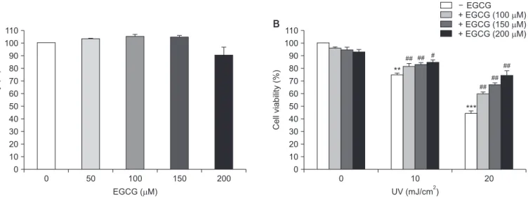
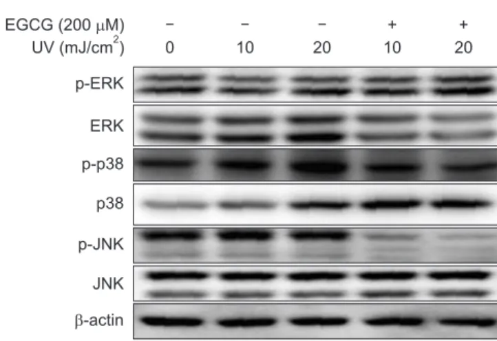
Green tea polyphenol (–)-epigallocatechin-3-gallate (EGCG) is a potent antioxidant with protective effects against neurotoxicity. However, it is currently unclear whether EGCG protects neuronal cells against radiation-induced damage. Therefore, the objective of this study was to investigate the effects of EGCG on ultraviolet (UV)-induced oxidative stress and apoptosis in PC12 cells. The effects of UV irradiation included apoptotic cell death, which was associated with DNA fragmentation, reactive oxygen species (ROS) production, enhanced caspase-3 and caspase-9 activity, and poly (ADP-ribose) polymerase cleavage. UV irradiation also increased the Bax/Bcl-2 ratio and mitochondrial pathway-associated cytochrome c expression. However, pretreatment with EGCG before UV exposure markedly decreased UV-induced DNA fragmentation and ROS production. Furthermore, the UV irradiation- induced increase in Bax/Bcl-2 ratio, cytochrome c upregulation, and caspase-3 and caspase-9 activation were each ameliorated by EGCG pretreatment. Additionally, EGCG suppressed UV-induced phosphorylation of p38 and rescued UV-downregulated phosphorylation of ERK. Taken together, these results suggest that EGCG prevents UV irradiation- induced apoptosis in PC12 cells by scavenging ROS and inhibiting the mitochondrial pathways known to play a crucial role in apoptosis. In addition, EGCG inhibits UV-induced apoptosis via JNK inactivation and ERK activation in PC12 cells. Thus, EGCG represents a potential neuroprotective agent that could be applied to prevent neuronal cell death induced by UV irradiation.

Keywords: Apoptosis, Epigallcatechin, PC12 cells, Radiation

Received November 3, 2020; Revised November 10, 2020; Accepted November 11, 2020

*Correspondence to: Ji-Yeon Jung, E-mail: jjy@jnu.ac.kr https://orcid.org/0000-0002-4419-8077

*Correspondence to: Won-Jae Kim, E-mail: wjkim@jnu.ac.kr https://orcid.org/0000-0001-6549-744X

† Su-Mi Woo, Yoon-Jung Kim, and Bangrong Cai contributed equally to this work.

Copyright © The Korean Academy of Oral Biology

CC

This is an open-access article distributed under the terms of the Creative Commons Attribution Non-Commercial License (http://creativecommons.org/licenses/by- nc/4.0/), which permits unrestricted non-commercial use, distribution, and reproduction in any medium, provided the original work is properly cited.

Original Article IJOB

(2)

cell carcinoma, and cutaneous malignant melanoma [1,2]. UV can be subdivided into UVA (315–400 nm), UVB (280–315 nm), and UVC (200–280 nm) components. Natural solar UVA and UVB radiation can penetrate the atmosphere and reach the Earth’s surface in amounts that have a significant impact on the biosphere. Although UVC cannot reach the Earth’s surface, it can be generated by artificial sources such as photocur- ing devices and germicidal lamps emitting UV irradiation. The harmful radiation, including UVB and UVC primarily, induces DNA damage at the maximal absorption peak of 245–290 nm [3]. Cell damage induced by UV radiation is associated with cell-cycle arrest, cell death, mutation, neoplastic transfor- mation, and immunosuppression, leading to photoaging and photocarcinogenesis [4]. UV irradiation induces apoptosis of neuronal cells. Thus, UV irradiation has been used as a tool to determine the underlying mechanism of apoptosis [5-7]. UV irradiation induces cell death via a variety of cellular mecha- nisms, including induction of nuclear DNA damage, activation of cell surface death receptors, formation of reactive oxygen species (ROS) accompanied by mitochondrial dysfunction, and release of pro-apoptotic factors such as cytochrome c [8-10].

Apoptosis, also known as programmed cell death, is a nor- mal developmental response to physiological stimulus [11,12].

In general, apoptosis is driven by the activation of a family of cysteine proteases called caspases, which cleave a set of critical cellular proteins to initiate apoptotic cell death. These caspases are expressed as proenzymes and activated by up- stream stimuli [13]. Mammalian caspases comprise at least 14 members. Caspases associated with apoptosis can be further subdivided into initiator caspases (-2, -8, -9, and -10) and effector caspases (-3, -6, and -7) [14]. Besides these cas- pases, members of Bcl-2 protein family also play a critical role in the regulation of apoptosis. The Bcl-2 family of proteins are functionally divided into anti-apoptotic (Bcl-2, Bcl-xL, Bcl-W, Mcl-1, and A1) and pro-apoptotic (Bax, Bcl-1s, Bid, Bad, Bim, and Bik) types [15]. Anti-apoptotic Bcl-2 and its homologs are anchored in the mitochondrial membrane to stabilize the transmembrane potential [16]. Mitochondria-mediated apop- totic pathway leads to the initiation of cell death by altering the mitochondrial permeability, resulting in pore formation, fol- lowed by release of cytochrome c and oxygen free radicals into the cytosol [16].

Tea polyphenols are natural plant flavonoids found in leaves and stems of tea plant. Green tea polyphenols exhibit a variety of pharmacological properties, including anti-inflammatory, anticarcinogenic, and antioxidant effects [17-19]. Green tea

polyphenols comprise (–)-epigallocatechin-3-gallate (EGCG), (–)-epigallocatechin, (–)-epicatechin, (+)-gallocatechin, and catechin. Among these polyphenols, EGCG is a major com- ponent exhibiting various biological activities [20-22]. EGCG prevented neuronal cell death and improved cerebral function after traumatic brain injury in rats [23]. Many studies have also investigated the molecular mechanisms underlying their neu- roprotective function in various neurodegenerative diseases [24,25]. Therefore, EGCG might have protective effects against UV irradiation-induced cell death in nervous tissues and cells.

However, whether EGCG can protect against UV radiation-in- duced damage in neuronal cells has yet to be reported. The rat pheochromocytoma cell line PC12, which displays phenotypic characteristics of both adrenal chromaffin cells and sympa- thetic neurons, is a useful system for exploring neuroprotective drugs [26,27]. Therefore, PC12 cells were used in this study to evaluate the neuroprotective effect of EGCG on UV irradiation- induced apoptosis in PC12 cells and investigate the underlying protective mechanism.

Materials and Methods Materials and Methods

1. Cell culture, UV irradiation and EGCG treatment

Rat pheochromocytoma PC12 cells were maintained in RPMI 1640 medium containing 10% horse serum, 5% fetal bovine serum (Gibco BRL, Gaithersburg, MD, USA) inactivated by heat, 100 U mL –1 of penicillin, and 100 mg mL -1 of streptomy- cin in a humidified atmosphere containing 5% CO 2 at 37℃. The culture medium was removed and the cells were washed with phosphate-buffered saline (PBS), followed by irradiation with UV using a UVP CL-1000 ultraviolet crosslinker (Transillumina- tors, Upland, CV, USA). The dose of UV irradiation is indicated in the Figure legend. EGCG was purchased from Sigma-Aldrich (St. Louis, MO, USA) and dissolved with sterile distilled water.

The original culture medium was then added back to cells in both treated and control groups.

2. Cell viability based on 3-(4,5-dimethylthiazol-2- yl)-2,5-diphenyltetrazolium bromide (MTT) assay

Cell viability was determined by MTT assay, which is based

on the reduction of MTT into a visible dark-blue formazan

product in viable cells with active mitochondria, thus provid-

ing an indirect measure of cell viability [28]. Briefly, PC12 cells

were plated into 96-well cell culture plates and exposed to

(3)

UV alone or pretreated with different concentrations of EGCG.

Next, a final concentration of MTT (0.1 mg/mL) was added to the culture medium at the respective time points and incu- bated at 37℃ for 4 hours. The reaction product of MTT was dissolved in dimethylsulfoxide. Optical density was then mea- sured at reference wavelengths of 570 nm and 630 nm on an ELx800 UV ELISA reader (BioTek Instruments, Winooski, VT, USA). The background absorbance of medium in the absence of cells was subtracted.

3. Agarose gel electrophoresis for assessment of DNA fragmentation

Oligonucleosomal fragmentation of genomic DNA was as- sessed using an Apopladder kit (TaKaRa Shuzo, Kyoto, Japan) according to the manufacturer’s instructions. Briefly, cells were lysed with 200 µL of cell lysis buffer and centrifuged at 1,100 g for 10 minutes. The supernatant was incubated at 56℃ for 1 hour after adding 20 µL of 10% sodium dodecyl sulfate (SDS) and 20 µL of proteinase K (20 mg/mL). Next, 1 µg of RNase was added to the reaction and incubated at 37℃ for 1 hour.

DNA was extracted and precipitated overnight at –20℃ in a precipitant mixture containing 0.95 mL of ethanol and centri- fuged at 12,000 g for 15 minutes at 4℃. The DNA pellet was re-suspended in 20 µL of Tris acetate-EDTA buffer. An aliquot of each sample was electrophoresed at 80 V for 2 hours on 1.5% agarose gels containing SYBR TM (safe DNA gel stain, Bi- onerr, Daejeon, Korea). DNA bands were visualized with a UVP transilluminator/Polaroid camera system (UVP Laboratories, Upland, CA, USA).

4. 4′,6-diamidino-2-phenylindole (DAPI) staining

PC12 cells at a density of 1.0 × 10 5 cells/mL were plated into 24-well plate overnight and pretreated with 200 mM EGCG for 24 hours. The medium was removed and the cells were washed with PBS solution twice, followed by irradiation with UV at 10 mJ/cm 2 or 20 mJ/cm 2 . These cells were further incubated at 37℃ in a humidified atmosphere containing 5%

CO 2 for additional 24 hours. After removal of the culture me- dium, the cells were washed twice with PBS and fixed with ice-cold methanol for 15 minutes at room temperature. The fixed cells were washed again with PBS and stained with 10 µg/mL of DAPI for 10 minutes in the dark. After washing twice with PBS, the cells were examined under a fluorescence mi- croscope (Leica Microsystems, Wetzlar, Germany) to observe

the morphology of the nucleus.

5. Detection of ROS production

ROS production was monitored with a fluorescence spec- trometer (Hitachi F-4500; Hitachi, Tokyo, Japan) using 2’, 7’

-dichlorofluorescin diacetate (DCF-DA). Briefly, cells were plated onto a 96-well cell culture plate and treated with UV radiation or EGCG. DCF-DA (25 µM) was then added to the media and cultured at 37℃ for 10 minutes. Emission was measured at a wavelength of 530 nm.

6. Western blot analysis

The PC12 cells were washed twice with PBS, and mixed with a cell lysis buffer (1% NP-40, 500 mM Tris-HCl, pH 7.4, 150 mM NaCl, 5 mM EDTA, 1 mM Benzamid, and 1 µL/mL trypsin inhibitor) containing an Xpert Protease Inhibitor Cocktail Solution and an Xpert Phosphatase Inhibitor Cocktail Solu- tion (GenDEPOT, Katy, TX, USA). Cell lysates were incubated at 4℃ for 20 minutes and centrifuged at 12,000 × g for 20 minutes at 4℃. Supernatants were collected and the protein concentrations were determined by BSA protein assay (Thermo Fisher Scientific, Rockford, IL, USA). Protein extracts (50 µg) were boiled for 5 minutes with SDS-sample buffer and then resolved via 8–15% polyacrylamide gel electrophoresis. Pro- teins were electroblotted onto a nitrocellulose membrane and blocked with 5% skim milk (Becton, Dickinson and Company, Franklin Lakes, NJ, USA) in Tris-buffered saline containing 0.1% Tween 20 (TBS-T) for 1 hour, followed by incubation with anti-poly (ADP-ribose) polymerase (PARP), anti-Bax, anti-Bcl-2, anti-Cyt c, anti-cleaved caspase-9, anti-cleaved caspase-3, anti-p-ERK, anti-ERK, anti-p-p38, anti-p38, anti- p-JNK, anti-JNK (Cell Signaling Technology, Danvers, MA, USA) and anti- β-actin antibodies (Santa Cruz Biotechnology, Dallas, TX, USA) for 4 hours at room temperature. Blots were subsequently washed three times in TBS-T for 5 minutes each and incubated with specific horseradish peroxidase (HRP)- conjugated secondary antibodies (Sigma, St. Louis, MO, USA) at room temperature for 1 hour. Bound antibodies were visu- alized using an Immobilon Western Chemiluminescent HRP Substrate (Millipore, Burlington, MA, USA).

7. Measurement of caspase activity

Caspase activity was assayed using a caspase-3 activity

(4)

assay kit (Calbiochem, San Diego, CA, USA) according to the manufacturer’s instructions. Briefly, PC12 cells were grown on 100 mm 2 cell culture dishes and irradiated with UV alone or pretreated with EGCG for the indicated time. After removing media from culture dishes, cells were collected, washed with PBS, and resuspended in cell lysis buffer, followed by incuba- tion on ice for 10 minutes. The lysates were centrifuged at 14,000 rpm for 20 minutes. Supernatants were collected and

the protein concentrations were determined with BCA assay (Pierce, Rockford, IL, USA). The cell lysate (50 µL) was then mixed with a reaction buffer containing DEVE-pNA substrate (200 µM) and incubated at 37℃ for 24 hours. Absorbance was then measured at 405 nm on an ELx800 UV ELISA reader.

0 110 100 90 80 70 60 50 40 30 20 10

Cell viability (%)

EGCG ( M)  0

50 100 150 200 0

110 100 90 80 70 60 50 40 30 20 10

Cell viability (%)

UV (mJ/cm ) 2 0

20 10

EGCG + EGCG

+ EGCG (150 M) + EGCG (200 M) 

 (100 M) 

**

## ## #

***

##

##

##

A B

Fig. 1. Effect of (–)-epigallocatechin-3-gallate (EGCG) on the viability of ultraviolet (UV)-irradiated PC12 cells. (A) PC12 cells were treated with different doses of UV irradiation with or without pretreatment with 200 µM EGCG for 24 hours (B) and irradiated with 10 or 20 mJ/cm 2 UV in the presence of 200 µM EGCG for the indicated time. Cell viability was estimated by 3-(4,5-dimethylthiazol-2-yl)-2,5-diphenyltetrazolium bromide (MTT) assay. Results are presented as mean ± standard error of the mean of three independent experiments.

**p < 0.01, ***p < 0.001 compared to the control; # p < 0.05, ## p < 0.01 compared to UV-irradiated group.

EGCG (200 M) UV (mJ/cm )

 2

0 10 20

+ 10

+ 20

A

EGCG ( ) EGCG (200 M)

0 10 20

UV irradiation (mJ/cm ) 2

B

M

Fig. 2. Effect of (–)-epigallocatechin-3-gallate (EGCG) on ultraviolet (UV) irradiation-induced apoptosis in PC12 cells. (A) DNA was prepared from cells ir- radiated with UV at 10 or 20 mJ/cm 2 with or without pretreatment with 200 µM EGCG for 24 hours, followed by electrophoresis and visualization with SYBR.

(B) Changes in nuclear morphology were observed using 4′,6-diamidino-2-phenylindole (DAPI) staining assay. The cells fixed with ice-cold methanol after

treatment were stained with 10 µg/mL of DAPI for 10 minutes in the dark and examined under a florescence microscope after washing twice with phosphate-

buffered saline. Results are representative of three independent experiments (×40 magnification).

(5)

Results Results

1. Effects of EGCG on cell viability in UV-irradiated PC12 cells

The MTT assay was used to determine the cytotoxic effects of UV irradiation on PC12 cells with or without EGCG pretreat- ment (200 µM) at the indicated time course. Cell viability was dramatically decreased after UV irradiation. The survival rate of PC12 cells was about 50% at 24 hours after irradiation at a dose of 100 mJ/cm 2 (Fig. 1A). However, pretreatment with EGCG significantly rescued the decrease in cell viability caused by UV irradiation in a dose-dependent manner (Fig. 1B).

2. EGCG ameliorates apoptosis in UV-irradiated PC12 cells

UV irradiation at 10 or 20 mJ/cm 2 for 24 hours induced DNA fragmentation in PC12 cells, resulting in a DNA ladder pattern, which was suppressed by pretreatment with 200 µM EGCG (Fig. 2A). To further analyze the effect of EGCG on the apopto- sis of PC12 cells induced by UV irradiation, changes in nuclear morphology were detected via DAPI staining after UV irradia- tion at both 10 and 20 mJ/cm 2 with or without EGCG pre- treatment (200 µM). As expected, UV exposure increased the number of condensed and fragmented nuclei with enhanced fluorescent intensities in PC12 cells compared with control

(untreated). However, pretreatment with EGCG obviously re- duced the number of condensed and fragmented nuclei in UV- irradiated PC12 cells (Fig. 2B). This result demonstrated that EGCG ameliorated UV-induced apoptosis of PC12 cells.

UV (mJ/cm ) EGCG (200 M)

2

 300

200

100

ROS (% of control)

0

10 10 20 20

***

***

### ###

Fig. 3. Effect of (–)-epigallocatechin-3-gallate (EGCG) on reactive oxygen species (ROS) production in ultraviolet (UV)-irradiated PC12 cells. PC12 cells were pretreated with 200 µM EGCG for 24 hours prior to UV irradia- tion. The culture medium was removed and cells were washed and irradi- ated with UV at 10 or 20 mJ/cm 2 for 24 hours. Cells were then incubated with 2’, 7’-dichlorofluorescin diacetate (DCF-DA) solution for 10 minutes.

After washing with 1× Hanks’ Balanced Salt Solution (HBSS) buffer, the intracellular levels of ROS were measured as DCF fluorescence. Results are representative of three independent experiments. Data are expressed as mean ± standard error of the mean.

***p < 0.001 compared to the control; ### p < 0.001 compared to UV-irradiat- ed group.

UV (mJ/cm ) EGCG (200 M)

2

 1.5

1.0

Bcl-2/Bax ratio 0.5

0.0

10 10 20 20

* *

# #

EGCG (200 M) UV (mJ/cm )

 2

Bax

Bcl-2

CytC

-actin

0 10 20

+ 10

+ 20

A B

Fig. 4. Effect of (–)-epigallocatechin-3-gallate (EGCG) on the expression of apoptosis-related proteins in ultraviolet (UV)-irradiated PC12 cells. (A) Protein levels of Bcl-2, Bax, and cytochrome c (CytC) in PC12 cells irradiated with UV at 10 or 20 mJ/cm 2 for 24 hours, with or without 200 µM EGCG, were deter- mined by western blot. (B) Bcl-2 and Bax expression was measured by densitometry and the relative level of protein expression was normalized to β-actin.

Results are representative of three independent experiments. Data are expressed as mean ± standard error of the mean.

*p < 0.05 compared to the control; # p < 0.05 compared to UV-irradiated group.

(6)

3. EGCG reduces ROS production in UV-irradiated PC12 cells To investigate the scavenging effects of EGCG on ROS pro-

duction, the ROS levels were measured using DCF-DA in UV- irradiated PC12 cells with or without pretreatment with 200 µM EGCG. UV irradiation at a dose of 10 to 20 mJ/cm 2 enhanced

UV (mJ/cm ) EGCG (200 M)

2

 4.0

3.0

2.0

Cleaved caspase-3 1.0

0.0

0 10 10 20 20

**

*

# #

A B

EGCG (200 M) UV (mJ/cm )

 2

Cleaved caspase-3

Cleaved caspase-9

-actin

0 10 20

+ 10

+ 20

UV (mJ/cm ) EGCG (200 M)

2

 800

600

400

200

Caspase-3 activity (%)

0

0 10 10 20 20

***

***

#

#

D

UV (mJ/cm ) EGCG (200 M)

2

 16

12

8

Cleaved P ARP 4

0

0 10 10 20 20

**

**

#

#

F E

EGCG (200 M) UV (mJ/cm )

 2

PARP

-actin

0 10 20

+ 10

+ 20

C

UV (mJ/cm ) EGCG (200 M)

2

 2.0

1.0

Cleaved caspase-9

0.0

0 10 10 20 20

*

*

#

# 1.5

0.5

Fig. 5. Effect of (–)-epigallocatechin-3-gallate (EGCG) on caspase activation in ultraviolet (UV)-irradiated PC12 cells. (A-C) Protein levels of cleaved cas- pase-3 and caspase-9 in PC12 cells irradiated by UV at 10 or 20 mJ/cm 2 with or without 200 µM EGCG pretreatment for 24 hours were measured by west- ern blot. (D) Caspase-3 activity in 10 or 20 mJ/cm 2 UV-irradiated PC12 cells with or without 200 µM EGCG pretreatment for 24 hours. Enzymatic activity of caspase-3 protease was determined by incubating 50 µg of total protein with DEVD-pNA substrate (200 µM). (E, F) Protein levels of cleaved poly (ADP-ribose) polymerase (PARP) in 10 or 20 mJ/cm 2 UV-irradiated PC12 cells for 24 hours with or without pretreatment with 200 µM EGCG were measured by Western blot. Results are representative of three independent experiments. Data are expressed as mean ± standard error of the mean.

*p < 0.05, **p < 0.01, ***p < 0.001 compared to the control; # p < 0.05 compared to UV-irradiated group.

(7)

the generation of ROS in a dose-dependent manner. In con- trast, EGCG significantly inhibited ROS production compared to that of UV-irradiated PC12 cells (Fig. 3). These results suggest that EGCG scavenges the ROS induced by UV irradiation in PC12 cells.

4. EGCG regulates the expression of Bcl-2 family and cytochrome c in UV-irradiated PC12 cells

The role of mitochondrial-dependent apoptotic pathways in UV irradiation-induced apoptosis was investigated by analyz- ing the expression of Bax and Bcl-2 as Bcl-2 family proteins and the cytochrome c levels via Western blot analysis. UV ir- radiation at 10 or 20 mJ/cm 2 for 24 hours significantly reduced the expression of Bcl-2, an inhibitor of cytochrome c released from mitochondria into cytoplasm. The expression of Bax and cytochrome c was upregulated by UV irradiation (10 or 20 mJ/

cm 2 ). However, pretreatment with 200 µM EGCG increased the expression of Bcl-2. It also ameliorated the upregulation of Bax and cytochrome c induced by UV (Fig. 4A). The Bax-to- Bcl-2 ratio is a significant parameter determining apoptosis.

A high ratio denotes a low apoptotic threshold for cytochrome c release from mitochondria to cytosol, whereas a low ratio indicates a high apoptotic threshold [29]. The Bcl-2:Bax ratio was decreased about 50% by UV irradiation. However, pre- treatment with EGCG significantly restored the Bcl-2/Bax ratio similar to that of the control group (Fig. 4B).

5. EGCG inhibits the activation of caspase-3 and caspase-9 in UV-irradiated PC12 cells

Caspases play an essential role in inducing and regulating apoptosis [30]. The decreased Bcl-2/Bax ratio and subse- quent release of cytochrome c into cytosol usually result in the activation of caspases, including initiator caspase-8 and cas- pase-9 and their downstream effector caspase-3 [31]. There- fore, it was further examined whether the activation of cas- pases played a role in the apoptosis of PC 12 cells induced by UV irradiation. As shown in Fig. 5A-C, the expression of both caspase-3 and caspase-9 was significantly increased by UV at 10 or 20 mJ/cm 2 compared with the control group. However, it was obviously reduced after pretreatment with 200 µM EGCG.

In addition, the caspase-3 activity that was increased in PC12 cells irradiated by UV at 10 or 20 mJ/cm 2 for 24 hours was blocked by pretreatment with 200 µM EGCG (Fig. 5D). Activa- tion of caspase-3 is monitored by effective cleavage of PARP

as its substrate [32]. The cleavage of PARP was detected by western blot in UV-irradiated PC12 cells pretreated with 200 µM EGCG. As shown in Fig. 5E and 5F, the concentration of cleaved PARP was increased by UV irradiation. Such increase was blocked by pretreatment with 200 µM EGCG. However, the expression of caspase-8 in UV-irradiated PC12 cells was not affected by EGCG pretreatment (data not shown).

6. EGCG ameliorates UV-induced apoptosis by inhibiting phosphorylation of p38 in PC12 cells

The mitogen-activated protein kinase (MAPK) pathways play an important role in regulating apoptosis [33]. The role of MAPK pathway, including ERK, p38, and JNK in UV-induced apop- tosis of PC12 cells was assessed. UV irradiation significantly increased the phosphorylation of p38 and JNK, but decreased that of ERK (Fig. 6). In addition, whether the inhibitory effect of EGCGs on apoptosis was associated with changes in MAPKs was investigated. Cells were incubated with 200 µM EGCG for 1 hour, exposed to UV 10 or 20 mJ/cm 2 irradiation, and har- vested 24 hours later. The UV-induced phosphorylation of p38 and p-JNK was suppressed by EGCG pretreatment, whereas UV-downregulated phosphorylation of ERK was rescued (Fig.

6). These findings suggest that EGCG ameliorated UV-induced apoptosis via JNK inactivation and ERK activation in PC12 cells.

0 10 20

+ 10

+ 20 EGCG (200 M)

UV (mJ/cm )

 2

p-ERK

ERK

p-p38

p38

p-JNK

JNK

-actin

Fig. 6. Effect of (–)-epigallocatechin-3-gallate (EGCG) on mitogen-acti-

vated protein kinase (MAPK) activation in ultraviolet (UV)-irradiated PC12

cells. Phosphorylated MAPK (p-ERK, p-p38, and p-JNK) in PC12 cells ir-

radiated by UV at 10 or 20 mJ/cm 2 with or without 200 µM EGCG pretreat-

ment for 24 hours were measured by Western blot analysis.

(8)

Discussion Discussion

UV irradiation induces a variety of biological effects, includ- ing the induction of programmed cell death. It not only causes DNA damage by generating ROS, but also induces nonspecific oxidative damage known to result in mitochondrial impairment and activation of death receptors [9]. Activation of caspase, calpain, JNK, and mitochondrial apoptosis is involved in neuro- nal cell death induced by UV irradiation [7,10,11]. In the pres- ent study, UV irradiation resulted in DNA fragmentation, aug- mented ROS production, increased the expression of cleaved caspase-3 and caspase-9, enhanced caspase-3 activity, and cleaved PARP. In addition, the Bcl-2/Bax ratio and cytochrome c release into cytosol were increased in UV-irradiated PC12 cells, while such increases were blocked by pretreatment with EGCG. Increased ROS production in the mitochondria is known to induce formation of mitochondrial permeability tran- sition pore that perturbs mitochondrial structure and function, resulting in the release of cytochrome c, which subsequently induces apoptosis [12]. Taken together, these results suggest that UV irradiation induces apoptosis of PC12 cells by gener- ating oxidative stress via caspase-dependent mitochondrial pathway, consistent with previous studies involving neuronal cells [9,10,34].

EGCG is a major catechin in green tea that has neuroprotec- tive and neurorestorative effects [35,36]. Our group has previ- ously reported that EGCG rescued lipopolysaccharide-impaired adult hippocampal neurogenesis by suppressing toll-like re- ceptor 4-nuclear factor- κB signaling pathway in mice [37]. In the present study, EGCG rescued cell viability decreased by UV irradiation and blocked DNA fragmentation and chromosome condensation, indicating that EGCG prevents UV-induced apoptosis in PC12 cells.

It has been demonstrated that UV irradiation strongly stimu- lates the synthesis of ROS such as superoxide anion radical, hydroxyl radical, hydrogen peroxide, and peroxynitrite, which are highly reactive and destructive oxidants [38]. EGCG is a potent antioxidant. Its antioxidant effects have been exten- sively studied in diverse cells [39-41]. A recent study has re- ported that the antioxidant and free-radical scavenging effects of EGCG are responsible for its neuroprotective effects in rats and cytotoxicity in PC12 cells [42]. In the present study, UV ir- radiation dose-dependently increased the ROS generation in PC12 cells. However, treatment with EGCG markedly reduced ROS production induced by UV irradiation, suggesting that EGCG has an antioxidant effect in UV-induced neuronal cells.

This finding also demonstrates that UV irradiation-induced apoptosis might be driven by oxidative stress, while the ROS- scavenging effects of EGCG might play an important role in protection against cell death induced by UV irradiation. Our re- sults are consistent with a previous study reporting the antioxi- dant effects of EGCG mediated via suppression of 1-methy- 4-phenyl-pyridine-induced oxidative stress in PC12 cells [43].

Anti-apoptotic proteins (e.g., Bcl-2 and Bcl-xL) and pro- apoptotic proteins (e.g., Bax and Bid) belonging to Bcl-2 fam- ily are important modulators of cell death [29]. The altered anti-apoptotic-to-pro-apoptotic ratio of Bcl-2 proteins was significant in determining the occurrence of apoptosis [44]. In the present study, the Bcl-2-to-Bax ratio was increased sig- nificantly by EGCG pretreatment compared with that of UV ir- radiation. It has been reported that EGCG directly regulated the expression of Bcl-2 family proteins such as Bad, Bax, Mdm2, Bcl-2, and Bcl-xL, and Bcl-2 family plays an important role in regulating the release of cytochrome c [44]. cytochrome c re- lease, a hallmark of apoptosis, is an early event in UV-induced cell death [45]. Our data showed that UV irradiation increased the level of cytochrome c release in PC12 cells. Such increase was attenuated by EGCG pre-treatment, suggesting that EGCG modulated the expression of Bcl-2 family proteins and blocked the downstream effectors of mitochondrial apoptotic pathway.

Caspases transduce apoptotic signal cascade. Initiator

caspases cleave and activate effector caspases, which then

degrade other cellular targets, leading to cell death [15]. The

cytochrome c released into the cytosol during apoptosis binds

to Apaf-1, which then recruits and activates pro-caspase-9

or caspase-8 as initiator caspases, leading to the activation of

caspase-3 as one of the effector caspases in apoptotic cells

[46]. Activated caspase-3 also plays a central role in the ex-

ecution of the apoptotic program resulting in the cleavage of

PARP during cell death [46]. In the present study, the induc-

tion of apoptosis by UV irradiation was accompanied by an

increase in caspase-3 activity. However, these effects were

attenuated by EGCG pretreatment. In addition, the cleavage

of PARP as a later event in apoptosis was increased by UV ir-

radiation, while such increase was blocked by EGCG. These

results demonstrate that EGCG inhibits UV irradiation-induced

apoptosis by suppressing the activation of caspase-3, which

in turn suppresses the cleavage of PARP in PC12 cells. Taken

together, our results suggest that EGCG protects PC12 cells

against UV irradiation-induced apoptosis by scavenging ROS,

regulating the expression of Bcl-2 family proteins, and inhibit-

(9)

ing mitochondrial pathway-mediated cytochrome c release.

The activation of caspase-9 and caspase-3 plays a crucial role in UV irradiation-induced apoptosis of PC12 cells. Thus, EGCG and its derivatives represent potential as neuroprotective drugs for treating neurotoxicity, neural damage, and neurodegenera- tive diseases.

Acknowledgements Acknowledgements

This work was supported by a National Research Foundation

of Korea (NRF) grant funded by the Korea government (MSIT) (No. 2019R1A5A2027521 and No. 2019R1A2C1087744) and Chonnam National University (Grant number: 2018-3478).

Conflicts of Interest Conflicts of Interest

No potential conflict of interest relevant to this article was reported.

References References

1. Matsumura Y, Ananthaswamy HN. Toxic effects of ultraviolet radiation on the skin. Toxicol Appl Pharmacol 2004;195:298- 308. doi: 10.1016/j.taap.2003.08.019.

2. Melnikova VO, Ananthaswamy HN. Cellular and molecular events leading to the development of skin cancer. Mutat Res 2005;571:91-106. doi: 10.1016/j.mrfmmm.2004.11.015.

3. Masuma R, Kashima S, Kurasaki M, Okuno T. Effects of UV wavelength on cell damages caused by UV irradiation in PC12 cells. J Photochem Photobiol B 2013;125:202-8. doi: 10.

1016/j.jphotobiol.2013.06.003.

4. Jiang W, Ananthaswamy HN, Muller HK, Kripke ML. p53 pro- tects against skin cancer induction by UV-B radiation. Onco- gene 1999;18:4247-53. doi: 10.1038/sj.onc.1202789.

5. Subrahmanyam K, Rao KS. Ultraviolet light-induced unsched- uled DNA-synthesis in isolated neurons of rat brain of differ- ent ages. Mech Ageing Dev 1991;57:283-91. doi: 10.1016/

0047-6374(91)90053-3.

6. Griffiths HR, Mistry P, Herbert KE, Lunec J. Molecular and cel- lular effects of ultraviolet light-induced genotoxicity. Crit Rev Clin Lab Sci 1998;35:189-237. doi: 10.1080/1040836989123 4192.

7. Kulms D, Schwarz T. Molecular mechanisms of UV-induced apoptosis. Photodermatol Photoimmunol Photomed 2000;16:

195-201. doi: 10.1034/j.1600-0781.2000.160501.x.

8. Kulms D, Schwarz T. Molecular mechanisms involved in UV- induced apoptotic cell death. Skin Pharmacol Appl Skin Physi- ol 2002;15:342-7. doi: 10.1159/000064539.

9. McCollum AT, Nasr P, Estus S. Calpain activates caspase-3 during UV-induced neuronal death but only calpain is neces- sary for death. J Neurochem 2002;82:1208-20. doi: 10.1046/

j.1471-4159.2002.01057.x.

10. McCollum AT, Estus S. NGF acts via p75 low-affinity neu- rotrophin receptor and calpain inhibition to reduce UV neu-

rotoxicity. J Neurosci Res 2004;77:552-64. doi: 10.1002/

jnr.20184.

11. Green DR, Reed JC. Mitochondria and apoptosis. Science 1998;281:1309-12. doi: 10.1126/science.281.5381.1309.

12. Hengartner MO. The biochemistry of apoptosis. Nature 2000;

407:770-6. doi: 10.1038/35037710.

13. Cohen GM. Caspases: the executioners of apoptosis. Bio- chem J 1997;326(Pt 1):1-16. doi: 10.1042/bj3260001.

14. Wolf BB, Green DR. Suicidal tendencies: apoptotic cell death by caspase family proteinases. J Biol Chem 1999;274:20049- 52. doi: 10.1074/jbc.274.29.20049.

15. Kroemer G. The proto-oncogene Bcl-2 and its role in regu- lating apoptosis. Nat Med 1997;3:614-20. doi: 10.1038/nm 0697-614.

16. Reed JC. Double identity for proteins of the Bcl-2 family. Na- ture 1997;387:773-6. doi: 10.1038/42867.

17. Landis-Piwowar KR, Huo C, Chen D, Milacic V, Shi G, Chan TH, Dou QP. A novel prodrug of the green tea polyphenol (–)-epigallocatechin-3-gallate as a potential anticancer agent.

Cancer Res 2007;67:4303-10. doi: 10.1158/0008-5472.

CAN-06-4699.

18. Rashidi B, Malekzadeh M, Goodarzi M, Masoudifar A, Mirzaei H. Green tea and its anti-angiogenesis effects. Biomed Phar- macother 2017;89:949-56. doi: 10.1016/j.biopha.2017.01.

161.

19. Gao X, Lin X, Li X, Zhang Y, Chen Z, Li B. Cellular antioxidant, methylglyoxal trapping, and anti-inflammatory activities of cocoa tea (Camellia ptilophylla Chang). Food Funct 2017;8:

2836-46. doi: 10.1039/c7fo00368d.

20. Singh BN, Shankar S, Srivastava RK. Green tea catechin, epi-

gallocatechin-3-gallate (EGCG): mechanisms, perspectives

and clinical applications. Biochem Pharmacol 2011;82:1807-

21. doi: 10.1016/j.bcp.2011.07.093.

(10)

21. Eng QY, Thanikachalam PV, Ramamurthy S. Molecular under- standing of Epigallocatechin gallate (EGCG) in cardiovascular and metabolic diseases. J Ethnopharmacol 2018;210:296- 310. doi: 10.1016/j.jep.2017.08.035.

22. Steinmann J, Buer J, Pietschmann T, Steinmann E. Anti- infective properties of epigallocatechin-3-gallate (EGCG), a component of green tea. Br J Pharmacol 2013;168:1059-73.

doi: 10.1111/bph.12009.

23. Itoh T, Imano M, Nishida S, Tsubaki M, Hashimoto S, Ito A, Satou T. (–)-Epigallocatechin-3-gallate protects against neu- ronal cell death and improves cerebral function after traumatic brain injury in rats. Neuromolecular Med 2011;13:300-9. doi:

10.1007/s12017-011-8162-x.

24. Singh NA, Mandal AK, Khan ZA. Potential neuroprotective properties of epigallocatechin-3-gallate (EGCG). Nutr J 2016;

15:60. doi: 10.1186/s12937-016-0179-4.

25. Mandel S, Weinreb O, Amit T, Youdim MB. Cell signaling pathways in the neuroprotective actions of the green tea polyphenol (–)-epigallocatechin-3-gallate: implications for neurodegenerative diseases. J Neurochem 2004;88:1555-69.

doi: 10.1046/j.1471-4159.2003.02291.x.

26. Abu-Raya S, Blaugrund E, Trembovler V, Lazarovici P. Rasagiline, a novel monoamine oxidase-B inhibi- tor with neuroprotective effects under ischemic condi- tions in PC12 cells. Drug Dev Res 2000;50:285-90. doi:

10.1002/1098-2299(200007/08)50:3/4<285::AID-DDR11>

3.0.CO;2-8.

27. Greene LA, Tischler AS. Establishment of a noradrenergic clonal line of rat adrenal pheochromocytoma cells which re- spond to nerve growth factor. Proc Natl Acad Sci U S A 1976;

73:2424-8. doi: 10.1073/pnas.73.7.2424.

28. Salakou S, Kardamakis D, Tsamandas AC, Zolota V, Apostola- kis E, Tzelepi V, Papathanasopoulos P, Bonikos DS, Papapet- ropoulos T, Petsas T, Dougenis D. Increased Bax/Bcl-2 ratio up-regulates caspase-3 and increases apoptosis in the thy- mus of patients with myasthenia gravis. In Vivo 2007;21:123- 32.

29. Friedlander RM. Apoptosis and caspases in neurodegenera- tive diseases. N Engl J Med 2003;348:1365-75. doi: 10.1056/

NEJMra022366.

30. McIlwain DR, Berger T, Mak TW. Caspase functions in cell death and disease. Cold Spring Harb Perspect Biol 2015;7:

a026716. doi: 10.1101/cshperspect.a026716.

31. Boulares AH, Yakovlev AG, Ivanova V, Stoica BA, Wang G, Iyer S, Smulson M. Role of poly(ADP-ribose) polymerase (PARP) cleavage in apoptosis. Caspase 3-resistant PARP mutant in-

creases rates of apoptosis in transfected cells. J Biol Chem 1999;274:22932-40. doi: 10.1074/jbc.274.33.22932.

32. Zhang W, Liu HT. MAPK signal pathways in the regulation of cell proliferation in mammalian cells. Cell Res 2002;12:9-18.

doi: 10.1038/sj.cr.7290105.

33. Valencia A, Mor án J. Reactive oxygen species induce different cell death mechanisms in cultured neurons. Free Radic Biol Med 2004;36:1112-25. doi: 10.1016/j.freeradbiomed.2004.

02.013.

34. Levites Y, Amit T, Youdim MB, Mandel S. Involvement of protein kinase C activation and cell survival/ cell cycle genes in green tea polyphenol (–)-epigallocatechin 3-gallate neuro- protective action. J Biol Chem 2002;277:30574-80. doi: 10.

1074/jbc.M202832200.

35. Weinreb O, Mandel S, Amit T, Youdim MB. Neurological mechanisms of green tea polyphenols in Alzheimer’s and Parkinson’s diseases. J Nutr Biochem 2004;15:506-16. doi:

10.1016/j.jnutbio.2004.05.002.

36. Seong KJ, Lee HG, Kook MS, Ko HM, Jung JY, Kim WJ. Epi- gallocatechin-3-gallate rescues LPS-impaired adult hippo- campal neurogenesis through suppressing the TLR4-NF- κB signaling pathway in mice. Korean J Physiol Pharmacol 2016;

20:41-51. doi: 10.4196/kjpp.2016.20.1.41.

37. de Jager TL, Cockrell AE, Du Plessis SS. Ultraviolet light in- duced generation of reactive oxygen species. Adv Exp Med Biol 2017;996:15-23. doi: 10.1007/978-3-319-56017-5_2.

38. Basu A, Sanchez K, Leyva MJ, Wu M, Betts NM, Aston CE, Lyons TJ. Green tea supplementation affects body weight, lipids, and lipid peroxidation in obese subjects with metabolic syndrome. J Am Coll Nutr 2010;29:31-40. doi: 10.1080/0731 5724.2010.10719814.

39. Katiyar SK, Afaq F, Perez A, Mukhtar H. Green tea polyphenol (–)-epigallocatechin-3-gallate treatment of human skin inhib- its ultraviolet radiation-induced oxidative stress. Carcinogen- esis 2001;22:287-94. doi: 10.1093/carcin/22.2.287.

40. Yan J, Zhao Y, Suo S, Liu Y, Zhao B. Green tea catechins ameliorate adipose insulin resistance by improving oxidative stress. Free Radic Biol Med 2012;52:1648-57. doi: 10.1016/j.

freeradbiomed.2012.01.033.

41. He Y, Tan D, Mi Y, Bai B, Jiang D, Zhou X, Ji S. Effect of epigallocatechin-3-gallate on acrylamide-induced oxidative stress and apoptosis in PC12 cells. Hum Exp Toxicol 2017;36:

1087-99. doi: 10.1177/0960327116681648.

42. Ye Q, Ye L, Xu X, Huang B, Zhang X, Zhu Y, Chen X. Epigallo-

catechin-3-gallate suppresses 1-methyl-4-phenyl-pyridine-

induced oxidative stress in PC12 cells via the SIRT1/PGC-1 α

(11)

signaling pathway. BMC Complement Altern Med 2012;12:82.

doi: 10.1186/1472-6882-12-82.

43. Rolland SG, Conradt B. New role of the BCL2 family of pro- teins in the regulation of mitochondrial dynamics. Curr Opin Cell Biol 2010;22:852-8. doi: 10.1016/j.ceb.2010.07.014.

44. Bossy-Wetzel E, Newmeyer DD, Green DR. Mitochondrial cytochrome c release in apoptosis occurs upstream of DEVD- specific caspase activation and independently of mitochon- drial transmembrane depolarization. EMBO J 1998;17:37-49.

doi: 10.1093/emboj/17.1.37.

45. Li P, Nijhawan D, Budihardjo I, Srinivasula SM, Ahmad M, Alnemri ES, Wang X. Cytochrome c and dATP-dependent formation of Apaf-1/caspase-9 complex initiates an apoptotic protease cascade. Cell 1997;91:479-89. doi: 10.1016/s0092- 8674(00)80434-1.

46. Fan TJ, Han LH, Cong RS, Liang J. Caspase family prote-

ases and apoptosis. Acta Biochim Biophys Sin (Shanghai)

2005;37:719-27. doi: 10.1111/j.1745-7270.2005.00108.x.

수치

Fig. 1.  Effect of (–)-epigallocatechin-3-gallate (EGCG) on the viability of ultraviolet (UV)-irradiated PC12 cells
Fig. 4. Effect of (–)-epigallocatechin-3-gallate (EGCG) on the expression of apoptosis-related proteins in ultraviolet (UV)-irradiated PC12 cells
Fig. 5. Effect of (–)-epigallocatechin-3-gallate (EGCG) on caspase activation in ultraviolet (UV)-irradiated PC12 cells
Fig. 6. Effect of (–)-epigallocatechin-3-gallate (EGCG) on mitogen-acti- mitogen-acti-vated protein kinase (MAPK) activation in ultraviolet (UV)-irradiated PC12  cells

참조

관련 문서

Activated H-Ras expression in human fibroblast cell lines increases the activity of Ku80 to bind injuried DNA, reduces γ-H2AX expression by UV irradiation,

Figure 1 UV-Vis absorption and fluorescence spectra of Compound 4 Figure 2 UV-Vis absorption and fluorescence spectra of 2-Aminopyridine Figure 3 UV-Vis absorption

As its result, periodical background with unbalanced diversity in showing chronological illustration of western music and explanation about composers almost

Glutamate excitotoxicity induced by excessive activation of NMDA receptor causes various damage to cells, which leads to cell death.. In previous studies, increased ROS

PI3K inhibition decreased antioxidants/GD-induced apoptosis in A549 cells, and PI3K inhibitor LY294002 had inhibitory effect on antioxidants/GD-induced caspase-3

The combination treatment of PBA and 6-gingerol induced the cleavage of caspase-3, caspase-4, PARP, GRP78, p-eIF2α, and CHOP in DU145 cells and Western blot analysis for

Taken together, this present study show that VSMC proliferation is blocked by CPP343 treatment, and this anti-proliferative effect is correlated with cell-cycle

Taken together, these results suggested that latex containing the ficin inhibited the cell growth and induced apoptosis by caspase and Bcl-2 family signaling pathway in