Copyrightⓒ 2010, The Korean Academy of Oral Biology
129
Journal of Oral Biology
Effects of NaOCl on the Intracellular Calcium Concentration in Rat Dorsal Root Ganglion Neurons
Hae In Lee and Sang Woo Chun*
Dept. of Oral Physiology, College of Dentistry, Institute of Wonkwang Biomaterial and Implant, Wonkwang University, Iksan 570-749 (received September 15, 2010 ; revised September 20, 2010 ; accepted September 24, 2010)
Recent studies have implicated reactive oxygen species (ROS) as determinants of the pathological pain caused by the activation of peripheral neurons. It has not been elucidated, however, how ROS activate the primary sensory neurons in the pain pathway. In this study, calcium imaging was performed to investigate the effects of NaOCl, a ROS donor, on the intracellular calcium concentration ([Ca2+]i) in acutely dissociated dorsal root ganglion (DRG) neurons.
DRG was sequentially treated with 0.2 mg/ml of both protease and thermolysin, and single neurons were then obtained by mechanical dissociation. The administration of NaOCl then caused a reversible increase in the [Ca2+]i, which was inhibited by pretreatment with phenyl-N-tert- buthylnitrone (PBN) and isoascorbate, both ROS scavengers.
The NaOCl-induced [Ca2+]i increase was suppressed both in a calcium free solution and after depletion of the intracellular Ca2+ pool by thapsigargin. Additionally, this increase was predominantly blocked by pretreatment with the transient receptor potential (TRP) antagonists, ruthenium red (50µM) and capsazepine (10µM). Collectively, these results suggest that an increase in the intracellular calcium concentration is produced from both extracellular fluid and the intracellular calcium store, and that TRP might be involved in the sensation of pain induced by ROS.
Key words: reactive oxygen species, intracellular calcium concentration, dorsal root ganglion, transient receptor potential channel
서 론
통증은 일차적으로 구심성 신경말단에 존재하는 동통 감 수기의 활성화로 발생한다. 강한 열, 기계적 및 화학적 자 극이 통증을 유발할 수 있으며 이러한 자극은 유해감수기 에서 전기적 신호로 전환되어 일차 감각신경섬유를 통해 척수와 뇌로 통각정보를 전달한다(Kumazawa & Perl, 1978; Yoshimura & Jessell, 1989). 또한 유해감수기는 지 속적인 통증자극에 의해 역치가 넘는 자극에 대해서 정상 적인 경우보다 강하게 반응하며 역치하 자극에 대해서도 반응하는 말초감작(peripheral sensitization) 현상을 보인다 (Woolf & Thompson, 1991; Willis & Coggeshall, 2004).
활성산소종(reactive oxygen species; ROS)은 과산화수소 (H2O2), superoxide 음이온(O2·−
), 수산기(hydroxyl radical; ·OH), 산화질소(NO), 과산화질산염(peroxynitrite; ONOO−)을 포 함하는데 조직손상이나 증가된 유해반응과 관련되며(Levy
& Zochodne, 1998; Khalil & Khodr, 2001; Liu et al., 2004; Wang et al., 2004), 최근에는 전사인자 활성, 유전 자 발현, 세포 분화와 증식 등 세포조절물질로서의 역할이 보고되고 있다(Gonzalez et al., 2002; Baran et al., 2004;
Bubici et al., 2006). ROS는 미토콘드리아 세포호흡의 부 산물로 형성되거나 nicotine adenine diphosphate (NADPH), xantine oxidase (XO), 아라키돈산 산화효소 등으로부터 만 들어 진다(Zorov et al., 2006).
최근의 연구에서 ROS가 통증발생에 관여되었다는 보고 가 있다. H2O2의 피하투여에 의해 통증을 유발하였고(Watt et al., 2004), 척수신경 결찰(spinal nerve ligation)에 의한 신경병증성 통증에서 척수내의 ROS 생성 증가가 관찰되 었으며(Park et al., 2006), 항산화제의 투여로 수 시간동 안 지속되는 진통효과가 나타남이 보고되었다(Kim et al., 2004; Kim et al., 2006). 또한 ROS는 일차구심신경을 활 성화시킴에 의해 통증을 일으키며 동통성 말초 신경손상
*Corresponding author: Sang-Woo Chun, Department of Oral Physiology, College of Dentistry, Wonkwang University, 344- 2, Shinyong-Dong, Iksan 570-749, Korea
Tel: +82-63-850-6932, E-mail: [email protected]
에 직접 관련이 된다(Adelson et al., 1996; Wagner et al., 1998; Pop-Busui et al., 2006).
고농도의 capsaicin 투여로 C 신경섬유가 탈감작되거나 손상된 마우스에서 chlorine과 과산화수소에 의해 유발되 는 호흡기 반사가 나타나지 않음이 보고되었는데(Ruan et al., 2005) 이는 비강의 삼차신경 C 섬유가 활성산소를 탐 지하는 수용체를 가지고 있음을 의미한다. C 신경섬유는 다양한 기계적, 화학적 자극에 대한 수용체를 가지고 있는 데 대표적으로 transient receptor potential (TRP) 통로, 산 민감성 통로(Wemmie et al., 2006), 퓨린성 수용체(Khakh
& North, 2006), 전압의존성 소디움, 포타슘 통로(Lai et al., 2003)를 들 수 있다. 특히 TRP 통로는 일차구심신경의 유 해 감수기, 세포체, 축색말단에 존재하며 활성산소와의 관 련성이 많이 보고되어 있다(Sawada et al., 2008; Spicarova
& Palecek, 2008).
따라서 이 연구에서는 ROS의 일종인 NaOCl이 실제로 일차 구심신경에 작용하여 통증을 유발할 수 있는지 확인 하고 그 기전을 알아보고자 fura-2를 이용한 형광칼슘영상 방법을 이용하여 후근신경절 세포의 세포내 칼슘농도에 대 한 NaOCl의 효과를 알아보았고 또한 NaOCl에 의한 세포 내 칼슘농도의 증가는 어떤 기전에 의하여 일어나는지를 조사하였다.
실험재료 및 방법
후근신경절 절편 제작
생후 13일-20일 된 Sprague-Dawley 흰쥐를 암수 구별 없이 사용하였으며 이 연구는 원광대학교 동물실험 윤리 위원회의 승인을 얻었다(WKU09-076). 흰쥐를 ether로 마 취한 후 20% urethane (2 ml/Kg)을 복강 내 투여하였다.
흉추에서부터 천추까지 척추제거술(laminectomy)을 시행한 후 척수 양측 측면을 따라 노출된 후근신경절 부위를 적 출하였다. 조직절편기(vibratome 752M, Campden, UK)의 고정대에 agar block을 먼저 고정한 후 후근신경절을 위 치시킨 후 녹는점이 낮은 agar를 이용하여 고정하였다. 95%
O2-5% CO2를 공급하면서 두께 300 µm의 후근신경절 절편 을 얻었는데, 절단 중 계속 온도 조절기model 765, Campden, UK)를 이용하여 용액의 온도를 1-2oC 정도로 낮게 유지 시켰다. 후근신경절 절편은 32oC의 인공 뇌척수액 용액에 1시간 정도 보관하여 정상상태로 회복시켰다.
단일 세포의 분리
조직절편기를 이용하여 얻은 후근신경절 절편을 인공 뇌 척수액에 실온에서 1시간 이상 보관한 후 단백분해 효소 인 protease (Sigma, 0.2 mg/ml)로 35oC에서 30-50분 동안 처리하고, thermolysin (Sigma, 0.2 mg/ml)에 같은 온도로 15분간 처리하였다. 효소처리가 끝난 절편은 효소가 없는
인공 뇌척수액에 보관한 후 이를 지름 1 cm의 유리판 위 에서 작은 피펫속으로 흡입, 분출하는 과정을 반복하여 단 일세포를 분리하였다.
실험용액
절편 제작에 사용했던 절단용액의 조성(mM)은 252 Su- crose, 2.5 KCl, 0.1 CaCl2, 2 MgCl2, 10 glucose, 26 NaHCO3, 1.25 NaH2PO4 등으로 구성되었으며, 칼슘농도 변화를 기록 하기 위한 인공뇌척수액의 조성은 117 NaCl, 3.6 KCl, 2.5 CaCl2, 1.2 MgCl2, 1.2 NaH2PO4, 25 NaHCO3, 11 Glucose 이었고 95% O2-5% CO2를 공급하여 pH를 7.4로 유지하 였다. 실험에 사용한 NaOCl, phenyl-N-tert-buthylnitrone (PBN), isoascorbate, thapsigargin, ruthenium red 등은 Sigma사(USA)에서 구입하였고, capsazepine은 Tocris사 (UK)에서 구입하였으며, fura-2 AM은 Molecular Probes사 (USA)에서 구입하여 사용하였다. NaOCl, PBN, isoascorbate 등은 세포외 용액에 녹여 사용하였으며, thapsigargin, caps- azepine, fura-2 AM 등은 DMSO (dimethyl sulfoxide;
Sigma)에 먼저 녹인 후 최종농도로 실험직전에 인공뇌척 수액에 희석하여 사용하였다. 세포에 대한 실험용액의 적 용은 중력을 이용한 관류장치(BPS-4SG, Ala Scientific Instruments, USA)를 이용하여 기록용기 내 용액을 교환 하였다.
세포내 칼슘양 측정
세포내 칼슘양의 변화를 측정하기 위한 표식자로 Fura- 2/AM(Molecular Probes, USA)을 사용하였다. 척수절편 혹 은 단일세포로 분리된 후근신경절 세포를 인공 뇌척수액 에 Fura-2/AM (5 µM)을 첨가한 용액에서 실온에서 45분 간 배양하였다. Fura가 loading된 세포는 현미경(BX50WI, Olympus, Japan) 위의 기록용기에 옮겨 기록하였는데 360 과 380 nm에서 excitation시키고(F340, F380) 형광은 510 nm에서 측정하였다. 영상은 4초 마다 저장하였고 F340과 F380의 상대적인 비율로 세포내 칼슘양을 표시하였다.
실험자료의 분석
세포내 칼슘양의 변화는 MetaFluor software (Molecular devices, USA)를 이용하여 분석하였다. 약물처리군 사이에 통계적으로 유의한 차이가 존재하는지의 여부는 independent t-test를 이용하였고, p < 0.05에서 통계적으로 유의하다고 판정하였다. 통계자료의 값은 평균값 ± 표준오차(mean ± SEM)로 표시하였다.
결 과
세포내 칼슘양에 대한 NaOCl의 효과
후근 신경절 세포의 세포내 칼슘양의 변화는 300 µm로
절편을 만들어 시행하거나 단백분해 효소처리에 의해 단 일세포로 분리된 세포에서 fura-2를 이용하여 측정하였다.
절편을 이용하여 기록하였을 때는 세포외 용액의 흐름에 의하여 상하로의 미세한 움직임이 발생하여 안정적인 결 과를 얻기가 어려웠으며 반응의 크기도 단일세포로 분리 된 세포에 비해 작게 기록되어 이 연구는 주로 후근 신경 절을 단일세포로 분리한 후 시행하였다.
단일세포로 분리된 후근신경절 세포체의 크기는 5-40 µm 로 다양하였다(Fig. 1A). 통각을 전달하는 신경세포는 Aδ 와 C 신경섬유로 직경이 작은 신경섬유이다. 따라서 이 연 구에서는 10 µm 이하의 작은 세포체만을 선택하여 세포내 칼슘양의 변화를 측정하였다. Fura-2가 10 µM 포함된 세포 외 용액에서 30분간 배양하여 loading한 후 세포내 칼슘양 의 변화를 민감하게 감지하는지를 확인하기 위하여 고농도 (65 mM)의 K+가 포함된 세포외 용액과 capsaicin이 포함된 용액으로 관류하였다. 두 가지 용액 모두에서 빠르고 큰 칼 슘양의 변화가 기록되어 이 세포는 NaOCl에 의한 칼슘양 의 변화를 측정하는데 적합한 상태라고 생각되었다(Fig. 1B).
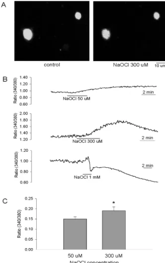
NaOCl 50µM(n = 7), 300 µM(n = 31)을 3분간 처리하였 을 때 세포내 칼슘양의 증가가 관찰되었는데 농도의 증가 에 따라 칼슘양의 증가가 현저하였다(0.153 ± 0.012, 0.186 ±
0.018) (p < 0.05). 세포내 칼슘양의 증가는 약물처리 30초 -2분 후부터 나타났는데 약물농도가 높으면 반응속도가 빠 른 경향을 보였으며 약물이 포함되지 않은 용액으로 재관 류시 5-20분 유지되다가 원래 상태로 회복되었다(Fig. 2B).
고농도의 NaOCl(1 mM)을 처리하였을 때는 대부분의 세 포에서 칼슘양의 증가를 보이다 빠르게 감소하는 파형을 보이는데 이러한 세포는 세포의 팽창에 의한 세포막이 파 열된 세포로 확인되었다.
대표적인 광범위 항산화제인 PBN 2 mM을 세포외액에 전처리한 후 NaOCl을 300 µM 투여 하였을 때는 NaOCl 에 의한 세포내 칼슘양의 증가가 억제 되었다(0.104 ± 0.018, n = 12) (p < 0.05). 또한 세포막을 통과하지 못하는 항산화 제인 isoascorbate 500 µM에 의해서도 NaOCl에 의한 변 화가 억제되었다(0.026 ± 0.008, n = 12, p < 0.01) (Fig. 3).
Fig. 1. Examples of fluorescent fura-2 imaging of a DRG slice and acutely dissociated DRG neurons. (A) A fluorescent imaging of a DRG slice (left) and dissociated DRG neurons (right). Original magnification, × 400. (B) When cells were treated with external high K+ solution in a dissociated DRG neurons, the increase of intracellular calcium concentration ([Ca2+]i) was observed (upper).
Capsaicin 5µM, a TRPV1 channel agonist, induced increase [Ca2+]i. Changes in intracellular Ca2+ concentration were monitored by ratio metric fluorescence imaging.
Fig. 2. [Ca2+]i responses by NaOCl in acutely dissociated DRG neurons. (A) Activation of Ca2+ influx by NaOCl 300µM before (left) and 3 min after wash-out (right). Original magnification,
× 400. (B) [Ca2+]i responses by NaOCl 50µM (upper), 300 µM (middle) and 1 mM (lower). Note that high concentration of NaOCl (1 mM) induced an abrupt decrease of [Ca2+]i indicating cell burst. (C) Mean NaOCl-induced peak [Ca2+]i responses by application of NaOCl 50 and 300µM. *denotes significant differ- ence from the 50µM group by paired t-test (*p < 0.05). Means ± SEM.
이러한 결과로 NaOCl에 의한 세포내 칼슘양의 증가는 주 로 활성산소종이 세포막에 존재하는 이온 수송 단백질에 영향을 미쳐 발생하였을 것으로 추측할 수 있다.
NaOCl에 의한 세포내 칼슘양 증가 기전
NaOCl에 의한 세포내 칼슘양의 증가가 어떤 기전에 의 하여 발생하는지를 확인하기 위하여 세포외 용액에서 칼슘 을 제거하고 칼슘 chelator인 EGTA 1 mM을 첨가한 용액 에서 NaOCl을 300 µM 투여하였다. 칼슘양의 변화는 −0.140
± 0.065 (n = 12)로 대조군에 비하여 유의하게 감소하였다 (p < 0.01). Thapsigargin은 세포내 칼슘저장소로 Ca2+을 reuptake 하는 과정을 차단함으로써 칼슘저장소내의 칼슘 을 고갈시킨다. Thapsigargin 1 µM을 전처리하고 NaOCl 을 추가적으로 투여하였을 때 0.010 ± 0.002 (n = 7, p < 0.01) 의 변화가 발생하였고, thapsigargin과 칼슘을 제거한 세포 외 용액을 같이 사용하였을 때에도 0.029 ± 0.032 (n = 8) 의 변화를 보여 대조군에 비해 유의하게 감소하였다(p < 0.01) (Fig. 4 Ab, Ac). 또한 칼슘을 제거하고 thapsigargin을 첨 가한 세포외 용액에서 세포내 칼슘양을 기록하다가 칼슘이 포함된 용액으로 교환하는 방법으로 저장고 의존성 통로의 존재를 확인하였는데 용액 교환 시 세포내 칼슘양의 증가 가 관찰되어(Fig. 4 Ad), 저장고 의존성 통로(store operated channel; SOC) 혹은 transient receptor potential 통로(TRP
channel)가 세포내 칼슘농도 조절에 관여하고 있음을 확 인하였다.
NaOCl이 TRP 수용체에 직접 작용하여 칼슘을 이동시키 는지 확인하고자 광범위 TRP 수용체 차단제 ruthenium red 50µM, TRPV1 수용체 차단제 capsazepine 10 µM을 전처 리한 후 NaOCl을 처리하였다. 각각 0.038 ± 0.007 (n = 17, p < 0.01), −0.047 ± 0.020 (n = 14, p < 0.01)의 변화를 보였 다(Fig. 5).
고 찰
후근신경절 뉴론은 통각 전달신경 섬유를 포함하는 일차 감각신경의 세포체가 존재하는 곳이다. 이 세포의 흥분성은 통증의 역치를 결정하는 첫 관문으로 많은 종류의 신경전 달물질이 이 신경세포의 시냅스 전달을 매개하고 있다. 말 초에서는 지속적인 통증자극에 의하여 역치가 넘은 자극에 대해서 정상적인 경우보다 더 강하게 반응하며 역치하 자
Fig. 3. Effects of antioxidants on NaOCl-induced [Ca2+]i responses.
(A) When cells were pretreated with 2 mM of PBN (upper) and 500µM of isoascorbate (lower), ROS scavengers, NaOCl failed to induce NaOCl-induced [Ca2+]i increase. (B) NaOCl-induced [Ca2+]i response under control condition and pretreatment of ROS scavengers. *denotes significant difference from the control by independent t-test (*p < 0.05, **p < 0.01). Means ± SEM.
Fig. 4. Effects of Ca2+ store depletion and a removal of extracelluar Ca2+ on NaOCl-induced [Ca2+]i responses. (A) NaOCl-induced [Ca2+]i increase was completely blocked by pretreatment with Ca2+
free solution (a), thapsigargin, Ca2+ ATPase inhibitor (b) and thapsigargin plus Ca2+ free solution (c). Cells were treated with thapsigargin in Ca2+ free solution to deplete the Ca2+ stores and Ca2+
was re-added (d). This resulted in a robust Ca2+ influx. (B) Mean NaOCl-induced [Ca2+]i responses in control and in neurons pre- treated with Ca2+ free solution, thapsigargin and thapsigargin plus Ca2+ free solution. **: Values are significantly different from the control by independent t-test (p < 0.01). Means ± SEM.
극에 대해서도 반응하는 말초감작(peripheral sensitization) 현상을 보인다(Woolf & Thompson, 1991; Willis &
Coggeshall, 2004).
NaOCl은 용액내에서 HOCl로 가수분해되며 HOCl은 myeloperoxidase (MPO)의 존재 하에 자연적으로 염소이 온과 과산화수소로 부터 합성되는데 최근의 보고에 의하 면 MPO는 미세교세포, 단핵세포, 대식세포, 호중성구 등 에서 많이 분포한다(Daugherty et al., 1994; Nagra et al., 1997). HOCl은 반응성이 매우 높아서 thiol group, thioether, amino group, heme group 등을 쉽게 산화시키며(Albrich et al., 1982; Winterbourn, 1985) 한편으로 높은 화학 반응 성에도 불구하고 쉽게 세포막을 통과하여 세포내 효소를 불활성화 시키고 미토콘드리아에서의 세포호흡을 억제하며 세포내 glutathione을 산화시킨다(Schraufstatter et al., 1990;
Eley et al., 1991; Vissers & Winterbourn., 1995; Pullar et al., 1999).
그러나 ROS는 세포독성과 관련된 역할 이외에 여러 가 지 생리적 기능을 조절하는 세포내 신호전달물질로도 작용 하고 있다(Droge, 2002). 최근에는 ROS가 통증발생에 관 여되어 있다고 보고되고 있는데, tirilazad (Khalil et al., 1999), superoxide dismutase (Wang et al., 2004), PBN (Kim et al., 2004), vitamin E (Kim et al., 2006)와 같은 다양한 항산화제를 전신투여 혹은 척수내로 투여함에 의 해 진통효과를 가짐이 보고되었다. 또한 ROS는 C 섬유
를 포함하는 일차구심신경을 활성화시킴에 의해 통증을 일 으키며 동통성 말초 신경손상에 직접 관련이 된다(Adelson et al., 1996; Wagner et al., 1998; Pop-Busui et al., 2006).
이러한 결과는 활성산소가 통증의 발생과 전달에 관여하고 있음을 의미하는 것으로 이 연구에서는 외부에서 투여한 NaOCl이 후근신경절 세포의 세포내 칼슘양을 증가시킴을 확인하였고(Fig. 2), 증가된 칼슘은 뉴론을 탈분극시켜 흥 분성을 증가시킬 수 있으며 이차전달물질로도 작용할 수 있다(Hirono et al., 1998; Chuang et al., 2000).
세포외 용액으로 고농도의 K+를 사용하면 안정막전압은 탈분극이 되고 이어 전압의존성 칼슘통로가 활성화되어 칼 슘의 세포내 유입이 발생한다. 또한 capsaicin을 처리하면 TRPV1 수용체를 활성화하여 Na+과 Ca2+등 양이온을 비 선택적으로 세포내로 이동시키는데 TRPV1 수용체는 통각 을 전달하는 C 신경섬유의 축삭, 세포체, 축삭말단에 많이 분포하는 것으로 알려져 있다(Lee et al., 2005; Spicarova
& Palecek, 2008). 이 연구에서도 고농도의 K+와 capsaicin 의 처리에 의해 빠르고 큰 칼슘유입이 관찰되어(Fig. 1) 이 실험에 사용한 세포와 fura-2의 농도 등이 NaOCl에 의한 칼슘양의 변화를 측정하는데 적절한 상태라고 생각된다.
이 연구에서 저농도의 NaOCl을 투여하였을 때 서서히 칼슘양의 증가를 유발하고 약물제거 후 5-15분 이후부터 원상태로 회복되는 가역적인 반응을 보였다(Fig. 2). 이는 통증에 관여하는 ROS의 작용은 영구적인 세포죽음을 일으 키는 것이 아닌 정상적인 세포내 신호조절물질로써의 작용 혹은 경미한 산화자극에 의한 일시적인 기능변화를 일으킨 상태라 할 수 있다. 그러나 고농도의 활성산소는 세포죽음 을 일으킬 수 있으며 이 연구에서도 1 mM의 NaOCl에 의 해 세포의 팽창에 의한 세포막이 파열이 관찰되었다(Fig. 2).
ROS는 세포막의 단백질에 직접 작용하여 산화작용을 일 으켜 효과를 나타낼 수 있다(Hong et al., 2006; Andrea et al., 2008). 후근신경절 세포에서 NaOCl에 의한 흥분성 의 증가가 직접적인 산화작용에 의한 것인지를 알아보기 위하여 세포막을 통과하지 못하는 항산화제인 isoascorbate 를 전처리 한 후 NaOCl를 투여하였을 때 NaOCl만 투여 하였을 때에 비해 유의한 감소를 보였다(Fig. 3). 이는 후 근신경절 세포에서 NaOCl에 의한 작용은 세포내 신호전 달 과정에 대한 영향보다는 세포막 수송단백질에 대한 직 접적인 산화작용에 의해 발생하였을 것으로 추측할 수 있 으며 이전에 보고된 활성산소종의 척수 후각세포에 대한 작용기전과는 차이가 있다(Lim & Chun, 2008).
ROS는 다양한 세포에서 세포내 칼슘농도의 증가를 일으 키는데, L형의 칼슘통로를 활성화 시키거나(Akaishi et al., 2004), TRPM2 (Wehage et al., 2002), ryanodine 수용체 (Favero et al., 1995), IP3 의존성 통로(Hu et al., 2000) 와 같은 칼슘방출통로를 조절하거나, 활면세포질세망 칼슘 펌프(smooth endoplasmic reticulum Ca2+ ATPase; SERCA 와 같은 칼슘펌프를 조절(Redondo et al., 2004)할 수 있 Fig. 5. Effects of transient receptor potential (TRP) channel block-
ers on NaOCl-induced [Ca2+]i responses. (A) NaOCl-induced [Ca2+]i increase was blocked by pretreatment with ruthenium red 50µM, a broad spectrum TRP antagonist (upper), capsazepine 10µM, a TRPV1 antagonist (lower). (B) Mean NaOCl-induced [Ca2+]i responses in control and in neurons pretreated with drugs.
**: Values are significantly different from the control by indepen- dent t-test (p < 0.01). Means ± SEM.
다. 세포내의 증가된 칼슘은 이차전달물질로 작용할 수 있고 Na+-Ca2+교환기전의 활성화(Hirono et al., 1998), 비 특이성 양이온 전도도의 증가(Chuang et al., 2000)를 유 발하여 막전압의 탈분극을 일으킬 수 있다. 이 연구에서 칼슘을 제거하고 thapsigargin을 첨가한 세포외 용액에서 세포내 칼슘양을 기록하다가 칼슘이 포함된 용액으로 교 환하는 방법으로 저장고 의존성 통로의 존재를 확인하였 다(Fig. 4 Ad). 이러한 세포내 칼슘의 고갈 후 밖으로부터 칼슘 유입을 일으키는 이온통로를 이전부터 저장고 의존 성 통로(SOC) 혹은 transient receptor potential 통로(TRP channel)로 명명하였고 최근 많이 연구되어지고 있다(Parekh and Penner, 1997).
실험적으로 연구자들이 TRP 통로를 구별하는 방법은 세 포내 칼슘 저장고를 고갈하였을 때 활성화되는 것을 기준 으로 하는데, 즉 thapsigargin이나 cyclopiazonic acid (CPA) 를 사용하여 전류가 활성화되거나 혹은 세포내 칼슘이 증 가하면 SOC가 존재한다고 하였다. 이 연구에서도 thapsigargin 을 전처리한 후 NaOCl을 처리하였을 때 세포내 칼슘양의 변화가 유의하게 감소한 것은 NaOCl이 SOC를 통하여 Ca2+
의 유입을 유발하였을 가능성을 배제할 수 없으며 차후의 연구에서 저장고 의존성 통로에 특이적으로 작용하는 차단 제를 사용하여 확인하고자 한다.
결론적으로 이 연구에서 NaOCl의 투여는 후근신경절 세 포에서 세포내 칼슘양을 증가시켰는데 이러한 결과는 주 로 NaOCl이 직접적으로 세포막에 작용하여 TRP 통로를 통하여 유발되었다고 할 수 있다. 또한 이러한 점은 ROS 가 이전의 역치하 입력신호에 대하여 더 쉽게 활동전압을 발생시킬 수 있어 통증의 정보전달과정에 중요하게 관여 할 수 있음을 시사한다.
감사의 글
이 논문은 2008년도 원광대학교의 교비지원에 의해서 수행되었음.
참 고 문 헌
Adelson, D.W, Wei, J.Y, Kruger, L. H2O2 sensitivity of afferent splanchnic C fiber units in vitro. J Neurophysiol. 1996;76:
371-80.
Akaishi T, Nakazawa K, Sato K, Saito H, Ohno Y, Ito Y.
Hydrogen peroxide modulates whole cell Ca2+ currents through L-type channels in cultured rat dentate granule cells. Neurosci Lett. 2004;356:25-8.
Albrich JM, McCarthy CA, Hurst JK. Biological reactivity of hypochlorous acid: implications for microbicidal mechanisms of leukocyte myeloperoxidase. Proc Natl Acad Sci USA.
1981;78:210-4.
Andrea P, Romanello M, Massimiliano B, Steinberg TH, Tell G. H2O2 modulates purinergic-dependent calcium signalling in osteoblast-like cells. Cell Calcium. 2008;43:457-68.
Baran CP, Zeigler MM, Tridandapani S, Marsh CB. The role of ROS and RNA in regulating life and death of blood monocytes. Curr Pharm Des. 2004;10:855-66.
Bubici C, Papa S, Pham CG, Zazzeroni F, Franzoso G. The NF- kappaB-mediated control of ROS and JNK signaling. Histol Histopathol. 2006;21(1):69-80.
Chuang SC, Bianchi R, Wong RK. Group I mGluR activation turns on a voltage-gated inward current in hippocampal pyramidal cells. J Neurophysiol. 2000;83:2844-53.
Daugherty A, Dunn JL, Rateri DL, Heinecke JW. Myeloperoxidase, a catalyst for lipoprotein oxidation, is expressed in human atherosclerotic lesions. J Clin Invest. 1994;94:437-44.
Droge W. Free radicals in the physiological control of cell function. Physiol Rev. 2002;82:47-95.
Eley DW, Eley JM, Korecky B, Fliss H. Impairment of cardiac contractility and sarcoplasmic reticulum Ca2+ATPase activity by hypochlorous acid: reversal by dithiothreitol. Can J Physiol Pharmacol. 1991;69:1677-85.
Favero TG, Zable AC, Abramson JJ. Hydrogen peroxide stimulates the Ca2+ release channel from skeletal muscle sarcoplasmic reticulum. J Biol Chem. 1995;270:25557-63.
Gonzalez C, Sanz-Alfayate G, Agapito MT, Gomez-Niño A, Rocher A, Obeso A. Significance of ROS in oxygen sensing in cell systems with sensitivity to physiological hypoxia.
Respir Physiol Neurobiol. 2002;22; 132(1):17-41.
Hirono M, Konishi S, Yoshioka T. Phospholipase C-independent group I metabotropic glutamate receptor-mediated inward current in mouse Purkinje cell. Biochem Biophys Res Commun. 1998;251:753-8.
Hong JH, Moon SJ, Byun HM, Kim MS, Jo H, Bae YS, Lee SI, Bootman MD, Roderick HL, Shin DM, Seo JT. Clinical role of phospholipase C1 in the generation of H2O2-evoked [Ca2+]i oscillations in cultured rat cortical astrocytes. J Biol Chem. 2006;281:13057-67.
Hu Q, Zheng G, Zweier JL, Deshpande S, Irani K, Ziegelstein RC. NADPH oxidase activation increases the sensitivity of intracellular Ca2+ stores to inositol 1,4,5-trisphosphate in human endothelial cells. J Biol Chem. 2000;275:15749-57.
Khakh BS, North RA. P2X receptors as cell-surface ATP sensors in health and disease. Nature. 2006;3;442(7102):
527-32.
Khalil Z, Khodr B. A role for free radicals and nitric oxide in delayed recovery in aged rats with chronic constriction nerve injury. Free Rad Biol Med. 2001;31:430-9.
Khalil Z, Liu T, Helme RD. Free radicals contribute to the reduction in peripheral vascular responses and the maintenance of thermal hyperalgesia in rats with chronic constriction injury. Pain. 1999;79:31-7.
Kim HK, Park SK, Zhou JL, Taglialatela G, Chung K, Coggeshall RE, Chung JM. Reactive oxygen species (ROS) play an important role in a rat model of neuropathic pain.
Pain. 2004,111:116-24.
Kim HK, Kim JH, Gao X, Zhou JL, Lee I, Chung K, Chung JM.
Analgesic effect of vitamin E is mediated by reducing central sensitization in neuropathic pain. Pain. 2006;122:53-62.
Kumazawa T, Perl ER. Excitation of marginal and substantia gelatinosa neurons in the primate spinal cord: indication of their place in dorsal horn functional organization. J Comp Neurol. 1978;177:417-34.
Lai J, Hunter JC, Porreca F. The role of voltage-gated sodium channels in neuropathic pain. Curr Opin Neurobiol. 2003;
13:291-7.
Lee YJ, Lee CH, Oh UT. Painful channels in sensory neurons.
Mol cells. 2005;20(3):315-24.
Levy D, Zochodne DW. Local nitric oxide synthase activity in a model of neuropathic pain. Eur J Neurosci. 1998;10:1846-55.
Lim SJ, Chun SW. Mechanisms of tert-buthyl hydroperoxide- induced membrane depolarization in rat spinal substantia gelatinosa neurons. Int J Oral Biol. 2008;33:117-23.
Liu D, Liu J, Sun D, Wen J. The time course of hydroxyl radical formation following spinal cord injury: the possible role of the iron-catalyzed Haber-Weiss reaction. J Neurotrauma. 2004;
21:805-16.
Nagra RM, Becher B, Tourtellotte WW, Antel JP, Gold D, Paladino T, Smith RA, Nelson JR, Reynolds WF. Immuno- histochemical and genetic evidence of myeloperoxidase involvement in multiple sclerosis. J Neuroimmunal. 1997;
78:97-107.
Parekh AB, Penner R. Store depletion and calcium influx.
Physiol Rev. 1997;77(4):901-30.
Park ES, Gao X, Chung JM, Chung K. Levels of mitochondrial reactive oxygen species increase in rat neuropathic spinal doesal horn neurons. Neurosci Lett. 2006;391:108-11.
Pop-Busui, R, Sima, A, Stevens, M. Diabetic neuropathy and oxidative stress. Diabetes Metab Res Rev. 2006;22:257-73.
Pullar JM, Winterbourn CC, Vissers MC. Loss of GSH and thiol enzymes in endothelial cell exposed to sublethal concentrations of hypochlorous acid. Am J Physiol. 1999;277:H1505-12.
Redondo PC, Salido GM, Rosado JA, Pariente JA. Effect of hydrogen peroxide on Ca2+ mobilisation in human platelets through sulphydryl oxidation dependent and independent mechanisms. Biochem Pharmacol. 2004;67:491-502.
Ruan T, Lin YS, Lin KS, Kou YR. Sensory transduction of pulmonary reactive oxygen species by capsaicin-sensitive vagal lung afferent fibres in rats. J Physiol. 2005;565:563-78.
Sawada Y, Hosokawa H, Matsumura K, Kobayashi S. Activation of transient receptor potential ankyrin 1 by hydrogen peroxide.
Eur J Neurosci. 2008;27:1131-42.
Schraufstatter IU, Browne K, Harris A, Hyslop PA, Jackson JH, Quehenberger O, Cochrane CG. Mechanisms of hypochlorite injury of target cells. J Clin Invest. 1990;85(2):554-62.
Spicarova D, Palecek J. The role of spinal cord vanilloid (TRPV1) receptors in pain modulation. Physiol Res. 2008;
57(3):S69-77.
Vissers MC, Winterbourn CC. Oxidation of intracellular glutathione after exposure of human red blood cells to hypochlorous acid. Biochem J. 1995;307(Pt1):57-62.
Wagner, R, Heckman, H.M, Myers, R.R. Wallerian degeneration and hyperalgesia after peripheral nerve injury are glutathione- dependent. Pain. 1998;77:173-9.
Wang ZQ, Porreca F, Cuzzocrea S, Galen K, Lightfoot R, Masini E. A newly identified role for superoxide in inflam- matory pain. J Pharmacol Exp Ther. 2004;309:869-78.
Watt, B.E, Proudfoot, A.T, Vale, J.A. Hydrogen peroxide poisoning. Toxicol Rev. 2004;23:51-7.
Wehage E, Eisfeld J, Heiner I, Jungling E, Zitt C, Luckhoff A.
Activation of the cation channel long transient receptor potential channel 2 (LTRPC2) by hydrogen peroxide. A splice variant reveals a mode of activation independent of ADP-ribose. J Biol Chem. 2002;277:23150-6.
Wemmie JA, Price MP, Welsh MJ. Acid-sensing ion channels:
advances, questions and therapeutic opportunities. Trends Neurosci. 2006;29(10):578-86.
Willis WD, Coggeshall RE. Sensory mechanisms of the spinal cord. Kluwer Academic/Plenum Publishers. New York, 2004.
Winterbourn CC. Comparative reactivities of various biological compounds with myeloperoxidase-hydrogen peroxide-chloride, and similarity of the oxidant to hypochlorite. Biochim Biophys Acta. 1985;840(2):204-10.
Woolf CJ, Thompson SWN. The induction and maintenance of central sensitization is dependent on N-methyl-D-aspartic acid receptor activation; implication for the treatment of post- injury pain hypersensitivity states. Pain. 1991;44:293-9.
Yoshimura M, Jessel TM. Membrane properties of rat substantia gelatinosa neurons in vitro. J Neurophysiol. 1989;62:109-18.
Zorov DB, Juhaszova M, Sollott SJ. Mitochondrial ROS- induced ROS release: an update and review. Biochim Biophys Acta. 2006;1757(5-6):509-17.
![Fig. 3. Effects of antioxidants on NaOCl-induced [Ca 2+ ]i responses.](https://thumb-ap.123doks.com/thumbv2/123dokinfo/5498957.451576/4.892.479.806.548.973/fig-effects-antioxidants-naocl-induced-ca-i-responses.webp)
