Wdpcp, a Protein that Regulates Planar Cell Polarity, Interacts with Multi‐PDZ Domain Protein 1 (MUPP1) through a PDZ Interaction
Won Hee Jang
1, Young Joo Jeong
1, Sun Hee Choi
1, Sung Su Yea
1, Won Hee Lee
2, Mooseong Kim
2, Sang-Jin Kim
3, Sang-Hwa Urm
4, Il Soo Moon
5and Dae-Hyun Seog
1*
1Department of Biochemistry, Inje University College of Medicine, Busan 614-735, Korea
2Department of Neurosurgery, Inje University College of Medicine, Busan 614-735, Korea
3Department of Neurology, Inje University College of Medicine, Busan 614-735, Korea
4Department of Preventive Medicine, Inje University College of Medicine, Busan 614-735, Korea
5Department of Anatomy & Dongguk Medical Institute, College of Medicine, Dongguk University, Gyeongju 780-714, Korea Received December 3, 2015 /Revised January 5, 2016 /Accepted January 5, 2016
Protein-protein interactions regulate the subcellular localization and function of receptors, enzymes, and cytoskeletal proteins. Proteins containing the postsynaptic density-95/disks large/zonula occlu- dens-1 (PDZ) domain have potential to act as scaffolding proteins and play a pivotal role in various processes, such as synaptic plasticity, neural guidance, and development, as well as in the pathophysi- ology of many diseases. Multi-PDZ domain protein 1 (MUPP1), which has 13 PDZ domains, has a scaffolding function in the clustering of surface receptors, organization of signaling complexes, and coordination of cytoskeletal dynamics. However, the cellular function of MUPP1 has not been fully elucidated. In the present study, a yeast two-hybrid system was used to identify proteins that inter- acted with the N-terminal PDZ domain of MUPP1. The results revealed an interaction between MUPP1 and Wdpcp (formerly known as Fritz). Wdpcp was identified as a planar cell polarity (PCP) effector, which is known to have a role in collective cell migration and cilia formation. Wdpcp bound to the PDZ1 domain but not to other PDZ domains of MUPP1. The C-terminal end of Wdpcp was essential for the interaction with MUPP1 in the yeast two-hybrid assay. This interaction was further confirmed in a glutathione S-transferase (GST) pull-down assay. When coexpressed in HEK-293T cells, Wdpcp was coimmunoprecipitated with MUPP1. In addition, MUPP1 colocalized with Wdpcp at the same subcellular region in cells. Collectively, these results suggest that the MUPP1-Wdpcp interaction could modulate actin cytoskeleton dynamics and polarized cell migration.
Key words :
MUPP1, PDZ domain, Planar cell polarity (PCP), scaffolding protein, Wdpcp
*Corresponding author
*Tel : +82-51-890-6974, Fax : +82-51-894-5801
*E-mail : [email protected]
This is an Open-Access article distributed under the terms of the Creative Commons Attribution Non-Commercial License (http://creativecommons.org/licenses/by-nc/3.0) which permits unrestricted non-commercial use, distribution, and reproduction in any medium, provided the original work is properly cited.
Journal of Life Science 2016 Vol. 26. No. 3. 282~288 DOI : http://dx.doi.org/10.5352/JLS.2016.26.3.282
Introduction
Many biological processes are regulated by protein-pro- tein interactions. Protein-protein interactions mediated by a variety of domains, functionally independent unit structures of protein, are critical for the formation of functional protein networks [10]. Postsynaptic density-95/disks large/zonula occludens-1 (PDZ) domain is one of those domains that me- diate protein-protein interactions [10, 24, 26]. PDZ do- main-containing proteins are generally soluble cytoplasmic proteins that act as scaffolds by linking the cell membrane
receptors via PDZ domains or other protein modules to actin cytoskeleton and actin-binding proteins or signaling proteins such as regulators of membrane trafficking, cell polarity and stabilization of cell surface proteins [8-10, 12, 24, 26]. PDZ domain-mediated interactions can play an important role in the assembly of large multi-molecular networks [26]. The im- portance of the PDZ-mediated multi-molecular networks is demonstrated by the fact that PDZ interactions are disrupted in pathological situations such as infectious diseases or can- cers [17, 22, 30].
PDZ domains are built of 80~100 amino-acid residues and
bound to the carboxyl (C)-termini of multiple binding part-
ners, including cell surface receptors, channel proteins, and
other adaptor proteins [7, 24, 28]. PDZ domain-binding part-
ners have one of two classes of consensus PDZ-binding mo-
tifs at their C-termini (class I: S/T-X-Φ, class II: Φ-X-Φ, where
Φ is a hydrophobic residue) [14]. PDZ domain-containing
proteins have single or multiple copies of PDZ domain [26,
29]. Multi-PDZ domain protein 1 (MUPP1), which possesses 13 PDZ domains, was first isolated as a protein that interacts with the C-terminus of the serotonin receptor type 2C (5-HT
2C) [29]. MUPP1 is expressed in the brain, enriched in synapse, especially in post-synaptic density (PSD) and tight junctions, and has been reported to interact with a variety of integral membrane proteins, including a synaptic adhe- sion molecule Cadm1 using its PDZ1-5 domain, junctional adhesion molecule-A (JAM-A) using PDZ9, neurexin 1 using PDZ2, and sodium channel Nav1.4, melatonin receptor (MT
1), Claudin-1, and γ-aminobutyric acid receptor 2 (GABAR2) using PDZ13 [1, 2, 3, 6, 13, 16, 18]. MUPP1 may act as a scaffold for the proper assembling and localization of its interacting proteins [11]. It also interacts with synaptic Ras GTPase-activating protein SynGAP, muskelin, and Ca
2+/ calmodulin-dependent kinase (CaMKII) to regulate neuronal signaling and dendritic spine morphology [2, 11, 19, 23].
To clearly define the scaffolding function of MUPP1, fur- ther identification of the interacting proteins of MUPP1 is required. We screened for proteins that interact with the amino (N)-terminal PDZ domain of MUPP1 through the yeast two-hybrid assay and identified Wdpcp, known to contain two WD40 domains and regulate cell polarity and polarized cell migration by modulation of the actin cytoske- leton through the interaction with septin 2 (Sept2) [4, 5, 8].
The MUPP1 and Wdpcp interaction suggests that MUPP1 may serve a critical scaffolding function for regulation of planar cell polarity and polarized cell migration.
Materials and Methods
Plasmid constructs
Full-length rat MUPP1 cDNA in the pCMV vector (a gift from Dr. H. Lubbert, Ruhr-Universitat, Denmark) was tag- ged with a FLAG-epitope at the N-terminus. Truncations of MUPP1 corresponding to different PDZ domains were pre- pared by PCR amplification using the appropriate primers.
The amplified fragments were subcloned into T-vector. The fragments were then EcoRI-restricted and subcloned into the
EcoRI site of pLexA. The correct orientation and in-framecloning of cDNA inserts were verified by restriction enzyme analysis and DNA sequencing. EGFP-fused Wdpcp was con- structed and used to visualize the intracellular localization in HEK-293T cells. General recombinant DNA techniques were performed according to standard protocol [25].
Screening of MUPP1-binding proteins by yeast two-hybrid assay
The Matchmaker LexA two-hybrid system was used for screening according to the manufacturer’s manual (Clontech, Palo Alto, CA, USA). In brief, the rat MUPP1 cDNA frag- ment corresponding to PDZ1 domain (amino acids 102-251) was fused to the DNA-BD region of the pLexA vector using PCR and the resulting plasmid was transformed into yeast strain EGY48 carrying the p8op-lacZ gene. Transformed EGY48 yeast cells containing the MUPP1 bait plasmid were transformed with the mouse brain cDNA library and grown on synthetic dextrose (SD) plates supplemented with glucose but with no histidine, tryptophan, or uracil (SD/-His/-Trp/
-Ura). The selection of positive clones was performed on an SD/-His/-Trp/-Ura/-Leu plate containing galactose, raffi- nose, X-gal, and BU salts. Plasmids from positive clones were analyzed by restriction digestion. Unique inserts were sequenced and protein sequence analysis was performed with the BLAST algorithm at the National Center for Biotechnology Information (NCBI). Sequence-verified clones were tested again for interaction with the bait in yeast by retransformation.
β-Galactosidase activity in liquid cultures of yeast The β-galactosidase activity of yeast was assayed as de- scribed previously [16]. Mid-log phase yeast cells were col- lected and permeabilized with 0.1% sodium dodecyl sul- phate (SDS) and chloroform. An excess amount of o-nitro- phenyl-β-D-galactoside (ONPG) was added to yeast lysate, the mixture was incubated at 30℃, and then the reaction was stopped by increasing pH to 11 by the addition of 1 M Na
2CO
3. The formation of the reaction product, o-nitro- phenol, was determined by measuring absorbance at 420 nm on a spectrophotometer and normalizing for the reaction time. The units of enzyme activity were calculated by the following equation: units = 1,000× [OD
420– 1.75x OD
550] / [reaction time (min) x culture volume (ml) x OD
600]. All ex- periments were independently performed at least three times.
Glutathione S-transferase (GST) pull-down assays
cDNA encoding the full length Wdpcp was cloned into
pET41a. The recombinant GST-Wdpcp fusion protein was
expressed in bacterial strain BL21 GOLD (Stratagene, La Jolla
CA, USA) after induction with 0.5 mM isopropyl thio-β-
D-galactopyranoside (IPTG) for 3 hr. The fusion proteins
were purified by attachment to glutathione-agarose beads (Sigma-Aldrich, St. Louis, MO, USA) according to the manu- facturer’s protocol. The mouse brain S2 fraction was in- cubated overnight at 4°C with the GST fusion protein-cou- pled glutathione beads. The beads were pelleted by cen- trifugation, washed three times with the extraction buffer (1% Triton X-100 in PBS containing 10 μg/ml each aprotinin, leupeptin, and pepstatin and 1 μM phenylmethanesulfonyl fluoride), and once with PBS. The bound proteins were elut- ed from the glutathione beads with 100 μl of Laemmli
’s load- ing buffer. The pulled-down proteins were analyzed by im- munoblotting with anti-MUPP1 antibody (BD SCIENCE, San Jose, CA, USA).
Cell culture and transfection
HEK-293T cells were cultured in Dulbecco's modified Eagle's medium supplemented with 10% fetal bovine serum, l-glutamine, and antibiotics. Transient transfections were done with the CaPO
4precipitation method.
Co-immunoprecipitation
Twenty-four hours after transfection with FLAG-MUPP1 and myc-Wdpcp constructs, HEK-293T cells were rinsed with ice-cold PBS twice and lysed with ice-cold lysis buffer [PBS containing 0.5% NP-40 and 1x protease inhibitor cock- tail set V (Calbiochem)] by gentle rotation for 30 min.
Lysates were centrifuged at 16,000× g for 10 min at 4°C. The supernatant was incubated with anti-FLAG M2 agarose beads (Sigma-Aldrich) for 2 hr at 4°C with constant shaking.
The beads were collected by centrifugation at 2,000× g for 30 sec and washed 5 times with ice-cold PBS containing 0.5%
NP-40. The immunoprecipitated proteins were analyzed by immunoblotting.
Immunocytochemistry
HEK-293T cells grown on poly-D-lysine-coated coverslips were transfected with EGFP-Wdpcp and MUPP1 constructs.
Twenty-four hours after transfection, cells were washed with PBS, fixed with 4% paraformaldehyde in PBS for 5 min, and permeabilized with 0.2% Triton X-100 in PBS for 10 min.
After blocking with 5% normal goat serum in PBS for 30 min, cells were incubated with anti-MUPP1 antibody diluted 1:500 in PBS containing 1% bovine serum albumin (BSA) and 0.05% Tween-20 overnight at 4°C. After washing 3 times with PBS, cells were incubated with Dylight 594-conjugated goat anti-rabbit IgG antibody (Jackson ImmunoResearch
Labs, West Grove, PA, USA) diluted 1:800 for 40 min. After washing 3 times with PBS, the cells were mounted with Fluoromount (DAKO). Fluorescence images were acquired on Zeiss LSM510 META confocal laser scanning microscope (Carl Zeiss, Oberkochem, Germany).
Results
Identification of MUPP1 interacting proteins by yeast two-hybrid screening
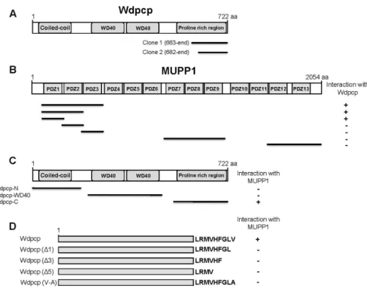
To identify the binding proteins of MUPP1, we screened a mouse brain cDNA library through the yeast two-hybrid assays using the N-terminal region of MUPP1 containing PDZ1 domain as bait (Fig. 1B). From 9×10
6colonies screened, we obtained two positive clones. The clones possessed cDNA fragments of Wdpcp (Fig. 1A). To determine whether the other PDZ domains of MUPP1 also interact with Wdpcp, we constructed various PDZ domain-containing fragments of MUPP1. Yeast two-hybrid assays with Wdpcp showed that, among the PDZ domains tested, only the PDZ1 domain of MUPP1 is required for binding (Fig. 1B). Wdpcp is a mul- ti-domain protein comprised of one coiled-coil domain and two WD40 repeated domains [4]. To identify the domain of Wdpcp required for interaction with MUPP1, various frag- ments of Wdpcp were constructed and tested for interaction with MUPP1. As shown in Fig. 1C, the short C-terminal re- gion of Wdpcp was required for interaction with MUPP1.
The C-terminus of Wdpcp contains a putative class II PDZ-binding motif [4, 14]. Therefore, we investigated whether the putative C-terminal motif of Wdpcp mediates the interaction with MUPP1. For this purpose, the C-termi- nal deletion and substitution mutants of Wdpcp were con- structed and co-transfected into yeast cells with pLexA- MUPP1. As shown in Fig. 1D, the MUPP1 and Wdpcp inter- action was impaired by the C-terminal deletion or the sub- stitution of the last C-terminal residue of Wdpcp. These re- sults indicate that MUPP1 and Wdpcp interact with each other through its PDZ domain and PDZ-binding motif, re- spectively, consistent with the previously described class II PDZ interaction [26]. Next, we compared the binding of Wdpcp to MUPP1 with those of two known MUPP1 interact- ing proteins, muskelin and neurexin 1 [15, 16]. A yeast two-hybrid assay (Fig. 2A) and a quantitative β-galactosi- dase assay (Fig. 2B) showed that Wdpcp has a binding affin- ity comparable to muskelin and neurexin 1.
Further, we examined the MUPP1 and Wdpcp interaction
Fig. 1. Identification of the protein interacting with MUPP1 by yeast two-hybrid screening. (A) Schematic diagram of Wdpcp. Wdpcp contains the coiled-coil domain, WD40 domains, and proline-rich region. Clone 1 and clone 2 were isolated from the yeast two-hybrid screen. (B) Minimal Wdpcp binding region in MUPP1. Different truncations of MUPP1 were constructed by PCR and tested in the yeast two-hybrid assay for interaction with Wdpcp. +, interaction with Wdpcp; -, no interaction with Wdpcp.
aa, the amino acid residue number. (C, D) Specific interaction of MUPP1 with the C-terminal region of Wdpcp. Several different truncated forms and deletion and substitution mutants of Wdpcp were constructed by PCR and tested in the yeast two-hybrid assay for interaction with MUPP1. +, interaction with MUPP1; -, no interaction with MUPP1.
A B
Fig. 2. Interaction between MUPP1 and Wdpcp. (A) Wdpcp and proteins containing class II PDZ-binding motif (muskelin and neurexin 1) were fused to the pLexA DNA binding domain. Wdpcp interacted with MUPP1. +, interaction with MUPP1;
-, no interaction with MUPP1. (B) The strength of interaction between MUPP1 and Wdpcp were compared with those of muskelin and neurexin 1 quantitatively using β–galactosidase activity in yeast two-hybrid reporter assay.
at the protein level using GST pull-down experiments.
Recombinant GST-Wdpcp fusion protein was expressed in
E. coli. The purified GST fusion protein was allowed to inter-act with mouse brain lysates. Immunoblot analysis revealed
that MUPP1 interacted with GST-Wdpcp, but not with GST,
and the C-terminal deletion of Wdpcp impaired the inter-
A B
C
Fig. 3. MUPP1 and Wdpcp were co-immunoprecipitated and co-localized in mammalian cells. (A) Proteins in the mouse brain lysate were allowed to bind to GST alone, GST- Wdpcp, GST-Wdpcp (△1), GST-Wdpcp (△5), and GST-Neurexin 1 fusion proteins. The elution fractions were resolved by SDS-PAGE and immunoblotting was performed using anti-MUPP1 antibody.
(B) HEK-293T cells were transiently transfected with FLAG-MUPP1 and myc-Wdpcp plasmids as indicated. Cell lysates were incubated with monoclonal anti-FLAG M2 agarose beads to immunoprecipitate MUPP1. Immunoblots were subsequently probed with anti-FLAG and anti-myc antibodies. Wdpcp was specifically co-immunoprecipitated with MUPP1. (C) Twenty- four hours after transfection with MUPP1 and EGFP-Wdpcp, HEK-293T cells were subjected to immunoflourescent staining using anti-MUPP1 and Dylight 594-conjugated secondary antibodies. EGFP-Wdpcp and MUPP1 co-localize largely in cells.
action (Fig. 3A).
MUPP1 is associated with Wdpcp in cells
To examine the interaction between MUPP1 and Wdpcp in mammalian cells, HEK-293T cells were co-transfected with constructs expressing FLAG-MUPP1 and myc-Wdpcp.
Cell lysates were immunoprecipitated with a monoclonal an- tibody against the FLAG epitope, followed by immunoblot analysis with anti-myc and anti-FLAG antibodies. Fig. 3B shows that Wdpcp was co-precipitated with MUPP1 (Fig.
3B). This result indicates that MUPP1 specifically interacts with Wdpcp in cells. To determine whether MUPP1 and Wdpcp co-localize in cells, MUPP1 was co-transfected with EGFP-Wdpcp into HEK-293T cells. Confocal microscopic im- ages of EGFP-Wdpcp and MUPP1 showed that MUPP1 and Wdpcp co-localized at the same region in cells (Fig. 3C).
These findings indicate that MUPP1 interacts with Wdpcp in cells.
Discussion
In the present study, we have shown that the scaffold
protein MUPP1 interacts with Wdpcp. Using the PDZ1 do- main containing region of MUPP1 as bait, we identified Wdpcp in a yeast two-hybrid screen. The C-terminal end region of Wdpcp can interact with PDZ1 domain of MUPP1.
Furthermore, using a combination of GST pull-down assay and co-immunoprecipitation, we confirmed that MUPP1 in- teracted with Wdpcp at the protein level. Moreover, when MUPP1 and Wdpcp were co-expressed in mammalian cells, they co-localize at the same region in cells.
Specific protein-protein interactions are important for the
localization and clustering of cell surface receptors at the
specific subcellular site [26]. The PDZ domain is one of most
abundant interaction modules found in the proteins that act
as scaffolds of multi-protein complexes through specific in-
teraction of their PDZ domains to the C-terminal binding
motif of interacting proteins, thereby linking the interacting
proteins to cytoskeletal proteins or other protein modules
[10, 24]. Therefore, PDZ domain-containing proteins usually
form large multimeric protein complexes [24, 26]. Interest-
ingly, MUPP1 contains 13 PDZ domains and plays an im-
portant role as a scaffold of protein complexes via PDZ do-
mains and binding protein’s modules [29]. Wdpcp has G-L-V
seqeunce at C-terminus, which is similar to the class II PDZ-binding motif [4]. In this study, we demonstrated through domain analysis and deletion analysis that the C-terminal three amino acids of Wdpcp act as a binding mo- tif required for specific interaction with the PDZ1 domain of MUPP1.
Wdpcp has a short N-terminal coiled-coil domain consist- ing of five heptad repeats and two WD40 domains [4]. The WD40 domain has been identified in many proteins, and forms a β-propeller structure that provides surfaces for pro- tein-protein interaction [27]. WD40 domain-containing pro- teins have been shown to serve as scaffolds for assembly of multi-protein complexes. Wdpcp is localized in the actin cytoskeleton and interacts with Sept2, also found in actin filaments [21]. Septins are highly conserved GTP-binding proteins which form filamentous cytoskeleton by hetero-oli- gomerization [20]. In recent report, Wdpcp-deficient cells showed a disruption in the actin cytoskeleton required for planar cell polarity and polarized cell migration [5]. Wdpcp appears to modulate the actin cytoskeleton by mediating Sept2 interaction with actin filaments [5]. Therefore, it is speculated that the MUPP1-Wdpcp interaction may indicate the formation of a multi-scaffold for regulation of cell polar- ity and cell migration by modulating the actin cytoskeleton.
Our findings provide insight into the possible regulation of maintaining cell polarity and polarized cell migration by MUPP1-Wdpcp complex through PDZ domain-mediated interaction. Further experiments are needed to examine the effect of MUPP1-Wdpcp interaction on Sept2 interaction with actin filament. Studies on the possibilities mentioned above and identification of other MUPP1 interacting proteins may help to shed light on regulation of maintaining cell po- larity and polarized cell migration.
Acknowledgments
The research was supported by the Basic Science Research Program through the National Research Foundation of Korea (NRF) by the Ministry of Science, ICT and Future Planning (NRF-2012R1A1A2020689).
References
1. Adachi, M., Hamazaki, Y., Kobayashi, Y., Itoh, M., Tsukita, S., Furuse, M. and Tsukita, S. 2009. Similar and distinct properties of MUPP1 and Patj, two homologous PDZ do- main-containing tight-junction proteins. Mol. Cell. Biol. 29,
2372-2389.
2. Balasubramanian, S., Fam, S. R. and Hall, R. A. 2007.
GABAB receptor association with the PDZ scaffold Mupp1 alters receptor stability and function. J. Biol. Chem. 282, 4162-4171.
3. Becamel, C., Figge, A., Poliak, S., Dumuis, A., Peles, E., Bockaert, J., Lubbert, H. and Ullmer, C. 2001. Interaction of serotonin 5-hydroxytryptamine type 2C receptors with PDZ10 of the multi-PDZ domain protein MUPP1. J. Biol.
Chem. 276, 12974-12982.
4. Collier, S., Lee, H., Burgess, R. and Adler, P. 2005. The WD40 repeat protein fritz links cytoskeletal planar polarity to frizzled subcellular localization in the Drosophila epi- dermis. Genetics 169, 2035-2045.
5. Cui, C., Chatterjee, B., Lozito, T. P., Zhang, Z., Francis, R.
J., Yagi, H., Swanhart, L. M., Sanker, S., Francis, D., Yu, Q., San Agustin, J. T., Puligilla, C., Chatterjee, T., Tansey, T., Liu, X., Kelley, M. W., Spiliotis, E. T., Kwiatkowski, A. V., Tuan, R., Pazour, G. J., Hukriede, N. A. and Lo, C. W. 2013.
Wdpcp, a PCP protein required for ciliogenesis, regulates directional cell migration and cell polarity by direct modu- lation of the actin cytoskeleton. PLoS Biol. 11, e1001720.
6. Dooley, R., Baumgart, S., Rasche, S., Hatt, H. and Neuhaus, E. M. 2009. Olfactory receptor signaling is regulated by the post-synaptic density 95, Drosophila discs large, zona-occlu- dens 1 (PDZ) scaffold multi-PDZ domain protein 1. FEBS J. 276, 7279-7290.
7. Doyle, D. A., Lee, A., Lewis, J., Kim, E., Sheng, M. and MacKinnon, R. 1996. Crystal structures of a complexed and peptide-free membrane protein-binding domain: molecular basis of peptide recognition by PDZ. Cell 85, 1067-1076.
8. Field, C. M. and Kellogg, D. 1999. Septins: cytoskeletal poly- mers or signaling GTPases? Trends Cell. Biol. 9, 387-394.
9. Garner, C. C., Nash, J. and Huganir, R. L. 2000. PDZ do- mains in synapse assembly and signalling. Trends Cell. Biol.
10, 274-280.
10. Gomperts, S. N. 1996. Clustering membrane proteins: It's all coming together with the PSD-95/SAP90 protein family.
Cell 84, 659-662.
11. Guillaume, J. L., Daulat, A. M., Maurice, P., Levoye, A., Migaud, M., Brydon, L., Malpaux, B., Borg-Capra, C. and Jockers, R. 2008. The PDZ protein mupp1 promotes Gi cou- pling and signaling of the Mt1 melatonin receptor. J. Biol.
Chem. 283, 16762-16771.
12. Guillemot, L., Foglia, A., Paschoud, S., Pulimeno, P. and Citi, S. 2008. The cytoplasmic plaque of tight junctions: a scaffolding and signalling center. Biochim. Biophys. Acta.
1778, 601-613.
13. Hamazaki, Y., Itoh, M., Sasaki, H., Furuse, M. and Tsukita, S. 2002. Multi-PDZ domain protein 1 (MUPP1) is con- centrated at tight junctions through its possible interaction- with claudin-1 and junctional adhesion molecule. J. Biol.
Chem. 277, 455-461.
14. Hirbec, H., Perestenko, O., Nishimune, A., Meyer, G., Nakanishi, S. and Henley, J. M. 2002. The PDZ proteins PICK1, GRIP and Syntenin bind multiple glutamate receptor
초록:Planar cell polarity 조절단백질 Wdpcp와 multi-PDZ domain protein 1 (MUPP1)의 PDZ 결합
장원희
1․정영주
1․최선희
1․예성수
1․이원희
2․김무성
2․김상진
3․엄상화
4․문일수
5․석대현
1*
(1인제대학교 의과대학 생화학교실, 2인제대학교 의과대학 신경외과학교실, 3인제대학교 의과대학 신경과학교실,
4인제대학교 의과대학 예방의학교실, 5동국대학교 의과대학 해부학교실)
단백질-단백질 결합은 수용체 단백질, 효소, 세포 골격 단백질의 세포내 위치 결정 및 기능 조절에 중요한 역할 을 한다. Postsynaptic density-95/disks large/zonula occludens-1 (PDZ) 도메인을 가진 단백질들은 시냅스 가소 성, 신경세포 성장과 분화뿐만 아니라 많은 질병의 병태생리에 중요하게 관여하는 scaffold 단백질로 작용한다.
Multi-PDZ domain protein 1 (MUPP1)은 13개 PDZ 도메인을 가지는 단백질로서 세포막 수용체 군집화, 신호전 달 복합체 구성, 세포 골격 조정에 대한 매개 역할을 하는 것으로 알려지고 있지만 MUPP1의 세포 내 기능은 아직 명확히 밝혀지지 않았다. 본 연구에서 MUPP1의 아미노 말단 PDZ 도메인과 결합하는 새로운 단백질을 규명하기 위하여 효모 two-hybrid 방법을 이용하였고 Wdpcp (전에 Fritz로 알려짐)이 MUPP1과 결합하는 것을 확인하였 다. Wdpcp는 planar cell polarity (PCP) effector로서 세포 이동과 섬모형성에 관여하는 것으로 알려져 있다.
Wdpcp는 MUPP1의 첫 번째 PDZ 도메인과 결합하지만, 다른 PDZ 도메인과는 결합하지 않았다. 또한 MUPP1와 Wdpcp의 결합에서 Wdpcp의 C-말단부위가 결합에 필수적임을 효모 two-hybrid 방법으로 확인하였다. 이러한 단백질간 결합은 glutathione S-transferase (GST) pull-down assay, 공동면역침강, HEK-293T 세포에서의 발현위 치를 통하여 추가적으로 확인하였다. 이러한 결과들은, MUPP1과 Wdpcp 결합은 세포내 액틴 다이내믹스 (dynamics)와 세포이동 조절에 역할을 할 가능성을 시사한다.
subtypes. J. Biol. Chem. 277, 15221-15224.
15. Jang, W. H., Jeong, Y. J., Choi, S. H., Lee, W. H., Kim, M., Kim, S. H., Urm, S. H., Moon, I. S and Seog, D. H. 2015.
Muskelin Interacts with Multi-PDZ Domain Protein 1 (MUPP1) through the PDZ Domain. J. Life Sci. 25. 594-600.
16. Jang, W. H., Choi, S. H., Jeong, J. Y., Park, J. H., Kim, S.
J. and Seog, D. H. 2014. Neuronal cell-surface protein neu- rexin 1 interaction with multi-PDZ domain protein MUPP1.
Biosci. Biotechnol. Biochem. 78, 644-646.
17. Javier, R. T. 2008. Cell polarity proteins: common targets for tumorigenic human viruses. Oncogene 27, 7031-7046.
18. Kimber, W. A., Trinkle-Mulcahy, L., Cheung, P., Deak, M., Marsden, L. J. and Kieloch, A. 2002. Evidence that the tan- dem-pleckstrin-homology-domain-containing protein TAPP1 interacts with Ptd(3,4)P2 and the multi-PDZ-domain-con- taining protein MUPP1 in vivo. Biochem. J. 361, 525-536.
19. Krapivinsky, G., Medina, I., Krapivinsky, L., Gapon, S. and Clapham, D. E. 2004. SynGAP-MUPP1-CaMKII synaptic complexes regulate p38 MAP kinase activity and NMDA receptor-dependent synaptic AMPA receptor potentiation.
Neuron 43, 563-574.
20. Mostowy, S. and Cossart, P. 2012. Septins: the fourth com- ponent of the cytoskeleton. Nat. Rev. Mol. Cell Biol. 13.
183-194.
21. Park, T. J., Kim, S. K. and Wallingford, J. B. 2015. The planar cell polarity effector protein Wdpcp (Fritz) controls epi- thelial cell cortex dynamics via septins and actomyosin.
Biochem. Biophys. Res. Commun. 456. 562-566.
22. Pearson, H. B., Perez-Mancera, P. A., Dow, L. E., Ryan, A., Tennstedt, P., Bogani, D., Elsum, I., Greenfield, A., Tuveson, D. A., Simon, R. and Humbert, P. O. 2011. SCRIB expression
is deregulated in human prostate cancer, and its deficiency in mice promotes prostate neoplasia. J. Clin. Invest. 121, 4257-4267.
23. Pei, L., Teves, R. L., Wallace, M. C. and Gurd, J. W. 2001.
Transient cerebral ischemia increases tyrosine phosphor- ylation of the synaptic RAS-GTPase activating protein, SynGAP. J. Cereb. Blood Flow Metab. 21, 955-963.
24. Ponting, C. P., Phillips, C., Davies, K. E. and Blake, D. J.
1997. PDZ domains: targeting signalling molecules to sub- membranous sites. Bioessays 19, 469-479.
25. Sambrook, J., Fritsch, E. F. and Maniatis, T. 1989. Molecular cloning: a laboratory manual. Cold Spring Habor Laboratory, Cold Spring Habor, New York.
26. Sheng, M. and Sala, C. 2001. PDZ domains and the organ- ization of supramolecular complexes. Annu. Rev. Neurosci.
24, 1-29.
27. Smith, T. F., Gaitatzes, C., Saxena, K. and Neer, E. J. 1999.
The WD repeat: a common architecture for diverse functions.
Trends Biochem. Sci. 24. 181-185.
28. Songyang, Z., Fanning, A. S., Fu, C., Xu, J., Marfatia, S. M.
and Chishti, A. H. 1997. Recognition of unique carboxyl-ter- minal motifs by distinct PDZ domains. Science 275, 73-77.
29. Ullmer, C., Schmuck, K., Figge, A. and Luëbbert, H. 1998.
Cloning and characterization of MUPP1, a novel PDZ do- main protein. FEBS Lett. 424, 63-68.
30. Zhan, L., Rosenberg, A., Bergami, K. C., Yu, M., Xuan, Z., Jaffe, A. B., Allred, C. and Muthuswamy, S. K. 2008.
Deregulation of scribble promotes mammary tumorigenesis and reveals a role for cell polarity in carcinoma. Cell 135, 865-878.