• 검색 결과가 없습니다.

Effects of Torilis japonica Extract on DNCB-induced Contact Dermatitis in BALB/c Mice

N/A
N/A
Protected

Academic year: 2021

Share "Effects of Torilis japonica Extract on DNCB-induced Contact Dermatitis in BALB/c Mice"

Copied!
17
0
0

로드 중.... (전체 텍스트 보기)

전체 글

(1)

pISSN 1738-6640 eISSN 2234-4020 http://dx.doi.org/10.6114/jkood.2016.29.4.061

Original Article / 원저

Effects of Torilis japonica Extract on DNCB-induced Contact Dermatitis in BALB/c Mice

Kyou-Young Lee

1)

·Eun-Hye Song

2)

·Chul-Hee Hong

1)*

1 Dept. of Oriental Opthalmology and Otolaryngology and Dermatology, College of Korean Medicine, Sangji University

2 Dept. of Pharmacology, College of Korean Medicine, Sangji University

사상자 추출물이 DNCB로 유도된 BALB/c 마우스의 접촉성 피부염에 미치는 효과

이규영

1)

·송은혜

2)

·홍철희

1)*

1 상지대학교 한의과대학 안이비인후피부과학교실 2 상지대학교 한의과대학 약리학교실

Abstract

목적: 본 연구에서는 TJ 열수추출물이 아토피 피부염에 미치는 효과를 확인하고자 하였다.

방법: 2, 4-dinitrochlorobenzen (DNCB)으로 BALB/c 마우스에 1차 감작 후 3주간 2차 감작을 시행하여 아토피 유사 피 부병변을 나타내는 접촉성 피부염을 유발한 뒤 동결건조 처리된 TJ 분말을 (PBS/EtOH/Cremophor=6:1:3)에 용해시켜 쥐의 등 피부에 10, 50 ㎎/㎖ 농도로 3주간 도포하였다.

결과: TJ 열수추출물은 아토피 피부 병변의 염증세포 침윤을 억제하였고 (10, 50 ㎎/㎖) 표피와 진피 두께를 회복시켰으며 (10, 50 ㎎/㎖), 혈청에서 히스타민 방출을 억제하였다 (10 ㎎/㎖). 또한 Th2 세포와 관련된 cytokine인 interleukin (IL)-4, IL-5, IL-13과 thymic stromal lymphopoietin (TSLP)의 mRNA 발현 양을 감소시켰고 (10, 50 ㎎/㎖), Th2 chemokine인 thymus- and activation-regulated chemokine (TARC or CCL17)의 mRNA의 발현 양을 감소시켰다 (10, 50 ㎎/㎖).

결론: TJ 열수추출물을 BALB/c 마우스에 외용하였을 때 항아토피 효과가 있음을 확인하였으며 따라서 TJ가 아토피 피부 염 치료의 외용제로 활용될 수 있을 것으로 사료된다.

Key words : Torilis japonica (TJ); Type 2 helper T (Th2); Atopic dermatitis (AD); Allergic contact dermatitis (ACD)

ⓒ 2016 the Society of Korean Medicine Ophthalmology & Otolaryngology & Dermatology

This is an Open Access journal distributed under the terms of the Creative Commons Attribution Non-Commercial License (http://creativecommons.org/license/by-nc/3.0/) which permits unrestricted non-commercial use, distribution, and reproduction in any medium, provided the original work is properly cited.

(2)

Ⅰ. Introduction

Allergic contact dermatitis (ACD) is a common occupational and environmental health issue. In common with other forms of allergy, this disease progresses in two phages; an initial stage during which sensitization is acquired, and if the sensitized individual is exposed subsequently, at the same or a different skin site, to the inducing chemical allergen, then a more vigorous secondary immune response will be provoked at the point of contact. This in turn initiates the cutaneous inflammatory reaction that is defined clinically as ACD

1)

.

2,4-dinitrochlorobenzene (DNCB) is an acute allergy-inducing substance. Atopic dermatitis (AD) mice model commonly uses chemical antibodes such as DNCB, 2,4-dinitrofluorobenzene and 2,4,6-trinitrochlorobenzene in order to cause atopy-like skin lesions. DNCB-induced mice model is frequently used for the investigation of AD

2,3)

. In the present study, acute ACD was induced by applying DNCB for 3 weeks of short period after first challenge, so skin lesions were atopy-like which is Th2 cell dominant.

AD, also called atopic eczema and allergic disorders, is a chronic inflammatory skin disease associated with cutaneous hyperreactivity to immunoglobulin E (IgE)-mediated sensitization

4)

. AD appears during babyhood and childhood but can last or start in adult

5)

. The prevalence of AD is 10–20% in children and 1–3% in adults

6)

. The

pathogenesis of AD can be attributed to complex interactions dependent on heritable and environmental factors

7,8)

.

The T cell plays important roles in allergic disorders

9)

. T helper cells, known as CD4+T cells, are subdivided into the type 1 helper T (Th1) and the type 2 helper T (Th2), which produce different profiles of cytokine

10)

. Pathognomonic features of AD include eczematous skin lesions with inflammatory cell accumulation, Th1/ Th2 balance skewed toward Th2

11)

. Th2 cells produce Th2 cytokines such as interleukin (IL)-4, IL-5, IL-13 and elevate serum IgE levels

11,12)

. IL-4 and IL-13 stimulate IgE production in B cell

13,14)

. IgE released from B cells binds to FcεRI, the high-affinity receptor for IgE of mast cell

15)

. Binding of an antigen by FcεRI-bound specific IgE leads to FcεRI clustering, which in turn induces downstream signaling and ultimately the release of mediators. The antigen complex IgE−

FcεRI on the surface of the mast cell induces mast cell degranulation

16)

. Mast cell releases histamine, leukotrienes, prostaglandins, chemokines and pro-inflammatory cytokines

17,18)

. IL-5 plays an important role in infiltration of eosinophil

19)

. The histology of AD is characterized by a dermal inflammatory infiltration containing eosinophils and mast cells

20)

. AD exhibits a compromised barrier function that leads to the activation of keratinocytes. Barrier disruption promotes the production of thymus- and activation-regulated chemokine (TARC or CCL17) from keratinocytes

21)

. Thymic stromal lymphopoietin (TSLP) is highly expressed in the epidermis of the skin lesions of AD patients and keratinocytes

22,23)

. Overexpression of TSLP by

Corresponding author : Chul-Hee Hong, School of Oriental Medicine, Sangji University, St. 80, Wonju city, Gangwon, 220-717, South Korea.

(Tel : 033-741-9266, E-mail : [email protected])

Recieved 2016/10/6

Revised 2016/11/3

Accepted 2016/11/10

(3)

keratinocytes results in an AD-like inflammatory phenotype in mice

24)

.

Corticosteroids and anti-histamines are drugs commonly available to treat AD

25,26)

. However, these drugs are accompanied by side effects that may include hypertension, osteoporosis, blurred vision, and dizziness

27)

. Calcineurin inhibitors also have been used topically and are considered quite safe

28)

, but prolonged application of these drugs is reported to cause skin cancer

29)

. Thus, development of alternative therapeutic agents is necessary for long-term therapy of AD.

The fruits of Torilis japonica (TJ) is 'Sa-Sang-Ja', which is traditional medical herbs prescribed as anti-fungal, anti-allergenic, antibacterial and sedative agent

30)

. In addition, several studies have demonstrated that TJ possess anti-inflammatory

31)

, anti-angiogenic

32)

, anti-invasive

33)

, and anti-microbial

34)

effects. Although these biological effects of TJ have been investigated, there were no reports of AD. In this study, we examined the inhibitory effect of TJ on DNCB-induced contact dermatitis (CD) animal models, and through acute CD skin lesions effects on AD were investigated.

Ⅱ. Materials and Methods

1. Materials for Experiments 1) Reagents

1-Chloro-2, 4-dinitrobenzene, dexamethasone, acetone and all other chemicals were purchased from Sigma Chemical Co. (St. Louis, MO, USA).

Histamine ELISA kits were purchased from Enzo

Life Sciences (East Farmingdale, NY, USA).

Power SYBR® Green Master Mix was purchased from applied biosystems (Foster City, CA, USA).

Glyceraldehyde 3-phosphate dehydrogenase (GAPDH) and IL-4, IL-5, IL-13, TARC, TSLP oligonucleotide primers were purchased from Bioneer (Daejeon, South Korea).

2) Preparation of aqueous extract of Torilis japonica (TJ)

TJ was purchased from Daehyo chamchowon (Suwon, Gyeong-gi, Korea). The dried fruit of TJ (400g) was extracted with 2800 ㎖ boiling distilled water for 2 h, and the extract was then concentrated under low pressure. After filtration, an aqueous solution of the extract was concentrated in a rotary evaporator, freeze-dried for five days, and stored in aliquots at 4 ℃. The yield of dried extract from starting crude materials was about 8.2% (w/w) for 32.8g dry weight.

3) Animals

BALB/c mice (female, 20-25g, 6 weeks old) were obtained from Daehan Biolink (Daejeon, South Korea). Animals were housed in an animal room at 23 ℃ with a relative humidity of 55%, a 12 h light/dark cycle. The mice were given an unlimited amount of water and food throughout the duration of the experiment. Animal care and experimental procedure were performed according to “the guide for the Care and Use of Laboratory Animal”, as adopted and promulgated by the

“Institutional Animal Care Committee, Sang ji

University” (Reg. No. 2016-03).

(4)

2. Experiments methods

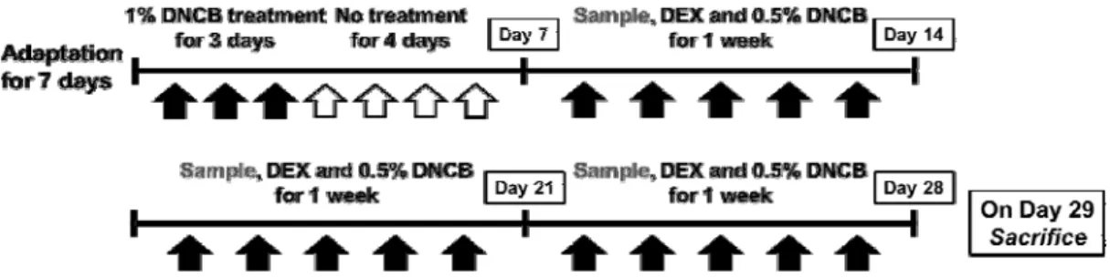
1) Induction of CD skin lesions in BALB/c mice

Animals were randomized into five groups (n=6 per group): Normal (mice treated with vehicle alone), DNCB (mice sensitized with DNCB, negative control), DNCB+DEX (mice sensitized with DNCB and treated with 10 µM dexamethasone, positive control) and DNCB+TJ 10 and 50 (mice sensitized with DNCB and treated with 10 and 50 ㎎/㎖ TJ).

After a 7 day adaptation period, the hair on the upper back was shaved and the experiment was immediately started. To induce CD skin lesions which were AD-like, mice were sensitized topically with 100 ㎕ of 1% DNCB in corn oil/acetone (1:4, v/v) for three days, followed by no treatment for four days. The same volume of vehicle(acetone/corn oil) was applied to the Normal group. After the first challenge, the treatment was repeated with 100 ㎕ of 0.5%

DNCB for 21 days. The DEX group and TJ group were respectively topically treated with 100

㎕ of DEX (10 µM, dissolved in PBS/EtOH/Cremophor=6:1:3) and 100 ㎕ of TJ (10, 50 ㎎/㎖, dissolved in PBS/EtOH/ Cremophor

Fig. 1. Schematic representation of the experimental design

=6:1:3) 4 h after DNCB treatment once a day, whereas Normal and DNCB groups were treated with vehicle (PBS/EtOH/Cremophor). Mice were sacrificed on day 29 of the experiment(Fig. 1).

Skin tissues from the back of the mice were excised and subjected to histological analysis, real time polymerase chain reaction examination.

2) Evaluation of dermatitis severity

Clinical dermatitis severity was tested using the method described by Yamamoto and colleagues

35)

. The severity of dermatitis was evaluated at experiment start day and end day. The development of erythema/hemorrhage, scarring/

dryness, edema, and excoriation/erosion was scored as follow: 0, none; 1, mild (<20%); 2, moderate (20-60%); and 3, severe (>60%). The sum of the individual scores was used as the dermatitis score.

3) Histological analysis of skin lesions

Upon euthanasia, skin samples from the dorsal

area were collected. The samples were fixed in

10% buffered formalin, embedded in paraffin,

sectioned into 4 ㎛ slices, and stained with

hematoxylin and eosin. Pathological changes

(epidermal, dermal hyperplasia, hyperkeratosis,

(5)

dermal edema, parakeratosis, vesicular formation, and inflammation) were assessed and compared between these groups. The images of the stained slides were observed using a DM IL LED microscope (Leica, Wetzlar, Germany) and photographed using a DFC295 (Leica, Wetzlar, Germany). Digital images were taken from each slide (2 per group), and measured using Leica Application Suite (Leica, Wetzlar, Germany).

4) Histamine assay

Blood from the orbital sinus was collected from each mouse at the end of the experiment. Serum was obtained by centrifugation at 1700 g for 30 min and stored at -80 ℃ until analysis. Serum levels of histamine in mice were measured using commercially available EIA kits (Enzo Life Sciences, Farmingdale, NY, USA) according to the manufacturer’s recommendations.

5) Quantitative real-time polymerase chain reaction (qRT-PCR)

The dorsal skin tissues were homogenized, and

Gene Primer Sequences (5’-3’)

IL-4 Left GGCTTCCAAGGTGCTTC

Right CAGGCATCGAAAAGCC

IL-5 Left TTCCTGAAGGCTGAGGTTACAG

Right AACATGCACAAAGCCTTGGG

IL-13 Left AGAGGATATTGCATGGCCTCTG

Right TGCTTTGTGTAGCTGAGCAG

TSLP Left TCCCCGACAAAACATTTGCC

Right GGACTTCTTGTGCCATTTCCTG

TARC Left TGCCATCGTGTTTCTGACTG

Right ATGGCCTTGGGTTTTTCACC

GAPDH Left AAATGGTGAAGGTCGGTGTG

Right AATCTCCACTTTGCCACTGC

Table 1. Primer Sequences Used for qRT-PCR Ampification

total RNA was isolated using the Easy-Blue®

Reagent according to the manufacturer’s instructions Kits (Intron Biotechnology, Seongnam, South Korea). Total RNA was quantified using an Epoch® micro-volume spectrophotometer system (BioTek Instruments;

Winooski, VT, USA). In brief, total RNA was converted to cDNA using a high-capacity cDNA reverse transcription kit (Applied Biosystems;

Foster City, CA, USA) and thermocycler (Gene Amp® PCR system 9700; Applied Biosystems) with the following program: initiation for 10 min at 25 ℃, followed by incubation at 50 ℃ for 90 min and at 85 °C for 5 min. Real-time PCR analysis was conducted using a Step One Plus®

Real-time PCR system (Applied Biosystems).

SYBR® Green master mix and primers were used for PCR analysis of GAPDH, IL-4, IL-5, IL-13, TARC or CCL17 and TSLP. The PCR cycling parameters were as follows: 10 min at 95 ℃; 40 cycles of 5 s at 95 ℃ and 45 s at 60 ℃; and a final melting curve of 15 s at 95 ℃, 1 min at 60

℃, and 15 s at 95 ℃. All primer sequences are

(6)

shown in Table 1. Gene expression was calculated according to the comparative threshold cycle (Ct) method.

6) Statistical analysis

All the values reported have been expressed as the mean ± standard deviation (SD). Data were analyzed using one-way analysis of variance (ANOVA) with Dunnett’s test, and p-values <

0.05 were considered statistically significant. All statistical analyses were performed using GraphPad Prism (version 5.00 for Windows, San Diego, CA, USA).

Ⅲ. Results

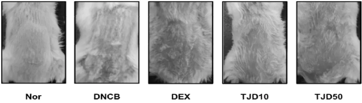
1. Effects of TJ on clinical features of skin lesions in DNCB-induced CD

Clinically, topical exposure of DNCB results in skin symptoms of itching, erythema, and hemorrhage after a week, then it rapidly progresses into edema, superficial erosion, deep excoriation, scaling, and dryness of the skin within two weeks following the repeated treatment with DNCB

11,36)

. As shown in Fig. 2, repeated topical application of DNCB induced

Fig. 2. Effects of TJ on clinical features of skin lesions in DNCB induced CD

erythema, superficial erosion, excoriation, scaling, and dryness. However, treatment with TJ inhibited these skin symptoms. Representative photographic images of mice from different treatment groups at the time of a sacrifice. Nor:

Normal control; DNCB: DNCB treated; DEX:

Dexamethasone treated with DNCB; TJD 10: TJ 10 ㎎/㎖ treated with DNCB; TJD 50: TJ 50 ㎎/㎖

treated with DNCB.

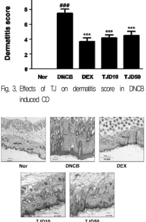
2. Effects of TJ on dermatitis score in DNCB-induced CD

Skin conditions were evaluated at experiment start day and end day using a dermatitis scoring method. The dermatitis scores of DNCB group increased compared with those of the Normal group. However, in DEX and TJ treated mice, the dermatitis scores were lower than DNCB group(Fig. 3).

Dermatitis scores were measured at experiment start day and end day. The dermatitis score was defined as the sum of scores graded as 0 (none), 1 (mild), 2 (moderate), 3 (severe) for each of the four symptoms (erythema/hemorrhage, scarring/

dryness, edema, and excoriation/erosion). Data are

presented as mean±standard deviation (SD; n=6).

(7)

Data were analyzed using one-way analysis of variance followed by Dunnett’s post hoc test.

***P < 0.001 versus DNCB group; ### P < 0.001 versus Normal group.

3. Effects of TJ on histological analysis in DNCB-induced CD

In addition, we examined application of TJcould affect the interrupted skin barrier using H&E staining. Histological features of the skin lesions showed epidermal, dermal hyperplasia and

Fig. 3. Effects of TJ on dermatitis score in DNCB induced CD

Fig. 4. Effects of TJ on histopathological feature of skin lesions in DNCB induced CD

Tissues were excised, fixed in 10% formaldehyde, embedded in paraffin, and sectioned. The sections were stained with hematoxylin and eosin (H&E) (magnification, 4x).

accumulation of inflammatory cells in the dermis/epidermis of DNCB group compared with Normal group. In contrast, TJ treated group exhibited less of these changes in DNCB-induced mice(Fig. 4).

4. Effects of TJ on histamine release in DNCB-induced CD

Mast cells play a key role in allergic reactions

37)

. Mast cells secrete inflammatory mediators such as histamine

38)

. Histamine, a major component of mast cells, is a symptom inducing substance in AD

36)

. Histamine is involved in induction of itching and edema

39)

. In order to evaluate whether TJ can modulate degranulation of mast cells, histamine assay was performed using EIA kit. The histamine release in the serum markedly increased in the DNCB group compared with Normal group, whereas TJ treatment significantly suppressed histamine levels. Treatment with DEX also attenuated the changes in histamine levels(Fig. 5).

Fig. 5. Effects of TJ on histamine release in DNCB-induced CD

Histamine concentrations were measured in mouse serum by ELISA. Data are presented as mean±standard deviation (SD;

n=6). Data were analyzed using one-way analysis of variance followed by Dunnett’s post hoc test. *P < 0.05 and **P < 0.01 versus DNCB group; ### P < 0.001 versus Normal group.

(8)

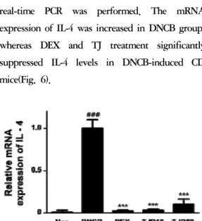
5. Effects of TJ on the mRNA expression of IL-4 in DNCB-induced CD

IL-4 promotes differentiation of Th2 cells and IgE class switching by B cells. Its receptor has been associated with AD

40)

. To investigate the effect of TJ on the mRNA expression of IL-4, real-time PCR was performed. The mRNA expression of IL-4 was increased in DNCB group, whereas DEX and TJ treatment significantly suppressed IL-4 levels in DNCB-induced CD mice(Fig. 6).

Fig. 6. Effects of TJ on mRNA expression of IL-4

Total RNA was prepared for the real-time PCR analysis of IL-4 gene expression from skin tissues. Real-time PCR analysis was conducted using a Step One Plus® Real-time PCR system. Data are presented as mean±standard deviation (SD; n=6). Data were analyzed using one-way analysis of variance followed by Dunnett’s post hoc test. ***P < 0.001 versus DNCB group; ###

P < 0.001 versus Normal group.

6. Effects of TJ on the mRNA expression of IL-5 in DNCB-induced CD

IL-5 produced by Th2 cells, induces eosinophil differentiation, activation, mobilization, and survival

41)

. Eosinophils are important mediators of the inflammatory process in AD, so agents that block IL-5 might be developed as therapeutics

42)

. In this study, the mRNA levels of IL-5 was increased in DNCB group compared with Normal group, but TJ inhibited the mRNA levels of IL-5

in DNCB induced CD mice(Fig. 7).

Fig. 7. Effects of TJ on mRNA expression of IL-5

Total RNA was prepared for the real-time PCR analysis of IL-5 gene expression from skin tissues. Data are presented as mean±standard deviation (SD; n=6). Data were analyzed using one-way analysis of variance followed by Dunnett’s post hoc test. ***P < 0.001 versus DNCB group; ### P < 0.001 versus Normal group.

Fig. 8. Effects of TJ on mRNA expression of IL-13

Total RNA was prepared for the real-time PCR analysis of IL-13 gene expression from skin tissues. Data are presented as mean±standard deviation (SD; n=6). Data were analyzed using one-way analysis of variance followed by Dunnett’s post hoc test. ***P < 0.001 versus DNCB group; ### P < 0.001 versus Normal group.

7. Effects of TJ on the mRNA expression of IL-13 in DNCB-induced CD

IL-13 is known as a major stimulator of

inflammation and tissue remodeling at sites of

Th2 inflammation, such as in asthma

43)

. To

investigate the effects of TJ on the mRNA

expression of IL-13, real-time PCR was

performed. Our data indicated that the mRNA

(9)

levels of IL-13 was increased in DNCB group compared with Normal group, but TJ inhibited the mRNA levels of IL-13 in DNCB induced CD mice(Fig. 8).

8. Effects of TJ on the mRNA expression of TSLP in DNCB-induced CD

TSLP represents a key molecule at the epithelial cell-dendritic cell (DC) interface to initiate allergic inflammation

44)

. To investigate the effect of TJ on the mRNA expression of TSLP, real-time PCR was performed. In this study, the TSLP was significantly higher in the DNCB-induced group than in the Normal group. In the TJ-treated group, it was significantly lower than that observed in the DNCB-induced group(Fig. 9).

Fig. 9. Effects of TJ on mRNA expression of TSLP

Total RNA was prepared for the real-time PCR analysis of TSLP gene expression from skin tissues. Data are presented as mean±standard deviation (SD; n=6). Data were analyzed using one-way analysis of variance followed by Dunnett’s post hoc test. ***P < 0.001 versus DNCB group; ### P < 0.001 versus Normal group.

9. Effects of TJ on the mRNA expression of TARC in DNCB-induced CD

Keratinocytes are inflammatory sites of epidermis which induce abnormal expression of cytokines and chemokines including Th2

chemokines such as TARC

45)

. Therefore, we examined the effects of TJ on the mRNA expression of TARC. As shown in Fig. 10, the TARC was increased in the DNCB-induced group compared with Normal group. The DEX and TJ-treated groups exhibited less of an increase in the levels of TARC mRNA.

Fig. 10. Effects of TJ on mRNA expression of TARC

Total RNA was prepared for the real-time PCR analysis of TARC gene expression from skin tissues. Data are presented as mean±standard deviation (SD; n=6). Data were analyzed using one-way analysis of variance followed by Dunnett’s post hoc test. ***P < 0.001 versus DNCB group; ### P < 0.001 versus Normal group.

Ⅳ. Discussion

ACD is a type Ⅳ, delayed-type reaction which is caused by skin contact with allergens that activate antigen-specific T cells in a sensitized individual. Langerhans and dermal dendritic cells uptake the allergen and migrate to the regional lymph nodes to activate antigen-specific T cells.

These T cells then proliferate and enter the circulation and site of exposure. When reexposed to the allergen, antigen-specific T cells are activated through the release of cytokines and induce an inflammatory process

46)

.

AD is characterized by frequent itching, severe

(10)

drying and eczematous skin lesions with infiltration of immune cells

47)

. AD is associated with genetic factors, environmental factors, and psychological factors, epidermal barrier defects and immune system dysregulation

48,49)

.

The topical corticosteroids or calcineurin inhibitors are widely used to treat AD patients, but these drugs cause serious side effects

27,28)

. Therefore, we need to develop alternative and augmentative treatment substances. Several studies have demonstrated that TJ has various biological properties such as anti-microbial activity and anti-inflammation effects. In the present study, we examined anti-atopic effects of TJ in DNCB-induced BALB/c mice.

DNCB-induced mice model was very similar to human skin lesions

45)

. Treatment with TJ on the dorsal skin suppressed these skin symptoms(Fig.

2, 3). DNCB-induced mice showed increased eosinophil infiltration and number of mast cells

50)

. Our histological data also showed that the dermis and epidermis were thicker in DNCB mice compared to normal mice, but TJ group was significantly reduced(Fig. 4).

Th2 cells increase IgE

13,14)

. Mast cells are key effectors in IgE-associated immediate allergic reactions including allergic disorders

37,51)

. Binding of an antigen by FcεRI-bound specific IgE leads to mast cell degranulation. Activated mast cell releases mediators such as histamine, cytokines, and chemokines

17)

.

Histamine is a major component of mast cells

52)

. Histamine is involved in induction of itching and edema

40)

. Histamine is detected commonly in AD skin lesions

53)

. AD patients indicated high levels of histamine in plasma

compared with healthy controls

54)

. In this study, the serum levels of histamine markdly increased following DNCB application, but TJ suppressed the serum levels of histamine in DNCB-induced CD mice(Fig. 5).

Th2 cells produce increased levels of Th2 cytokines. IL-13 and IL-4 are known to induce isotype switching to IgE synthesis. IL-4 was detected immunohistochemically in several skin lesions of AD patients

55)

, and that hyperreactivity plays a pathogenic role

56)

. The mRNA expression of IL-4 was increased in DNCB group, whereas TJ treatment significantly suppressed IL-4 levels in DNCB-induced CD mice(Fig. 6). IL-5 plays a crucial role in eosinophil promoting the production, development and survival

4,57)

. Overexpression of IL-5 is observed in eosinophil-associated diseases including AD

58)

. Our results showed that the mRNA levels of IL-5 was increased in DNCB group compared with Normal group, but TJ inhibited the mRNA levels of IL-5 in DNCB induced CD mice(Fig. 7). IL-13 is known as stimulator of inflammation at sites of Th2 inflammation, such as in asthma

44)

and plays a role in the pathogenesis of AD

59)

. Treatment of TJ downregulated DNCB-induced IL-13 mRNA expression in CD mice(Fig. 8).

TSLP mediates signaling by DCs that promotes

Th2-cell responses, and is highly upregulated in

atopic skin

60)

. TSLP released from keratinocytes

acts immediately on sensory neurons to trigger

robust itch-evoked scratching

61)

. TSLP indicates

high expression from epidermis in AD patient’s

skin

22,62)

. The TSLP was significantly higher in

the DNCB-induced group than in the Normal

group. In the TJ-treated group, it was

(11)

significantly lower than that observed in the DNCB-induced group(Fig. 9).

TARC is a member of the CC chemokine group that plays a significant role in recruiting Th2-type lymphocytes to the AD-like skin lesions

63,64)

. TARC is overproduced in epidermal keratinocytes and it is also believed to attract Th2 cells into skin from circulation blood

65,66)

. Our results indicated an increase in TARC mRNA levels in the DNCB-induced group compared to the levels in the Normal group. However, TJ-treated group exhibited less of an increase in the levels of TARC mRNA(Fig. 10).

TJ has been used as a traditional medicine for the treatment of inflammation and skin diseases such as eczema, trichophytia and pruritus of external genitals for external use. It is also prescribed as an internal medicine for impotence and infertility

67)

. Fumigation treatment of TJ is frequently applied to treat gynecologic symptoms

68)

. Since mainly it indicates the characteristical effect on skin diseases when used in external-use, we studied the effects of TJ extract on AD when used as an external application.

Limitations of this study include that firstly, concentration of the TJ extract was not a variety of settings. Because this study was the basic steps to find out whether TJ extract can be used as a therapeutic material for AD, the concentration was set by dividing only low concentration (10 ㎎/

㎖) and the high concentration (50 ㎎/㎖). The second is that assessment of the toxicity of the TJ extract was not performed. For reference, TJ is a non-toxic medicine

69)

, and previous researches using 3-(4,5-dimethylthiazol-2-yl)-2, 5 diphenyl-

tetrazolium bromide (MTT) assay indicated that TJ extract did not affect the cell viability

70,71)

. The third is that internal factors causing AD like genetic factors, environmental factors, and psychological factors have not been such considerations. The forth is that the therapeutic effects between external and internal-use of TJ extract were not compared. Lastly, it is related to the type of the study. This study is the animal experiments using BALB/c mice model. Data obtained through the animal experiments are often can not be directly converted to human clinical trials, and the results have the limitation that it can not be applied directly to the human beings.

In the present study, all results indicated that the inhibitory effect of TJ is better in the administration of 10 ㎎/㎖ dose of TJ than that of 50 ㎎/㎖ dose. These findings reveal that in concentration, 10 ㎎/㎖ is more optimal than 50

㎎/㎖. Further research is needed to determine optimum concentration of TJ.

In conclusion, TJ prevented infiltration of inflammatory cells(Fig. 4) and attenuated release of histamine from DNCB induced CD models(Fig.

5). TJ also suppressed expression of Th2-type cytokines and chemokine(Fig. 6-10). These results suggest that external use of TJ may have a potential therapeutic effects for AD by suppressing Th2-dominant allergic inflammation.

Ⅴ. Conclusions

In this study, we examined the potential role of

TJ on skin lesions and gene expression of

(12)

Th2-type cytokines, chemokine in DNCB induced CD in BALB/c mice.

1. TJ reduced clinical features of skin lesions in DNCB-induced CD mice.

2. TJ reduced dermatitis scores in DNCB- induced CD mice.

3. TJ reduced infiltration of inflammatory cells in DNCB-induced CD mice.

4. TJ reduced dermis and epidermis thickening in DNCB-induced CD mice.

5. TJ inhibited serum histamine release in DNCB-induced CD mice.

6. TJ suppressed mRNA level of IL-4 in DNCB-induced CD mice.

7. TJ suppressed mRNA level of IL-5 in DNCB-induced CD mice.

8. TJ suppressed mRNA level of IL-13 in DNCB-induced CD mice.

9. TJ suppressed mRNA level of TSLP in DNCB-induced CD mice.

10. TJ suppressed mRNA level of TARC/CCL4 in DNCB-induced CD mice.

Through these results we can conclude that external use of TJ may have a potential therapeutic effects for AD by suppressing Th2-dominant allergic inflammation.

※ 본 논문은 2016년도 상지대학교 박사학위논문으로 작성되었습니다.

References

1. Kimber L, Basketter DA, Gerberick GF,

Dearman RJ. Allergic contact dermatitis.

International immunopharmacology. 2002;2 (2-3):201-11.

2. Sung YY, Yang WK, Lee AY, Kim DS, Nho KJ, Kim YS, et al. Topical application of an ethanol extract prepared from illicium verum suppresses atopic dermatitis in NC/Nga mice.

J Ethnopharmacol. 2012;144:151-9.

3. Takeda K, Gelfand EW. Mouse models of allergic diseases. Curr Opin Immunol.

2009;21:660-5.

4. Leung DY, Boguniewicz M, Howell MD, Nomura I, Hamid QA. New insights into atopic dermatitis. The Journal of clinical investigation. 2004;113(5):651-7.

5. Leung DY, Bieber T. Atopic dermatitis.

Lancet. 2003;361(9352):151-60.

6. Nutten S. Atopic dermatitis: global epidemiology and risk factors. Annals of nutrition & metabolism. 2015;66 suppl 1:8-16.

7. Boguniewicz M, Leung DY. Atopic dermatitis: a disease of altered skin barrier and immune dysregulation. Immunological reviews. 2011;242(1):233-46.

8. Wang IJ, Guo YL, Weng HJ, Hsieh WS, Chuang YL, Lin SJ, et al. Environmental risk factors for early infantile atopic dermatitis.

Pediatric allergy and immunology : official publication of the European Society of Pediatric Allergy and Immunology. 2007;

18(5):441-7.

9. Hijnen D, De Bruin-Weller M, Oosting B,

Lebre C, De Jong E, Bruijnzeel-Koomen C, et

al. Serum thymus and activation-regulated

chemokine (TARC) and cutaneous T cell-

attracting chemokine (CTACK) levels in

(13)

allergic diseases: TARC and CTACK are disease-specific markers for atopic dermatitis.

The Journal of allergy and clinical immunology. 2004; 113(2):334-40.

10. Sher A, Coffman RL. Regulation of immunity to parasites by T cells and T cell-derived cytokines. Annual review of immunology.

1992;10:385-409.

11. Romagnani S. Immunologic influences on allergy and the TH1/TH2 balance. The Journal of allergy and clinical immunology.

2004;113(3):395-400.

12. Akdis CA, Akdis M, Simon D, Dibbert B, Weber M, Gratzl S, et al. T cells and T cell-derived cytokines as pathogenic factors in the nonallergic form of atopic dermatitis. The Journal of investigative dermatology. 1999;

113(4):628-34.

13. Hasbold J, Hong JS, Kehry MR, Hodgkin PD.

Integrating signals from IFN-gamma and IL-4 by B cells: positive and negative effects on CD40 ligand-induced proliferation, survival, and division-linked isotype switching to IgG1, IgE and IgG2a. Journal of immunology. 1999;

163(8):4175-81.

14. Izuhara K, Shirakawa T. Signal transduction via the interleukin-4 receptor and its correlation with atopy. International journal of molecular medicine. 1999;3(1):3-10.

15. Galli SJ, Tsai M. IgE and mast cells in allergic disease. Nature medicine. 2012;18(5):693-704.

16. Urb M, Sheppard DC. The role of mast cells in the defence against pathogens. PLoS pathogens. 2012;8(4):e1002619.

17. Weller K, Foitzik K, Paus R, Syska W, Maurer M. Mast cells are required for normal

healing of skin wounds in mice. FASEB journal : official publication of the Federation of American Societies for Experimental Biology. 2006;20(13):2366-8.

18. Amin K. The role of mast cells in allergic inflammation. Respiratory medicine. 2012;106 (1):9-14.

19. Kouro T, Takatsu K. IL-5- and eosino- phil-mediated inflammation: from discovery to therapy. International immunology. 2009;21 (12):1303-9.

20. Simon D, Braathen LR, Simon HU.

Eosinophils and atopic dermatitis. Allergy.

2004;59(6):561-70.

21. Onoue A, Kabashima K, Kobayashi M, Mori T, Tokura Y. Induction of eosinophil- and Th2-attracting epidermal chemokines and cutaneous late-phase reaction in tape-stripped skin. Experimental dermatology. 2009;18(12):

1036-43.

22. Soumelis V, Reche PA, Kanzler H, Yuan W, Edward G, Homey B, et al. Human epithelial cells trigger dendritic cell mediated allergic inflammation by producing TSLP. Nature immunology. 2002;3(7):673-80.

23. Takai T, Chen X, Xie Y, Vu AT, Le TA, Kinoshita H, et al. TSLP expression induced via Toll-like receptor pathways in human keratinocytes. Methods in enzymology. 2014;

535:371-87.

24. Yoo J, Omori M, Gyarmati D, Zhou B, Aye

T, Brewer A, et al. Spontaneous atopic

dermatitis in mice expressing an inducible

thymic stromal lymphopoietin transgene

specifically in the skin. The Journal of

experimental medicine. 2005;202(4):541-9.

(14)

25. Correale CE, Walker C, Murphy L, Craig TJ.

Atopic dermatitis: a review of diagnosis and treatment. American family physician. 1999;

60(4):1191-8, 1209-10.

26. Herman SM, Vender RB. Antihistamines in the treatment of dermatitis. Journal of cutaneous medicine and surgery. 2003;7(6):

467-73.

27. Stanbury RM, Graham EM. Systemic corticosteroid therapy--side effects and their management. The British journal of ophthalmology. 1998;82(6):704-8.

28. Segal AO, Ellis AK, Kim HL. CSACI position statement: safety of topical calcineurin inhibitors in the management of atopic dermatitis in children and adults. Allergy, asthma, and clinical immunology : official journal of the Canadian Society of Allergy and Clinical Immunology. 2013;9(1):24.

29. Carr WW. Topical calcineurin inhibitors for atopic dermatitis: review and treatment recommendations. Paediatric drugs. 2013;15 (4):303-10.

30. Kim GT, Lee SH, Kim YM. Torilis japonica extract, a new potential EMT suppressor agent by regulation of EGFR signaling pathways. International journal of oncology.

2014;45(4):1673-9.

31. Kim SM, Kim TH, Lee EB. Anti-inflammatory activities of Torilis japonica fruit. The Korean Society of Pharmacognosy. 1998;29(4):384-90.

32. Kim MS, Lee YM, Moon EJ, Kim SE, Lee JJ, Kim KW. Anti-angiogenic activity of torilin, a sesquiterpene compound isolated from Torilis japonica. International journal of cancer. 2000;87(2):269-75.

33. Kim MS, Baek JH, Park MT, Sohn TK, Kim SE, Lee JJ, et al. Anti-invasive activity of torilin, a sesquiterpene compound isolated from Torilis japonica. Oncology reports.

2001;8(2):359-64.

34. Cho WI, Choi JB, Lee K, Chung MS, Pyun YR. Antimicrobial activity of torilin isolated from Torilis japonica fruit against Bacillus subtilis. Journal of food science. 2008;73 (2):37-46.

35. Sone K, Yamamoto-Sawamura T, Kuwahara S, Nishijima K, Ohno T, Aoyama H, et al.

Changes of estrous cycles with aging in female F344/n rats. Experimental animals / Japanese Association for Laboratory Animal Science. 2007;56(2):139-48.

36. Park SJ, Lee HA, Kim JW, Lee BS, Kim EJ.

Platycodon grandiflorus alleviates DNCB- induced atopy-like dermatitis in NC/Nga mice. Indian journal of pharmacology.

2012;44(4):469-74.

37. Galli SJ, Tsai M, Piliponsky AM. The development of allergic inflammation. Nature.

2008;454(7203):445-54.

38. Petersen LJ, Mosbech H, Skov PS.

Allergen-induced histamine release in intact human skin in vivo assessed by skin microdialysis technique: characterization of factors influencing histamine releasability.

The Journal of allergy and clinical immunology. 1996;97(2):672-9.

39. Gschwandtner M, Mildner M, Mlitz V, Gruber F, Eckhart L, Werfel T, et al.

Histamine suppresses epidermal keratinocyte

differentiation and impairs skin barrier

function in a human skin model. Allergy.

(15)

2013;68(1):37-47.

40. Minami K, Kamei C. A chronic model for evaluating the itching associated with allergic conjunctivitis in rats. International immunopharmacology. 2004;4(1):101-8.

41. Guttman-Yassky E, Dhingra N, Leung DY.

New era of biologic therapeutics in atopic dermatitis. Expert opinion on biological therapy. 2013;13(4):549-61.

42. Hamelmann E, Gelfand EW. IL-5-induced airway eosinophilia--the key to asthma?

Immunological reviews. 2001;179:182-91.

43. Corren J. Anti-interleukin-5 antibody therapy in asthma and allergies. Current opinion in allergy and clinical immunology. 2011;11 (6):565-70.

44. Oh MH, Oh SY, Yu J, Myers AC, Leonard WJ, Liu YJ, et al. IL-13 induces skin fibrosis in atopic dermatitis by thymic stromal lymphopoietin. Journal of immunology. 2011;

186(12):7232-42.

45. Stein ML, Villanueva JM, Buckmeier BK, Yamada Y, Filipovich AH, Assa'ad AH, et al.

Anti-IL-5 (mepolizumab) therapy reduces eosinophil activation ex vivo and increases IL-5 and IL-5 receptor levels. The Journal of allergy and clinical immunology. 2008;121(6):

1473-83.

46. Gittler JK, Kreuger JG, Guttman-Yassky E.

Atopic dermatitis results in intrinsic barrier and immune abnormalities: implications for contact dermatitis. J Allergy Clin Immunol.

2013;131:300-13.

47. Jung M, Lee TH, Oh HJ, Kim H, Son Y, Lee EH, et al. Inhibitory effect of 5,6-dihydro- ergosteol-glucoside on atopic dermatitis-like

skin lesions via suppression of NF-kappaB and STAT activation. Journal of derma- tological science. 2015;79(3):252-61.

48. Kabashima K. New concept of the pathogenesis of atopic dermatitis: interplay among the barrier, allergy, and pruritus as a trinity. Journal of dermatological science.

2013;70(1):3-11.

49. Fonacier LS, Dreskin SC, Leung DY. Allergic skin diseases. The Journal of allergy and clinical immunology. 2010;125(2 Suppl 2):

S138-49.

50. Liu FT, Goodarzi H, Chen HY. IgE, mast cells, and eosinophils in atopic dermatitis.

Clinical reviews in allergy & immunology.

2011;41(3):298-310.

51. Kalesnikoff J, Galli SJ. New developments in mast cell biology. Nature immunology.

2008;9(11):1215-23.

52. Lindstedt KA, Kovanen PT. Isolation of mast cell granules. Current protocols in cell biology. 2006;Chapter 3(Unit 3):16.

53. Greaves MW. Antihistamines in dermatology.

Skin pharmacology and physiology. 2005;18 (5):220-9.

54. Imaizumi A, Kawakami T, Murakami F, Soma Y, Mizoguchi M. Effective treatment of pruritus in atopic dermatitis using H1 antihistamines (second-generation antihistamines):

changes in blood histamine and tryptase levels. Journal of dermatological science.

2003;33(1):23-9.

55. Tazawa T, Sugiura H, Sugiura Y, Uehara M.

Relative importance of IL-4 and IL-13 in

lesional skin of atopic dermatitis. Archives of

dermatological research. 2004;295(11):459-64.

(16)

56. Yamazaki F, Aragane Y, Maeda A, Matsushita K, Ueno K, Yudate T, et al. Overactivation of IL-4-induced activator protein-1 in atopic dermatitis. Journal of dermatological science.

2002;28(3):227-33.

57. Ackerman SJ, Bochner BS. Mechanisms of eosinophilia in the pathogenesis of hypereosinophilic disorders. Immunology and allergy clinics of North America. 2007;27(3):

357-75.

58. Corren J. Inhibition of interleukin-5 for the treatment of eosinophilic diseases. Discovery medicine. 2012;13(71):305-12.

59. Hamid Q, Naseer T, Minshall EM, Song YL, Boguniewicz M, Leung DY. In vivo expression of IL-12 and IL-13 in atopic dermatitis. The Journal of allergy and clinical immunology. 1996;98(1):225-31.

60. Wang YH, Liu YJ. Thymic stromal lymphopoietin, OX40-ligand, and interleukin-25 in allergic responses. Clinical and experimental allergy : journal of the British Society for Allergy and Clinical Immunology. 2009;39(6):798-806.

61. Moran TP, Vickery BP. The Epithelial Cell-Derived Atopic Dermatitis Cytokine TSLP Activates Neurons to Induce Itch.

Pediatrics. 2014;134 suppl 3:S160-1.

62. Alysandratos KD, Angelidou A, Vasiadi M, Zhang B, Kalogeromitros D, Katsarou-Katsari A, et al. Increased affected skin gene expression and serum levels of thymic stromal lymphopoietin in atopic dermatitis. Annals of allergy, asthma & immunology : official publication of the American College of Allergy, Asthma, & Immunology. 2010;105(5):

403-4.

63. Vestergaard C, Yoneyama H, Murai M, Nakamura K, Tamaki K, Terashima Y, et al.

Overproduction of Th2-specific chemokines in NC/Nga mice exhibiting atopic dermatitis-like lesions. The Journal of clinical investigation.

1999;104(8):1097-1105.

64. Saeki H, Tamaki K. Thymus and activation regulated chemokine (TARC)/CCL17 and skin diseases. Journal of dermatological science.

2006;43(2):75-84.

65. Shimada Y, Takehara K, Sato S. Both Th2 and Th1 chemokines (TARC/CCL17, MDC/

CCL22, and Mig/CXCL9) are elevated in sera from patients with atopic dermatitis. Journal of dermatological science. 2004;34(3):201-8.

66. Kakinuma T, Nakamura K, Wakugawa M, Mitsui H, Tada Y, Saeki H, et al. Thymus and activation-regulated chemokine in atopic dermatitis: Serum thymus and activation- regulated chemokine level is closely related with disease activity. The Journal of allergy and clinical immunology. 2001;107(3):535-41.

67. College of Oriental Medicine Compilation Committee of Herbalogy. Herbalogy. Seoul:

Younglimsa. 2006:630-1.

68. Park SM. The documentary examining of gynecologic symptoms and medical substances of fumigation treatment. Master's Thesis Kyung-San University. 2002:57.

69. Sin MG. Clinical Herbalogy. Younglimsa.

1994:197, 271, 590.

70. Koo BS, Hwang EI, So SH, Lee SK, Han GH,

Kim NM. Effect of Torilis Fructus on

Procollagen Biosynthesis and Activity of

Matrix Metalloproteinase-I(MMP-1) in Human

(17)

Dermal Fibroblast. Kor. J. Pharmacogn.

2007;38(4):350.

71. Choi JE, Park BK, Jin MR. Anti-inflammatory and Antioxidant Effects of Water Extracts of Sasangja-tang(SSJ) and Gami-sasangja-tang (GSJ). Collection of dissertations Institute of Oriental Medicine Daejeon University. 2015;

23(2):7-8.

수치

Fig. 1. Schematic representation of the experimental design
Table 1. Primer Sequences Used for qRT-PCR Ampification
Fig. 2. Effects of TJ on clinical features of skin lesions in DNCB induced CD
Fig. 4. Effects  of  TJ  on  histopathological  feature  of  skin  lesions in DNCB induced CD
+3

참조

관련 문서

Effects of Scutellaria baicalensis Extract on Skin Lesion of Contact Dermatitis induced by DNFB in Mice.. Beodul Yang, Sura Kim, Chan Hun Choi 1 , Hyun Woo Jeoung 1 ,

Fig. Effect of RJWE on clinical skin features of DNCB- applied BALB/c mice. Effect of RJWE on change of body weight in DNCB- applied BALB/c mice. Effect of RJWE on change

Therapeutic Effects of Yijungtang on Atopic Dermatitis- like Skin Lesions of NC/Nga Mouse Induced by Mite Antigen.. The Journal of Korean Oriental

DJE: 95% ethanolic extract of Dioscorea japonica rhizomes, CON: vehicle control group, oxazolone: oxazolone-treated group, oxazolone-dexa: oxazolone and 0.5%

Abstract The purpose of this study was to examine the effects of Lespedeza Caneata (LC) extracts on atopic dermatitis.. Skin thickness is a symptom of atopic dermatitis, and in

Rhemanniae radix extract on allergic contact dermatitis on Mice induced by DNCB.. The Korean Journal of Oriental

Objectives : The purpose of this study is to investigate effects of Syzygium aromaticum (SAE) spread on the allergic contact dermatitis caused

Repeated treatment with DNCB made epidermal spongiosis, edema and dermal inflammatory cellular infiltration in ACD group (B).. Effects of SR on symptom score