J Korean Neurosurg Soc/Volume 30/June, 2001 777 KISEP Case Reports J Korean Neurosurg Soc 30::::777-780, 2001
전두골에 발생한 콜레스테롤 육아종 1례
- 증 례 보 고 -
부산대학교 의과대학 신경외과학교실
이상원·차승헌·박동준·송근성·최창화·이영우
= Abstract =
Cholesterol Granuloma of Frontal Bone
---
- A Case Report ----
Sang Weon Lee, M.D., Seung Heon Cha, M.D., Dong June Park, M.D., Geun Sung Song, M.D., Chang Hwa Choi, M.D., Young Woo Lee, M.D.
Department of Neurosurgery, Pusan National University, School of Medicine, Pusan, Korea
holesterol granuloma of frontal bone is a rare disease which usually occurs at the lateral part of the supraorbital ridge. This expanding lesion grows slowly and extends into the orbit and anterior cranial fossa. The most common symptom is proptosis. This granuloma is composed of a granulomatous reaction surrounding cholesterol crystals.
Surgical treatment involves aspiration of contents and stripping or curettage of the lining which is highly successful.
We experienced a case of cholesterol granuloma of frontal bone with huge intracranial extension, which was cured by surgical removal. The clinical features, radiologic, and pathologic finding were discussed and the pertinent literatures were reviewed.
KEY WORDS:Cholesterol granuloma・Frontal bone・Proptosis.
서 론
콜레스테롤 육아종(cholesterol granuloma)은 추체첨부 (petrous apex), 상악골, 관골 등 두개골과 안면골의 다양 한 부위에서 발생이 가능하며5) 특히 측두골 추체의 병변에 대해서는 널리 알려져 있다.
전두골의 콜레스테롤 육아종은 드문 질환으로 누선와 (lacrimal fossa)에 인접한 안와상연(supraorbital ridge) 이 특정 발생부위로3) 재발성 출혈로 의해 종괴가 점진적으 로 커져 안와 및 전두개와를 침범하여 증상을 야기한다.
최근 본 교실에서 거대 두개내 종괴를 보이는 전두골 콜 레스테롤 육아종 1례를 치험하여 문헌고찰과 함께 보고하 고자 한다.
증 례
환자는 34세 여자로 내원 6개월 전부터 좌안의 점진적인
안구돌출(progressive proptosis), 시력장애, 안통을 주소 로 내원하였다. 신경학적 및 이학적 검사에서 좌안에 안구 돌출, 상방향 주시장애가 관찰되었으며 동공은 정상크기로 등대(isocoric)하였으며 대광반사는 양측 모두 정상반응을 보였다.
방사선 소견:두개골 단순촬영에서 좌측 안와 상연에 골 용해성 병변이 관찰되었으며 이는 동측의 관골돌기에 이르 고 있었다(Fig. 1). 두부 및 안와 컴퓨터 단층촬영에서 좌측 전두엽 부위에 약 6×6×7cm 크기의 균질성의 저밀도 음 영으로 보이는 종괴가 관찰되었으며, 좌측 안와상벽에 골 결손 부위를 통하여 안와내로 침습하여, 안구를 하방으로 전위시키고 있었다. 조영증강에서는 조영증강이 되지 않았 다(Fig. 2). 자기공명영상에서는 이 종괴는 T1 및 T2 강조 영상에서 모두 고신호강도를 보였고, 두개강내에서는 축외 (extraaxial), 안와내에서는 근추체외강(extraconal space) 에 위치하고 종괴와 인접하고 있는 전두골의 비후가 관찰 되었으며, 내판의 상승이 관찰되었다. Gadolinium 조영증강
CCCC
전두골에 발생한 콜레스테롤 육아종 1례
J Korean Neurosurg Soc/Volume 30/June, 2001 778
시 조영증강이 되지 않았다(Fig. 3).
수술소견:수술은 앙와위에서 전두부에 피부절개후 병 소부위 주위로 좌측 전두골 부위에 개두술을 시행하였으며 경막외에 위치한 병변은 낭성종괴로 전두골과 심한 유착이 관찰되었고 암황색(dark yellow color)의 액체를 흡인후 피막(capsule)을 경막으로부터 박리 시켰다. 안와상벽의 골 결손부위를 통하여 안와내로 침범되어 있었으며 종괴를 안 와골막(periorbita)에서 박리시킨후 제거하였다. 안와상벽 의 골결손 부위는 절개된 전두골의 내판에서 골편을 채취하 여 골이식하였다.
조직학적 소견:조직학적소견에서 hemosiderin pigment 와 많은 cholesterol cleft가 관찰되며, 염증세포와 이물거 세포 (foreign body giant cell) 및 출혈소견이 관찰되었다 (Fig 4).
수술후 경과:수술후 좌측 안구돌출은 사라졌으며 점진 적인 시력회복을 보였고 신경학적인 결손 없이 1개월후 퇴 원하였으며 2년후 검사한 CT에서 재발의 증거는 관찰되지 않았다(Fig. 2-D).
고 찰
콜레스테롤 육아종은 육아종성 침윤을 특징으로 하는 골
용해성 병변으로 전두골에 발생한 콜레스테롤 육아종은 1902년 Denig가 처음으로“subperiosteal blood cyst”로 보고하였으며 Hanbery와 Rayport4)가 전두골에 발생한 원인불명의 골파괴성 병변에 대해 콜레스테롤 육아종이란 용어를 사용하였다. 이후“chronic hematic cyst”로 보고 된 경우도 있었으나6)10) hematic cyst란 안와내에 한정된 병변을 의미하므로 전두골에 발생한 경우에는 콜레스테롤 육아종이란 용어를 사용하고있다.
전두골의 콜레스테롤 육아종은 주로 중년남성에서 호발 하며 가장 흔한 증상은 점진적 안구돌출로 종괴의 안와내 침범에 기인한다3). 그 외 안와 주변통(periorbital pain), 시력저하, 그리고 복시가 나타날 수 있으며 복시는 상방향
Fig. 2. Preoperative CT scan images(A-C). A:Coronal CT scan demonstrates bony defect with extension of the mass into the orbit and anterior cranial fossa. B:Axial CT scan shows 6×6×
7cm sized hypodense homogeneous cystic mass with mass effect in the left frontal lobe. C:There is no contrast enhan- cement. Postoperative CT scan D:Shows no evidence of rec- urrence.
i i ( ) i
AA
AA BBBB
C C
CC DDDD
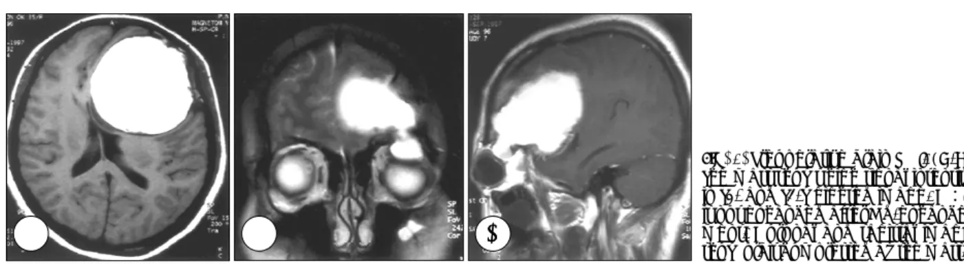
Fig. 3. Preoperative brain MRI. (A-B) The mass shows high signal intensity in T1- and T2-weighted image. C:It is not enhanced after Gd-enhance- ment. Coronal and sagittal image show orbit compressed by the mass.
AA
AA BBBB CCCC
Fig. 1. Plain radiograph shows osteolytic lesion(arrow) in left supraorbital ridge extending to the fronto-zygomatic suture.
이상원·차승헌·박동준·송근성·최창화·이영우
J Korean Neurosurg Soc/Volume 30/June, 2001 779
주시시 잘 생긴다. 이학적 소견으로 안구돌출, 안구의 하 방전위, 안검하수, 촉지성 종괴, 안와상연의 골결손 부위가 촉지되기도하며 종괴 방향으로의 안구운동장애가 있을 수 있다3)8).
두개골 단순촬영상 잘 국한된 골용해성 병변으로 주위에 경화증(sclerosis)이 없는 것이 특징이다5). 골용해성 병변 (osteolytic lesion)은 동측 전두골의 관골돌기까지 침범하 는 경우가 많다.
컴퓨터 단층촬영에서 전두골의 판간층에 중심을 둔 등밀 도 또는 저밀도 음영의 잘 국한된 균질성의 연부조직으로 나타나며 전두골 외판의 파괴로 안와의 상이측 사분면(su- perotemporal quadrant)을 침범하여 안구돌출과 안구의 하방전위를 야기시킨다. 드물게 내판이 파괴되어 전두개와 나 측두와의 경막외로 확대되어 두개강내를 침범하기도 한
다3)5)8). 컴퓨터 단층촬영에서의 특징적인 소견은 종괴주위
골이 주름진 윤곽(rugged contour)을 보인다는 점이며 이 는 높은 골흡수를 의미하고 인접 골에서의 경화증은 일반 적으로 관찰되지 않는다3)5).
자기공명영상에선 만성혈종의 소견으로 T1, T2 강조영 상 및 inversion recovery sequence에서 헤모글로빈 분 해산물의 상자성 효과(paramagnetic effect)에 의해 모두 고신호강도로 나타난다5)8). T1 강조영상에서 저신호강도를 보이는 예도 보고된바 있다2).
콜레스테롤 육아종의 조직학적 소견은 이물거세포(fo- reign body giant cell)로 둘러싸인 많은 cholesterol cleft 가 특징이다. 유피종(dermoid)과의 뚜렷한 차이점으로 상 피성분(epithelial element)이 없다는 것으로 술후 재발이 드문 것도 이에 기인한다5)10).
전두골의 콜레스테롤 육아종은 출혈에 의해 발생하며 출혈의 불완전한 흡수로 혈액과 그 분해산물에 대한 이물 반응(foreign body reaction)이 병인으로 알려져 있다3). 추체에 발생하는 콜레스테롤 육아종은 함기공동(aerated cavity)의 환기장애로 인한 점막부종과 출혈이 그 원인으 로 알려져 있으나1) 함기공동 형성이 되지 않은 전두골에 서는 이와는 다른 유발인자가 있어야 된다5). 출혈의 원인 에 대해서는 아직 명확하진 않지만 외상이 가장 가능한 원 인으로 제시되고 있으며 남자에서 호발한다는 것과 중노 동의 직업비율이 높은 점이 이를 뒷받침 해주고 있다5). 그 러나 본 례에서와 같이 뚜렷한 외상의 병력이 없는 경우 가 대부분이었다3). 그 외 콜레스테롤 육아종이 호발할 수 있는 판간층내의 이상(anomaly)이 또다른 원인으로 제시 되고 있다4).
골흡수는 프로스타글란딘(prostaglandin, PG) 특히 PGE2
에 기인하는 것으로 알려져있다7). 혈종내의 응집된 혈소판 에서 PG이 생성되며 골흡수로 새로운 판간정맥(diploic ve- ssel)이 노출되고 이는 계속적인 출혈의 원인이 된다8).
병변의 점진적인 확장은 tissue plasminogen activator 와 섬유소원 분해산물(fibrinogen degradation product)의 이차적인 섬유소 용해작용(fibrinolytic activity)에 의한 재 발성 출혈에 기인한다9).
치료는 수술적 제거로 내용물 흡인과 피막을 주위 골과 경막에서 박리 시켜 제거하며 대부분 성공적인 결과를 기 대할 수 있고 조직학적으로 상피성분이 없어 재발이 드 물다3)5)10).
결 론
전두골의 콜레스테롤 육아종은 드문 질환으로 외상이나 전두골 판간층(frontal bone diploe)의 이상(anomaly)에 의한 출혈로 발생하며 종괴의 크기가 점진적으로 증가하여 안와나 두개강을 침범하여 증상을 야기한다. 최근 본 교실 에서 안와상벽의 골결손을 동반한 안와 및 전두개와의 거 대종괴를 보인 콜레스테롤 육아종 1례를 치험하여 문헌고 찰과 함께 보고하는 바이다.
•논문접수일:2000년 6월 2일
•심사완료일:2001년 4월 17일
•책임저자:이 상 원
602-739 부산광역시 서구 아미동 1가 10번지 부산대학교 의과대학 신경외과학교실
전화:051) 240-7257, 전송:051) 244-0282 E-mail:[email protected]
Fig. 4. Photograph of the wall of cholesterol granuloma. There are numerous cholesterol crystal clefts, inflammatory infiltration, and zone of chronic hemorrhage. H & E, Original magnific- ation ×40.
전두골에 발생한 콜레스테롤 육아종 1례
J Korean Neurosurg Soc/Volume 30/June, 2001 780
References
1) Amedee RG, Marks HW, Lyons GD:Cholesterol granuloma of the petrous apex. Am J Otol 8:48-55, 1987
2) Dickey JB, Mullenix CD, O’Grady RB:Atypical magnetic resonance findings in an orbitofrontal cholesterol granuloma.
Ophthal Plast Reconstr Surg 8(3):215-220, 1992
3) Eijpe AA, Koornneef L, Verbeeten B Jr, Peeters FL, Zonnevert FW:Cholesterol granuloma of the frontal bone:CT Diag- nosis. J Comput Assist Tomogr 14(6):914-917, 1990 4) Hanbery JW, Rayport M:Unilateral exophthalmos due to
orbitofrontal cholesterol gramuloma. Am J Surg 89:1144- 1162, 1955
5) Isaacson JE, Sismanis A:Cholesterol granuloma cyst of the petrous apex. Ear Nose Throat J 75(7):425-429, 1996 6) Kersten RC, Kersten JL, Bloom HR, Kulwin DR:Chronic
hematic cyst of the orbit . Role of magnetic resonance imaging in diagnosis. Ophthalmology 95:1549-1553, 1988
7) Lund VJ:Anatomical consideration in the aetiology of fronto- ethmoidal mucoceles. Rhinology 25:83-88, 1987
8) McNab AA, Wright JE:Orbitofrontal cholesterol granuloma.
Ophthalmology 97(1):28-32, 1990
9) Pearson PA, Rakes SM, Bullock JD:Clinicopathologic study of hematic cysts of the orbit. Am J Ophthalomol 102(6):804- 805, 1986
10) Wiot JG, Pleatman CW:Chronic hematic cyst of the orbit.
AJNR 10(5 Suppl):S37-39, 1989