Acetaminophen (N-acety1-p-aminophenol, paracetamol) is widely used as an over-the-counter analgesic and antipyretic drug. Intake of a over dose of acetaminophen may result in severe hepatic necrosis. In this study, we investigated the liver damage in mice using single dose (300
Acetaminophen 유도 간 손상에 대한 조릿대 애엽 추출물의 보호 효과
장선일1, 윤용갑2, 박광현3, 설광화4, 권태오5,6
1전주대학교 대체의학대학 대체건강관리학부, 2원광대학교 한의과대학 방제학교실,
3전북대학교 의학전문대학원 생화학교실, 4중국 연변대학교 의과대학,
5원광대학교 의약자원연구소, 6원광대학교 생명자원과학대학
ABSTRACT
Protective Effects of Sasa borealis Bamboo Browse Extract on Acetaminophen-induced Liver Damage in Mouse Model
Seon Il Jang1, Young-Gab Yun2, Kwang-Hyun Park3, Guanghua Xie4, Tae-Oh Kwon5,6
1School of Alternative Medicine & Health Science, College of Alternative Medicine, Jeonju University ,
2Department of Oriental medical prescription, Wonkwang University,
3Department of Biochemistry and Institute for Medical Sciences, Chonbuk National University Medical School,
4Department of General Surgery, Affiliated Hospital of Yanbian University College of Medicine ,
5Medicinal Resources Research Institute, Wonkwang University,
6College of Life Science and Natural Resources, Wonkwang University
1)
•교신저자 : 권태오
•전북 익산시 신용동 원광대학교 생명자원과학대학
•Tel : 063-850-6681 E-mail : [email protected]
•접수 : 2008/ 12/ 02 수정 : 2008/ 12/ 10 채택 : 2008/ 12/ 15
mg/kg) of acetaminophen and the possible protective effects of administration (50-200 mg/kg body weight) of SB-Ex on acetaminophen-induced liver damage in mice. The alanine aminotransferase (ALT), and aspartate aminotransferase (AST) activities were determined in the plasma of mice. The effect of SB-Ex on lipid peroxidation product thiobarbituric reacting substances (TBARS) and some antioxidant enzymes superoxide dismutase (SOD), catalase, d-aminolevulinate dehydratase (σ-ALA-D) activities, and gluthathione peroxidase (GPx), were also evaluated in the mouse liver homogenate. Acetaminophen caused liver damage as evident by statistically significant increased in plasma activities of AST and ALT. There were general statistically significant losses in the activities of SOD, catalase, σ-ALA-D, and GPx and an increase in TBARS in the liver of acetaminophen-treated group compared with the control group.
However, SB-Ex was able to counteract these effects. These results suggest that SB-Ex can act as hepatoprotectives against acetaminophen toxicity and is a good candidate for further evaluation as an effective chemotherapeutic agent.
Key word : Sasa borealis, Oxidative stress, Hepatotoxicity, Anti-oxidant, Acetaminophen
Ⅰ. 서 론
Acetaminophen (N-acety1-p-aminophenol, paracetamol)은 세계적으로 해열 및 진통에 활용 되는 일반 의약품이다. 일반적으로 사용되는 농 도의 acetaminophen은 비교적 안전하지만, 과량의 약물이 투입되면, 간 괴사(liver necrosis), 신경독 소(nephrotoxicity), 간경화(liver cirrhosis)를 유발 할 뿐만 아니라 사망에 이르는 약물로 잘 알려져 있다1,2). 이와 같은 acetaminophen의 독성기전은 대사과정에 의해 발생되는 산화적 스트레스가 주 원인인 것으로 보고되었다2). Acetaminophen의 대 사과정에서 발생된 N-acetyl-p-benzoquinoneimine (NAPQI)는 간 독성의 주요인자로 알려져 있는데,
NAPQI는 glutathione (GSH)의 고갈을 초래하여 세포막을 치명적으로 손상시켜 회복할 수 없게 하 여 사망에 이르는 것으로 알려졌다3,4). 한국을 비 롯한 동양에서는 전통적으로 간 손상에 따른 치료 약으로 약초를 주로 활용해왔다.
조릿대 (Sasa borealis)는 한국, 중국 및 일본을
비롯한 아시아에 주로 분포하며, 항염증
(anti-inflammatory), 항해열(anti-pyretic)과 이뇨 (diuretic properties) 촉진제로서 전통적으로 활용 되어 왔다5-7). 더불어 조릿대 잎은 고혈압 (hypertension), 동맥경화증(arteriosclerosis), 심혈 관계 질환(cardiovascular disease) 및 암 치료에 효과가 있는 것으로 보고 되었다(Shibata et al., 1975). 약용에 활용되는 부위는 동절기를 지난 조 릿대의 애엽으로 주로 차(tea)를 만들어 복용하였
으나, 약리학적 수준에서는 검증되지 않았다. 최근 에 애엽으로 부터 antioxidant flavone glycosides가 분리되어 간암 세포주인 HepG2에서 항산화 및 항 염작용이 있는 것으로 알려졌다8). 그러나 acetaminophen에 의한 간 독성에 대한 보호 작용 에 대해서는 전혀 알려진 바 없다.
따라서 본 연구는 조릿대의 애엽으로 부터 추출 된 가용성 물질이 acetaminophen 유도 BALB/c 모 델 마우스의 간 보호 작용을 알아보기 위하여, 지 질산화, alanine aminotransferase (ALT), aspartate aminotransferase (AST), σ-aminolevulinate dehydratase (σ-ALA-D), superoxide dismutase (SOD), catalase, glutathione peroxidase (GPx) 등의 활성을 조사한 결과를 보고하는 바이다.
Ⅱ. 재료 및 방법
1. 재 료 1) 시 약
Aminolevulinic acid, dithiothreitol (DTT), DL-dithiothreitol, thiobarbituric acid, mono &
dibasic potassium phosphate, acetic acid, ortho- phosphoric acid, trichloroacetic acid, superoxide dismutase, catalase, GPx catalyses, sodium chloride는 Sigma-Aldrich (St. Louis, MO, USA), AST & ALT kit는 영동제약(경기도, 한국)으로부 터 구입했다. Protein assay kit는 Bio-Rad사로 부 터 구입했다. 기타 시약은 Merck사 (Darmstadt, Germany)로 부터 구입했다.
2) 약물의 추출
실험에 사용한 조릿대 애엽은 전북 전주 약재 상으로 부터 구입하였으며, 약물의 제조는 조릿대 애엽을 한약 추출기 (미강약탕기, ME-727)에 주 입하고, 정수기(하나로, U-2000)에서 정수된 물 2,000 ml를 첨가하여 100℃에서 2시간 30분 동안 전탕하였다. 추출액은 동결 건조하여 37g을 얻었
고, -20℃에서 보관하면서 실험에 사용하였다.
3) 실험동물
무균환경에서 사육된 7주령의 암컷 BALB/c계 마우스는 중앙 동물실험실(서울)사로 부터 구입하 였다. 마우스는 1주일간 스트레스를 해소하기 위 해 1주일간 낮과 밤의 주기를 12시간씩 고정하여 사료(중앙 동물실험실)와 멸균 물을 공급하면서 사육한 후 실험동물 위원회의 실험 규정에 준하여 실험에 사용하였다.
2. 방 법 1) 약물투여
8주된 BALB/c계 마우스는 실험군당 5마리씩 나누어 수용하였으며, 음성 대조군은 단지 정제수 만 투여했고, 양성 대조군은 37℃ 정제수에 마우 스 kg 당 300 mg의 acetaminophen을 용해하여 feeding needle을 이용하여 1회 경구투여 하였다.
애엽 추출물 투여군은 마우스 kg 당 300 mg의 acetaminophen을 용해하여 feeding needle을 이용 하여 1회 경구투여한 후 30분 후에 애엽 추출물을 마우스 kg당 50 mg, 100 mg, 200 mg 등 3개 실험 군으로 나누어 7일 동안 하루에 1회씩 경구투여 하였다.
2) 혈 청
혈액은 마지막 약물을 투여하고 다음날 heparin 이 처리된 주사기를 이용하여 심장으로부터 채혈 한 후 4℃가 유지되는 원심분리기로 3,000 rpm으 로 원심 분리하여 AST와 ALT를 측정하기 위하 여 혈장(plasma)을 얻어 -20℃에 보관하여 AST와 ALT 활성 측정에 사용하였다.
3) 간 조직 준비
심장 채혈 후 신속히 복강을 절개하여 간 조직 을 적출하여 얼음상자에 유지하고 간 조직의 7배 가 되게 0.85% NaCl을 주입하여 간 조직을 마쇄 한 후 3,000 rpm으로 원심 분리하여 상층 액을 얻 었다. 먼저 상층 액은 단백질을 정량하고, lipid peroxidation, catalase, SOD, σ-ALA-D, GPx 등의
활성 측정에 사용하였다.
4) 단백질 정량
단백질 정량은 표준 단백질 우태아 혈청 albumin을 사용하여 Lowery 등9) 방법으로 측정하 였다.
5) Lipid peroxidation assay
간 조직 상층 액을 37℃에 1시간 동안 방치한 후 지질 산화를 측정하였다. Thiobarbituric acid reactive species (TBARS) 산물은 Ohkawa 등10)의 방법에 준하여 측정하였다.
6) Aspartate aminotransferase와 alanine aminotransferase assay
Aspartate aminotransferase (AST)와 alanine aminotransferase (ALT)는 혈장으로부터 영동제약 에서 제시한 방법에 준하여 측정하였다.
7) σ-Aminolevulinate dehydratase assay σ-ALA-D 활성의 측정은 porphobilinogen 형성 율 측정에 의한 Sassa11)의 방법에 준하여 실험하 였다. 시료를 37℃에서 1시간 동안 방치한 후 반 응산물은 Ehrlich-porphobilinogen salt에 대한 6.1×10⁴M-1 몰라 흡수계수를 가진 555 nm에서 측 정하였다.
8) Superoxide dismutase assay
Misra와 Fridovich12)의 방법에 따라 간 조직 상 층 액을 20배(0.85% NaCl) 희석하여 SOD 활성을 측정하였다. 요약하면, pH 10에서 신속하게 자동 산화되는 핑크색의 산물을 UV spectrophotometer (Beckman Coulter)를 이용하여 480 nm에서 그 흡광도를 측정하였다. 50% 효소를 억제하는 양을 효소의 1 unit로 계산하였고, SOD의 활성은 단백 질 1 mg 당 unit로 계산하였다.
9) Catalase assay
Catalase 활성은 Aebi13)의 방법에 준하여 측정 하였다. 요약하면, 간 조직 상층 액 10 ml을 quartz cuvette에 주입하고 인산완충액(pH 7.0)에 30 mM H2O2를 신속하게 주입하여 반응시켰다.
H2O2의 분해율은 120초 동안 240 nm에서 UV
spectrophotometer를 이용하여 측정하였다. Catalase 의 활성은 mmol H2O2/mg protein/min으로 계산 했다.
10) Glutathione peroxidase assay
GPx 활성은 Paglia와 Valentine14)의 방법에 따 라 cumen hydroperoxide에 의한 GSH의 산화되는 양을 측정하였다. glutathione reductase and NADPH 존재 하에 산화된 GSH는 NADPH에서 NADP+로 즉시 전환되기 때문에 340 nm UV spectrophotometer로 감소된 흡광도를 측정하여, 간 조직 상층 액의 GPx 활성을 NADPH/mg protein/min으로 계산하였다.
11) 통계처리
모든 실험 값은 평균±표준오차로 표시했으며, 통계분석은 ANOVA와 Student’s t-test로 처리했 으며, 유의성 한계는 p<0.05로 정하였다.
Ⅲ. 결과 및 고찰
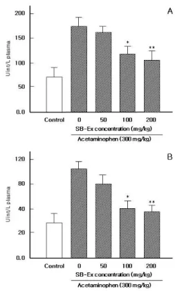
과량의 acetaminophen은 혈청의 AST와 ALT가 증가되는 것으로 알려졌다15-17). 일반적으로 acetaminophen 투여 후 72-96 시간에 간 독성이 최대로 나타나는데, 이러한 특성은 혈액의 응고덩 어리가 형성될 때까지 걸리는 시간 즉, prothrombin time (PT) 뿐만 아니라 transaminases와 bilirubin 의 농도가 증가하고 간 조직의 괴사가 진행된다한다
18). 이때 간 독성으로 잘 알려진 AST와 ALT가 증 가하고 이는 혈청내로 유입되는데, acetaminophen에 의한 간 독성의 기전은 P450 cytocrome C에 의존 적이라 알려졌다18). 본 연구의 결과 Fig. 1에서 나 타낸바와 같이 acetaminophen을 투여할 경우 물만 투여한 대조군에 비해 주요하게 AST와 ALT가 증가하였다. 그러나 acetaminophen을 투여하고 SB-Ex을 투여한 경우 농도에 의존적으로 유의하 게 감소되었다(p<0.05 : 100 mg/kg; p<0.01 : 200 mg/kg). 이러한 결과는 SB-Ex가 acetaminophen에
의한 간 독성을 해소시키는데 효능이 있다는 것을 시사해 주었다.
Fig. 1. Effect of SB-Ex on AST (A) and ALT (B) activity following acetaminophen administration in the plasma.
Control group received only filtered water, acetaminophen alone group received a single dose (300 mg/kg body weight) of acetaminophen dissolved in filtered water (37℃). SB-Ex treated groups received a single dose (300 mg/kg body weight) of acetaminophen dissolved in filtered water (37℃), 30 min later they received the different dose of SB-Ex (50-200 mg/kg body weight).
AST and ALT levels determined after 7 days ingestion.
Each column represents the mean ± S.D. from 5 mice.
*p<0.05 and **p<0.01 versus control group treated with acetaminophen alone.
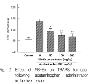
SB-Ex가 지질산화에 미치는 영향을 알아보기 위해서 간 조직에 형성된 thiobarbituric acid reactive species (TBARS) 양을 측정하였다. Fig.
2와 같이 acetaminophen 단독 투여 군은 물만 투여 한 대조군에 비해 매우 증가되었으나, acetaminophen 과 SB-Ex을 투여한 실험 군에서는 농도에 의존적 으로 감소되었다. 특히 100-200 mg/kg 투여 군에 서는 현저히 감소되었다(p<0.01). 이러한 결과는 SB-Ex가 acetaminophen에 의해 간 조직에 형성된 TBARS 형성 억제에 효능이 있음을 나타내 주었다.
Acetaminophen는 지질산화를 촉진시키는 TBARS을 형성하여 간 조직 내에 축적하는 것으로 알려져 있
다19-21). 지질산화와 관련되어 있는 산화적 스트레스
는 대표적으로 acetaminophen에 의해 유도되는 것 으로 알려져 있는데, SB-Ex는 acetaminophen에 의해 유발되는 지질산화를 억제하여 간 기능을 보 호하는 것이라 사료된다. 이러한 SB-Ex의 효과는 황금 성분의 일종인 baicalin과 유사한 작용을 하 는 것으로 볼 수 있다22). 최근에 SB-Ex에는 flavone glycosides 성분이 함유되어 있는 것으로 알려져 이 물질에 의한 항산화 효과가 있는 것이라 추정 된다8).
σ-ALA-D는 산화적 스트레스 산물로 알려진 반 능 산소 중간물질의 형성을 억제하는 물질로 알려 졌다23). 그런데, acetaminophen은 산화적 스트레스 를 유발하여 간 조직 내에 σ-ALA-D의 활성을 억 제시키는 것으로 보고되었다24,25). Acetaminophen 의 반응 대사산물로 알려진 NAPQI는 σ-ALA-D 의 SH- 그룹과 직접 반응할 수 있는 물질로 고농 도의 acetaminophen은 더욱 많은 NAPQI를 생산 해 결국 GSH를 고갈 시키는 것으로 알려졌다26). 우리는 acetaminophen 투여에 의한 σ-ALA-D의 활성과 SB-Ex의 효능을 알아 본 결과 Fig. 3과 같 이 acetaminophen 단독 투여 군은 물만 투여한 대 조군에 비해 현저히 감소되었으나, acetaminophen 과 SB-Ex을 투여한 실험 군에서는 농도에 의존적 으로 증가되었다. 특히 100-200 mg/kg 투여 군에 서는 유의하게 증가되었다(p<0.01). 이러한 결과는 SB-Ex가 acetaminophen에 의한 유발된 간 독성을 해소시키는데 효능이 있다는 것을 시사해 주었다.
Fig. 2. Effect of SB-Ex on TBARS formation following acetaminophen administration in the liver tissue.
Control group received only filtered water, acetaminophen alone group received a single dose (300 mg/kg body weight) of acetaminophen dissolved in filtered water (37℃). SB-Ex treated groups received a single dose (300 mg/kg body weight) of acetaminophen dissolved in filtered water (37℃), 30 min later they received the different dose of SB-Ex (50-200 mg/kg body weight).
Thiobarbituric acid reactive species (TBARS) levels determined after 7 days ingestion. Each column represents the mean ± S.D. from 5 mice. *p<0.05 and **p<0.01 versus control group treated with acetaminophen alone.
Fig. 3. Effect of SB-Ex on σ-ALA-D activity following acetaminophen administration in the liver tissue.
Control group received only filtered water, acetaminophen alone group received a single dose (300 mg/kg body weight) of acetaminophen dissolved in filtered water (37℃). SB-Ex treated groups received a single dose
(300 mg/kg body weight) of acetaminophen dissolved in filtered water (37℃), 30 min later they received the different dose of SB-Ex (50-200 mg/kg body weight).
Thiobarbituric acid reactive species (TBARS) levels determined after 7 days ingestion. Each column represents the mean ± S.D. from 5 mice. **p<0.01 versus control group treated with acetaminophen alone.
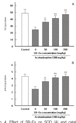
Fig. 4A와 같이 acetaminophen의 투여는 SOD 활성의 감소를 초래하였다. 그러나 SB-Ex을 투여 한 결과 농도에 의존적으로 현저히 증가되었다.
이러한 결과는 SB-Ex가 acetaminophen에 의한 유 발된 간 독성을 해소시키는데 효능이 있다는 것을 시사해 주었다. Acetaminophen 투여에 의한 SOD 의 감소는 cytochrome C P450을 경유해 형성된 NAPQI의 축적에 따라 O-이온을 감소시키는 NADPH의 억제와 관련되어있다27).
Catalase는 H2O2를 효율적으로 가수분해하는 생체 내 중요한 효소로서 항산화 작용이 있다28,29). Fig. 4B와 같이 acetaminophen에 의해 catalase는 간 조직 내에서 현저히 감소되었으나, SB-Ex 투 여에 의해 복원되는 결과를 얻었고 그 효과는 농 도에 의존적이었다. 이러한 결과는 SB-Ex의 flavone glycosides 성분이 함유되어 있는 것으로 알려져 이 물질에 의한 항산화 효과가 있는 것이 라 추정된다8).
Fig. 4. Effect of SB-Ex on SOD (A) and catalase (B) activity following acetaminophen administration in the liver tissue.
Control group received only filtered water, acetaminophen alone group received a single dose (300 mg/kg body weight) of acetaminophen dissolved in filtered water (37℃). SB-Ex treated groups received a single dose (300 mg/kg body weight) of acetaminophen dissolved in filtered water (37℃), 30 min later they received the different dose of SB-Ex (50-200 mg/kg body weight).
SOD and catalase activity levels determined after 7 days ingestion. Each column represents the mean ± S.D. from 5 mice. **p<0.01 versus control group treated with acetaminophen alone.
마지막으로 acetaminophen에 의해 손상 받은 간 조직에 SB-Ex가 미치는 영향을 알아보기 위하 여, GPx의 활성을 조사하였다. 그 결과 Fig. 5와 같이 acetaminophen에 의해 감소된 GPx의 활성을 SB-Ex 투여에 의해 증가됨을 확인 하였다. 이러 한 결과는 상기에서 기술한 SB-Ex의 항산화 작용 이 있다는 것을 사사해 주었다. Acetaminophen에
의한 간 조직 내의 낮은 농도의 GPx는 지질산화 를 촉진한다. 즉, goldthioglucose (GTG)에 의한 GPx의 억제는 간세포에 민감하게 작용하여 acetaminophen에 의해 유발된 간 독성을 억제하지 못하는 것으로 알려졌다30). 약용식물 유래 flavonoid 계열의 물질은 지질산화를 억제하고 GSH의 활성 을 증가시킴으로써 acetaminophen 독성을 억제하는 것으로 알려져 있다22,31). 그러므로 우리의 결과는 SB-Ex가 GSH를 증가시킴으로써 acetaminophen의 독성으로부터 간을 보호 할 수 있다는 사실을 제 공해주었다.
Fig. 5. Effect of SB-Ex on GPx activity following acetaminophen administration in the liver tissue.
Control group received only filtered water, acetaminophen alone group received a single dose (300 mg/kg body weight) of acetaminophen dissolved in filtered water (37℃). SB-Ex treated groups received a single dose (300 mg/kg body weight) of acetaminophen dissolved in filtered water (37℃), 30 min later they received the different dose of SB-Ex (50-200 mg/kg body weight).
GPx activity levels determined after 7 days ingestion.
Each column represents the mean ± S.D. from 5 mice.
**p<0.01 versus control group treated with acetaminophen alone.
이상의 결과를 종합해볼 때 SB-Ex는
acetaminophen에 의해 유발된 증가된 AST, ALT 와 지질산화를 억제하며, 저하된 σ-ALA-D, SOD, catalase 및 GPx의 활성을 증가시킴으로써 간 손상
을 보호하는 효과가 있었다. 따라서 acetaminophen 과 같은 약물에 의한 간 손상으로부터 SB-Ex는 간 기능 보호 작용에 활용할 수 있는 좋은 소재라 사료된다.
감사의 글
이 논문은 2006년도 원광대학교의 교비지원에 의하여 수행됨
참고문헌
1. Ray SD, Mumaw VR, Raje RR, Fariss MW.
Protection of acetaminophen-induced hepatocellular apoptosis and necrosis by cholesteryl hemisuccinole pretreatment. J Pharmacol Exp Ther. 1996;
279:1470-1483.
2. Webster PA, Roberts DW, Benson RW, Kearns GL. Acetaminophen toxicity in children: diagnostic confirmation using a specific antigenic biomarker.
J Clin Pharmacol. 1996;36:397-402.
3. Albano E, Rundgren M, Harvison PJ, Nelson SD, Moldeus P. Mechanisms of N-acetyl-p -benzoquinone-imine cytotoxicity. Mol Pharmacology.
1985;28:306-311.
4. Kyle ME, Miccadei S, Nakae D, Farber JL.
Superoxide dismutase and catalase protect cultured hepatocytes from the cytotoxicity of acetaminophen. Biochem Biophys Res Commun.
1987;149:889-894.
5. Yook CS. Colored Medicinal Plants of Korea.
Academy Book Publishing Company, Seoul.
1993;pp. 88-91.
6. Bae K., The Medicinal Plants of Korea.
Kyo-Hak Publishing Company, Seoul. 2000;
pp. 565-567.
7. Namba T. The Encyclopedia of Wakan-Yaku (Traditional Sino-Japanese Medicines) with Color Pictures, Vol. II. Hoikusha Publishing Company, Osaka, 1993;pp. 92-95.
8. Park HS, Lim JH, Kim HJ, Choi HJ, Lee IS.
Antioxidant flavone glycosides from the leaves of Sasa borealis. Arch Pharm Res. 2007;
30:161-166.
9. Lowry OH, Rosebrough NJ, Farr AL, Randall RJ. Protein measurement with the Folin phenol reagent. J Biol Chem. 1951;193:265.
10. Ohkawa H, Ohishi H, Yagi K. Assay for lipid peroxide in animal tissues by thiobarbituric acid reaction. Anal Biochem. 1979;95:351-358.
11. Sassa S. Delta-aminolevulinic acid dehydratase assay. Enzyme. 1982;28:133-145.
12. Misra HP, Fridovich I. The role of superoxide anion in the autoxidation of epinephrine and simple assay for superoxide dismutase. J Biol Chem. 1972;247:3170-3175.
13. Aebi H. Catalase in vitro. Methods Enzymol.
1984;105:121-126.
14. Paglia DE, Valentine WN. Studies on the quantitative characterisation of erythrocyte glutathione peroxidase. J Lab Clin Med.
1967;70:158-167.
15. Janbaz KH, Saeed SA, Gilani TA. Protective effect of rutin on paracetamol- and CCl4- induced hepatotoxicity in rodents. Fitoterapia.
2002;73:557-563.
16. Ali BH, Bashir AK, Rasheed RA. Effect of the traditional medicinal plants Rhazya stricta, Balanitis aegyptiaca and Haplophylum tuberculatum on paracetamol-induced hepatotoxicity in mice. Phytother Res. 2001;15:598-603.
17. Kozer E, Evans S, Barr J, Greenberg R, Soriano I, Bulkowstein M, et al. Glutathione,
glutathione-dependent enzymes and antioxidant status in erythrocytes from children treated with high-dose paracetamol. Br J Clin Pharmacol 2003;55:234-240.
18. Hinson JA, Pohl LR, Monks TJ, Gillele JR, Guengerich FP. 3-Hydroxyacetaminophen. A microsomal metabolites of acetaminophen.
Evidence against an epoxide as the reactive metabolite of acetaminophen. Drug Metab Dispos. 1980;8:289-294.
19. Younes M, Siegers CP. The role of Iron in the paracetamol and CCl4-induced lipid peroxidation and hepatotoxicity. Chem Biol Interact. 1985;55:327-334.
20. Katyare SS, Satav JG. Altered kinetic properties of liver mitochondrial membrane–
bond enzymes activities following paracetamol hepatotoxicity in rat. J Biosci. 1991;16:71-79.
21. Katikova O. Effect of mexidol on the homeostasis and lipid peroxidation in paracetamol poisoning.
Eksp Klim Farmakol. 2002;65:53-56.
22. Jang SI, Kim HJ, Hwang KM, Jekal SJ, Pae HO, Choi BM, Yun YG, Kwon TO, Chung HT, Kim YC. Hepatoprotective effect of baicalin, a major flavone from Scutellaria radix, on acetaminophen-induced liver injury in mice. Immunopharmacol Immunotoxicol.
2003;25:585-594.
23. Arnaiz SL, Llesuy S, Curtrın JC, Boveris A.
Oxidative stress by acute acetaminophen administration in mouse liver. Free Radical Biol Med. 1995;19:303-309.
24. Soares JCM, Folmer V, Rocha JBT. Influence of dietary selenium supplementation and exercise on thiol-containing enzymes in mice.
Nutrition. 2003;19:627-633.
25. Folmer V, Soares JC, Rocha JBT. Oxidative stress in mice is dependent on the free glucose content of the diet. Int J Biochem Cell Biol. 2002;34:1279-1285.
26. Bechara EJH. Oxidative stress in acute intermittent porphyria and lead poisoning may be triggered by 5-aminolevulinic acid.
Brazil J Med Biol Res. 1996;29:841-851.
27. Bessems JG, Vermeulen NP. Paracetamol (acetaminophen)-induced toxicity: molecular and biochemical mechanisms, analogues and protective approaches. Crit Rev Toxicol.
2001;31:55-138.
28. Mirochnitchenko O, Weisbrot-Lefkowitz M, Reuhl K, Chen L, Yang C, Inouye M.
Acetaminophen toxicity: opposite effects of two forms of glutathione peroxidase. J Biol Chem. 1999;274:10349-10355.
29. Rajesh B, Parames CS. The protein fraction of Phyllanthus niruri plays a protective role against acetaminophen induced hepatic disorder via its antioxidant properties. Phytother Res.
2006;l20:595-601.
30. Adamson GM, Harman AW. A role for the glutathione peroxidase/reductase enzyme system in the protection from paracetamol toxicity in isolated mouse hepatocytes. Biochem Pharmacol 1989;38:3323-3330.
31. Olaleye MT, Rocha BT. Acetaminophen- induced liver damage in mice: effects of some medicinal plants on the oxidative defense system. Exp Toxicol Pathol. 2008;
59:319-327.