1
**Corresponding Author
치의학박사, 박정철 교수, 충남 천안시 안서동 단국대학교 치과대학 부속 치과병원 치주과, 이메일: [email protected], 전화 : +82-41-550-1931, 펙스 : +82-303-3442-7364
Acknowledgements
이 논문은 2011년도 정부(교육부)의 재원으로 한국연구재단의 지원을 받아 수행된 기초연구사업임(No. 2011-0010739) 단국대학교 치과대학 부속치과병원 치주과학교실
조 인 우*, 임 성 준*, 신 현 승, 박 정 철**, Equal contribution*
콜라겐 유도 관절염 모델에서 동반된 치주염 유발시 EGCG가 치주염 치료에 미치는
효과에 관한 연구
Effect of Epigallocatechin-3-Gallate on the alveolar bone remodeling and arthritis in collagen-induced arthritis model in mice
Department of Periodontology,College of Dentistry, Dankook University In-Woo Cho*, Seong-Jun Yim*, Hyun-Seung Shin, Jung-Chul Park**, Equal contribution*
The aim of this study was to evaluate the effect of Epigallocatechin-3-Gallate (EGCG) on the alveolar bone metabolism in a collagen-induced arthritis (CIA) model in mice to enhance the understanding of rheumatoid arthritis (RA)-associated alveolar bone loss.
Following the induction of CIA in animals (mice, n=16), mandibles were retrieved for micro-computed tomography (micro-CT) and isolation of alveolar bone cells (ABCs). In vitro osteogenic potentials of ABCs were evaluated and the mRNA expression of downstream effector genes was assessed.
CIA was successfully induced in all animals, and micro-CT data showed that alveolar bone loss was significantly increased in the CIA group while the treatment of EGCG prevented the alveolar bone resorption. Osteogenesis by ABCs was significantly increased in the CIA+EGCG group in vitro. The analysis of mRNA expressions showed that osteoclastogenesis-associated genes were increased in CIA group while bone protecting genes were upregulated in EGCG treated group.
The results demonstrate that EGCG downregulated the alveolar bone resorption in a CIA model in mice, and upregulation of bone protecting genes appear to be involved. Further studies are warranted.
Key words : Rheumatoid Arthritis, Periodontitis, Epigallocatechin-3-Gallate (EGCG) ABSTRACT
모델에서동반된치주염유발시EGCG가치주염치료에미치는효과에관한연구
Ⅰ. 서론
치주 조직의 만성 염증상태는 치주 질환의 특징으로 서 치은 연하에 형성된 바이오필름 내의 세균과 숙주 간의 복잡한 면역 반응의 결과로 발생하게 된다1). 이 런 병원체와 숙주간의 지속적인 염증성 면역 반응에 의해 주변 연조직 및 경조직의 파괴가 일어나고2), 계 속적인 부착 소실 및 골흡수 진행 시 결국 지지 조직 의 파괴로 인해 치아 상실까지 이르며, 궁극적으로 환 자의 심미와 기능에 큰 영향을 끼치게 된다.
류마티스 관절염은 대표적인 만성 염증성 질환으로 관절활막을 침범하는 만성 자가면역질환으로 그 발병 원인은 아직 정확히 밝혀지지 않았다. 하지만 만성 염 증성 질환이라는 특징과 질환이 진행될 경우 골흡수를 동반한다는 측면에서 치주염과 진행 양상이 상당히 유 사한 것으로 사료 된다. 또한 최근 많은 연구에서 치주 염에 의해서 만성 관절염이 악화되거나 반대로 만성 관절염에 의해서 치주염이 발병하거나 악화되는 등의 인과 관계 내지는 연관성이 주목 받고 있으며3~5), 관절 염을 보유한 치주질환자는 치주염치료와 더불어 관점 염에 관한 평가 및 대처가 필요함이 제안되고 있다6). 이러한 관점에서 치주염과 관절염의 연관성을 규명하 기 위해 본 저자들은 백서 콜라겐 유도 관절염 모델을 통해서 전신적으로 유도된 관절염이 치주염을 동시에 유발함을 보인바 있다7).
일반적으로 치주염의 치료로는 비외과적 또는 외과 적 치료 방법이 있으며, 두 방식 모두 기계적, 화학적 방식을 통해 치주염의 원인이 되는 세균성 치태를 제 거하는 것이 주목적이다. 하지만 단순히 이러한 세균 만 제거하는 것으로는 치료에 한계가 있게 되는데 환 자의 염증 반응 및 골의 흡수 등의 대사 작용은 숙주의 전신 반응에 따라 조절되기 때문이다8, 9). 따라서, 치주 염의 궁극적인 치료를 위해서는 전신적인 면역 반응에 대한 적절한 이해가 선행되어야 할 것이다.
류마티스 관절염과 치주염 간의 상호 연관성 연구를
통해 확인된 바로는 전신적인 관절염을 억제한다면 기 존에 유도된 치주염의 진행이 중단되거나 이상적으로 는 파괴된 조직이 일부 재건될 것으로 기대할 수 있으 며 이는 치주염 치료의 새로운 패러다임을 제시할 수 있을 것이다. 현재 류마티스 관절염의 조절을 위해서 는 다양한 약물들이 사용되는데 기본적으로는 증상 완 화를 위해 소염진통제를 투약 하거나, 질환의 초기에 disease-modifying antirheumatic drugs (DMARDs)을 처방하는 것이 보편적이다10, 11). 하지만 치료 효과가 적거나 부작용이 있는 경우도 종종 있기 때문에 다양한 약물들이 계속 개발 중에 있다12). 이중 에 최근 많은 연구 대상이 되고 있는 녹차 추출물인(- )-epigallocatechin-3-gallate(EGCG)는 쉽게 구할수 있으며, 녹차 자체의 부작용이 보고된 바가 없 기 때문에 안전한 재료로 사료된다13, 14). 작용 기전으 로는 골모세포의 싸이토카인의 활동에 영향을 미쳐 골 형성을 촉진하고, 파골 세포의 활동을 억제하며, 섬유 모세포의 활동을 조절함으로써15~17), 염증시의 신체 면 역 반응에 긍정적인 효과가 보고되고 있으며 관절염의 치료를 위한 연구에서도18, 19)여러 가지 싸이토카인 및 염증 반응을 통하여 관절염의 치료에 유익함이 보고되 었다.
이러한 연구를 토대로 EGCG를 치과 분야에 적용 한 결과 미생물학적으로 치주질환의 유해균인 P.
Gingivalis의 감소에 효과가 있음이 보고 되었고20,
21), 동물 치주염 모델 실험에서 골흡수를 억제하는 효 과도 보고 되었다22, 23). 이에 본 연구에서는 콜라겐을 이용하여 관절염을 유도하고 이로 인해 치주염이 유도 된 백서에서 EGCG를 정기적으로 복용시킴으로서 치주 질환으로 인한 골흡수와 염증 반응에 대한 조절 효과를 관찰하고, host modulation을 이용한 치주 질환 치료제로서 EGCG의 가능성에 대해 평가해 보 고자 하였다.
Ⅱ. 재료 및 방법
실험 동물 및 관절염 유발 정도 평가
16마리의 생후 8주 백서를 4개의 군으로 나누어 실 험하였다. 1군은 콜라겐을 이용한 관절염을 유도한 군 (Collagen-induced arthritis, CIA)이고, 2군은 콜라겐을 이용하여 관절염을 유도한후 EGCG를 투여 한 실험군(CIA+EGCG)이다. 3군은 양성대조군으로 EGCG만 투여한 군이고 4군은 음성대조군으로 어떠 한 실험 약제도 적용하지 않았다. 실험 1, 2군의 백서 에서 관절염을 유도하기 위해 제2형 콜라겐(bovine type II collagen, Sigma-Aldrich, St. Louis, MO, USA) 100μg을 동량의 Freund’s complete adjuvant(Chondrex, SA, USA)와 잘 혼합하여 백서의 꼬리에 피하주사 하였다. 3주 후 동일한 제 2 형 콜라겐을 80μg 농도 시약으로 조제하여 꼬리에 추 가적으로 피하주사하였다24). 2, 3군은 EGCG (Sigma-Aldrich, St. Louis, MO, USA)를 10 mg/kg 용량으로 매일 1회 oral gavage 방식으로 8 주간 주입하였다25). 백서를 고형사료와 물을 제한 없 이 공급하면서 일정한 온도(25±5 C)와 습도 (50±
10 %) 및 12시간 낮과 밤의 주기를 유지하여 사육하 며 주 2회 관절염 심도를 평가하여 측정하였으며 같은 시점에 혈액 분석을 시행한 후 8주간 관찰한 후 희생 시켰다26). 희생된 동물은 체중 계측 및 유발된 관절염 의 심도를 관절염의 증후가 없는 경우를 0점, 한개의 발가락 부종을 동반한 홍조를 띄는 경우를 1점, 두개 의 관절에 이환된 경우를 2점, 3개 이상의 관절에 이 환된 경우를 3점, 발 전체와 모든 발가락에 심한 관절 염이 유발된 경우를 4점으로 기록하여 평가 하였다.
Micro-computed tomography (micro-CT) 를 활용한 치조골 파괴 분석과 조직학적 분석
8주 후 희생된 백서의 좌측 하악을 micro-CT (SkyScan, SCANCO USA, Southeastern, PA, USA)로 촬영한 후 3차원으로 재구성하여 분석하였다
27). 치조골 흡수의 양은 3차원으로 재건된 이미지상에 서 노출된 치근면의 면적을 계측해서 구하였다. 이후 조직학적 분석을 위해서 표본을 4% 포름알데히드로 고정한 다음 파라핀 처리를 위해 pH 8.0, 10%의 ethylenediaminetetraaetic acid(EDTA)로 탈회 시켰다. 제작된 파라핀 표본의 파라핀을 제거하고 탈수 시킨후 협설 수직면의 연속적 관찰을 위해 5μm 두께로 절단한 다음 haematoxylin와 eosin (H&E)으로 염 색하였다. 두개의 대구치로 제작된 시편 중 가장 중심 부위의 것을 조직학적 분석을 위해 선택하였다. 아울러 관절염 진행 정도 확인을 위해 백서의 앞발을 동일한 과정으로 조직 시편을 만들었다. 관찰은 두명의 숙련 된 술 자 가 광 학 현 미 경 (Olympus multiview microscope BH2, Tokyo, Japan)을 이용하여 진 행하였으며 관찰 영상을 이미지 분석 프로그램 (Image-Pro Plus, Media Cybernetic, Silver Spring, MD, USA)을 활용하여 분석하였다28).
치조골 유래 골세포의 골성 분화 분석
치조골 세포의 분리 및 골성 분화를 관찰하기 위해 우측 하악골을 분리하고 이를 파쇄하여 alveolar bone cell(ABC)을 채득한 뒤 이를 osteogenic medium에 배양하였으며, 세포를 35mm 배양 접시 에서 단위 면적당 1 x 10⁵가 되도록 한 후 subcon fluent 상태가 될때까지 배양하였다. 골성 분화를 관 찰하기 위한 배지는 α-MEM(Gibco BRL, Grand Island, NY, USA), 20% FBS(Gibco BRL), 2 mM L-glutamine(Gibco BRL), 100 μM L- ascorbic acid 2-phosphate, 10 nM dexamethasone(Sigma-Aldrich), 2 mM β- glycerophosphate(Sigma-Aldrich), 55 μM
모델에서동반된치주염유발시EGCG가치주염치료에미치는효과에관한연구 2-mercaptoethanol(Amersco), 100 U/ml
penicillin(Gibco BRL), 100 μg/ml strepto mycin(Gibco BRL)로 구성되어있으며 3일 주기로 교체해 주었다. 골성 분화 유도 4주후 alizarin red 염색을 이용하여 새롭게 형성되는 광화된 결절을 이미 지 분석 시스템(Image-Pro Plus)을 사용하여 계측 하였다29-31).
파골세포 분화도 분석
ABCs에 1α,25-dihydroxyvitamin D3(10-8 M, Enzo Life Sciences, Farmingdale, NY, USA)에 첨가하였다. 7일후 세포를 tartrate- resistant acid phosphatase(TRAP) 분석을 위 해 파골세포 표시자인 acid phosphatase kit (Sigma Chemical, St. Louis, MO, USA)로 염 색 및 고정하였다. 핵이 3개 이상인 TRAP에 양성을 띠는 다핵 세포를 파골세포로 파악하고 3회 측정하여 평균값으로 계측하였다32).
Reverse Transcription- Polymerase Chain Reaction (RT-PCR) 분석
파골세포성 분화 표본을 이용하여 RT-PCR을 시 행하였다. RNA의 전부를 TRIzol(Invitrogen, Carlsberg, CA, USA)을 이용하여 분리하였다.
RT-PCR는 1 mg의 RNA를 상품화된 기구(Cat.
No. 25081, Maxime RT Pre- Mix, Oligo dT primer, iNtRon Biotechnology, Daejon, Korea)를 이용하여 분석하였다. cDNA의 합성을 gene-specific primers를 이용하여 PCR로 증폭 시킨후, ethidium bromide 염색법과 gel- documentation system(Gel Doc XR, BIO- RAD, Hercules, CA, USA)을 활용하여 시각화 하
였다33). TRAP 및 RT-PCR을 이용하여여 rece ptor activator of nuclear factor κ B (RANKL), osteoprotegerin(OPG), osteoclast differentiating factor (ODF)를 평가하였다.
통계학적 분석
ANOVA를 이용하여 p<0.05의 유의성으로 각군을 분석하였다.
Ⅲ. 결과 관절염 심도
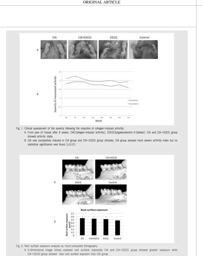
관절염을 유도한 군(1,2군)에서 유발된 관절염의 심 도를 측정한 결과 두 군 모두 관절염이 유발되었으며 1 군은 평균 지수 2.1의 관절염이 유발 되었으며 2군은 1 군보다 적은 평균지수 약 1.8의 관절염이 유발 되었다 (Figure 1A, B). 유발된 관절염은 두 군 모두 관찰 기 간동안 서서히 감소하는 것으로 관찰 되었으며, EGCG 를 투여한 2군에서는 1군에 비해 통계적 유의성은 없었 으나 상대적으로 큰 관절염 지수 감소를 보였다.
Micro CT 분석
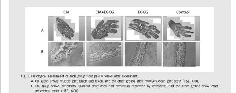
Micro CT 영상에서 1군에서는 백악질 및 치조골 의 심한 흡수가 일어나 손상된 백악질 및 치조골 외측 의 피질골이 흡수된 양상을 보였으며 2군 및 대조군에 서는 선명한 백악질이 존재하며, 치조골의 피질골이 잘 보존되어 있는것을 관찰할 수 있었다(Figure 2A). 노출된 치근의 양은 1에서 0.56 ㎟, 2군 0.53
㎟, 3군 0.51 ㎟, 4군은 0.47 ㎟로 측정되었다 (Figure 2B). 관절염을 유도한 군(1,2군)에서 다른 군보다 심한 골흡수가 일어나서 치근 노출량이 많았으
Fig. 1. Clinical assessment of the severity following the induction of collagen-induced arthritis.
A. Front paw of mouse after 8 weeks. CIA(Collagen-induced arthritis), EGCG(Epigallocatechin-3-Gallate). CIA and CIA+EGCG group showed arthritic state.
B. CIA was successfully induced in CIA group and CIA+EGCG group clinically. CIA group showed more severe arthritis index but no statistical significance was found (p>0.05).
Fig. 2. Root surface exposure analysis by micro-computed tomography.
A. 3-dimensional image shows exposed root surface, especially CIA and CIA+EGCG group showed greater exposure while CIA+EGCG group showed less root surface exposure than CIA group
B. Assessment of exposed root surface area. There were no statistically significant (p>0.05).
모델에서동반된치주염유발시EGCG가치주염치료에미치는효과에관한연구 며 EGCG를 처치한 2군이 1군에 비하여 골흡수량이
적은 것으로 계측되었으나 통계적 유의성은 없었다 (p>0.05).
조직학적 소견
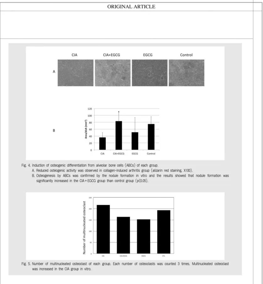
1군은 발가락의 관절의 융합 및 관절의 굴곡이 다수 관찰되는 반면에 2, 3, 4군에서는 굴곡이나 유합이 관찰되지 안으며 상대적으로 깨끗하게 유지된 관절을 관찰할 수 있었다. 콜라겐을 이용하여 관절염을 유발 한 1군의 경우 조직의 부종과 홍반 등의 전형적인 관 절염 소견과 관절 부위의 골흡수 양상을 관찰할 수 있 었다. 반면에 EGCG를 투여한 2,3군에서는 염증 소 견이 거의 관찰되지 않았으며 관절 부위의 골흡수양도 경미했다(Figure 3A).
치아 및 치조골 부위의 조직학적 소견으로는 관절염 유발군에서는 염증으로 인한 치주인대의 소실 및 많은 부분의 백악질에서 흡수 양상이 관찰 되었으며 EGCG를 투여한 2,3군과 대조군에서는 건전한 치주 인대와 백악질이 관찰되었다(Figure 3B).
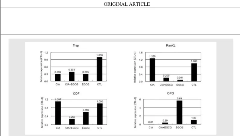
골성 분화 소견
ABCs의 배양 소견에서 1군은 현저하게 골성분화 가 저하된 것이 관찰되었으며(Figure 4A) , 골성분 화 분석 결과 1군에서는 36.1 area/dish(㎟)의 골 성분화가 관찰되었으며, 2군에서는 83.2 area/dish (㎟), 3군은 51 area/dish (㎟), 4군은 75.1 area/dish (㎟)가 각각 관찰되었다(Figure 4B).
파골세포 분화 분석
파골세포 배양 분석에서는 3개 이상의 핵을 보유한 파골세포가 216, 163, 152, 193개가 각각 관찰되었 다(Figure 5).
mRNA 분석
골재생을 평가하기 위한 관련 mRNA의 발현량 평 가를 위한 RT-PCR 분석 결과 TRAP 지수는 대조 군 1.000 대비 1, 2, 3군은 각각 0.296, 0.393, 0.295로 나타났다. RANKL 분석 결과 1, 2, 3군이
Fig. 3. Histological assessment of each group front paw 8 weeks after experiment.
A. CIA group shows multiple joint fusion and fexion, and the other groups show relatively clean joint state (H&E, X10).
B. CIA group shows periodontal ligament destruction and cementum resorption by osteoclast, and the other groups show intact periodontal tissue (H&E, X400).
각각 1.269, 0.209, 0.091의 수치를 보여 관절염을 유발한 군이 EGCG를 처치한 군보다 통계적으로 유 의하게 높은 결과를 보였다. ODF 수치는 1군은 1.097, 2군은 0.269, 3군은 0.596으로 측정되었 다. OPG 수치는 1군에서 0.03, 2군에서 0.39로 관 절염 유발후 EGCG를 투여한 군이 더 높게 나타났다 (Figure 6).
Ⅳ. 고찰
치주염과 류마티스 관절염은 많은 유사점을 갖는다.
이전 선행 연구에서 collagen 주입에 의한 류마티스 관절염 유도 백서에서 치조골의 파괴가 관찰이 되었고 이것은 전신적 면역 반응에 의해 유도된 골파괴 양상 으로 해석되었다7). 그 기전으로는 bone-coupling
Fig. 4. Induction of osteogenic differentiation from alveolar bone cells (ABCs) of each group.
A. Reduced osteogenic activity was observed in collagen-induced arthritis group (alizarin red staining, X100).
B. Osteogenesis by ABCs was confirmed by the nodule formation in vitro and the results showed that nodule formation was significantly increased in the CIA+EGCG group than control group (p<0.05).
Fig. 5. Number of multinucleated osteoclast of each group. Each number of osteoclasts was counted 3 times. Multinucleated osteoclast was increased in the CIA group in vitro.
모델에서동반된치주염유발시EGCG가치주염치료에미치는효과에관한연구
현상이 균형을 잃어 과도한 파골세포의 활성화와 반대 로 억제된 조골세포의 활성이 주된 것으로 관찰되었 고, 파골세포의 활성 증가는 RANK와 macrop hage colony-stimulating factor(M-CSF)의 증가와 부가적으로 지방성 분화가 bone coupling에 관여하여 세포 활성도에 영향을 끼친 것으로 보고되며
34), 본 연구에서도 동일한 반응이 관찰되었다.
이전의 많은 연구가 관절염 환자에게서 높은 치아 상실률과 깊은 치주낭 깊이가 관찰됨을 보고 하였다3). 이는 단순히 관절염으로 칫솔을 잡거나 치실을 사용하 기 어려움으로 인해 구강 위생 관리가 저하된 것을 우 선적으로 의심할 수 있으나 그 기저에는 전신적인 대 사의 불균형이 발생하여 그 효과가 더욱 증폭된 것으 로 해석되고 있다6, 7). 따라서 류머티스 관절염 환자에 서의 치주염 예방 및 진행 차단을 위해서는 host modulation의 관점에서 EGCG 의 전신적 투여가 치주염에 효과가 있을 것으로 예상되어 본 연구가 진 행 되었다.
실험 결과 1군에서 백서 콜라겐 유도 관절염 모델을
이용하여 성공적으로 관절염 및 골흡수를 동반한 치주 염을 유발할 수 있었으며 이는 앞서 보고된 연구와 유 사한 양상이었다7). Micro CT 분석에서 유도된 관절 염이 치조골의 흡수를 야기시켜 결과적으로 치근이 노 출량이 증가 되었다. 반면에 관절염 유도 후 EGCG 를 투여한 2군에서는 약간의 치조골 흡수로 인하여 1 군에 비하여 적은 치근 노출이 관찰되었다. 400배율 조직학적 소견에서도 관절염이 유발된 군에서는 심한 염증 소견과 피질골의 흡수를 관찰할 수 있었던 반면 대조군과 관절염 유도후 EGCG 투여한 2군에서는 염증 소견이 거의 없는 정상적인 골 외형을 관찰할 수 있었다. 이는 관절염 환자에게 EGCG를 투여할 경우 전신적으로 염증이 조절되어 전신적인 관절염 완화 효 과와 더불어 치조골에서의 골흡수도 줄일수 있어 결과 적으로 치주염의 치료에 효과가 있음을 시사하는 바이 며, 이는 wire 결찰로 유발된 치주염 동물실험에서 EGCG의 전신적 투여가 보여준 치주염 치료 효과와 유사한 양상상이며 파골세포 활성 감소와 교원질 파괴 의 감소로 인한 것으로 사료된다22).
Fig. 6. Reverse transcription-polymerase chain reaction (RT-PCR) analysis was performed to determine the mRNA expression associated with osteoclastogenic differentiation. Osteoclast differentiation factor (ODF) was significantly decreased in CIA+EGCG group, and osteoprotegerin (OPG) differentiation was significantly increased In EGCG group (p<0.05).
이를 규명하기 위하여 본 연구에서는 골대사에 직접 적인 작용을 하는 세포들의 배양 실험을 활용하였는 데, 치조골을 파쇄하여 골성분화 배양을 관찰한 결과 실험 2군에서 통계적으로 유의성 있는 골재생 증가가 관찰 되었으며, 파골세포 배양 분석에서는 보유한 핵 의 수와 상관 없이 실험 2군에서 많은 수의 파골세포 가 관찰 되었다. 이러한 결과 차이는 RT PCR 분석으 로 설명할 수 있는데, EGCG 처치군(3군)에서 RT PCR 결과에서 OPG의 발현이 6배 가까이 증가했다 는 점이다. OPG는 RANKL의 natural inhibitor 로써 RANKL과 결합하여 RANK의 발현을 억제하 는 것으로 밝혀졌으며35, 36), 이러한 작용으로 동물을 이용한 관절염 실험에서 OPG 투여시 관절의 염증이 완화되고 파골세포의 작용이 조절되어 골흡수가 감소 한다고 보고되었다37). EGCG의 세포간 작용을 알아 보기 위한 여러 연구에서도 투여 후 OPG의 생성이 촉진되어 파골세포 작용에 관여한다고 보고하고 있다
38~40). 이러한 작용으로 본 연구에서 실험 2군에서 관
절염 유도후 투여된 EGCG의 작용으로 골재생이 활
발이 일어나게 되고, 파골세포의 활성이 억제되어 치 조골 흡수가 감소된 것으로 사료된다.
본 연구의 한계점은 EGCG의 농도를 다양하게 적 용하지 못한 점이다. 하지만 EGCG의 농도에 따른 효 과를 평가한 많은 연구가 있었으며41, 42), 본 연구에서 는 이전 연구를 토대로 적정 농도를 결정하였고43), 그 결과 기대한 효과를 관찰할 수 있었으나, 그 농도의 차 이에 따른 효과를 확인하였다면 실제 임상 적용 시에 유용한 근거로 활용될 수 있었을 것이다.
또한 EGCG가 실제 관절염이나 치주염 환자에게 사용되기 위해서는 임상 연구가 필수적인데 이 부분에 관해서 EGCG의 인체 투여시에 대한 많은 연구가 이 미 보고 되었으며 골대사에 관여하는 세포들의 활성을 조절하기 위한 농도에서 인체에 특별한 부작용이 없는 것으로 알려졌다44~46). 이는 치주염 약물 치료의 가능 성으로 충분한 가치가 있을 것으로 판단되며 향후 임 상시험을 통한 후속 연구 등의 여러 연구가 필요할 것 으로 사료된다.
모델에서동반된치주염유발시EGCG가치주염치료에미치는효과에관한연구 1. Tonetti MS, Claffey N, European Workshop in
Periodontology group C. Advances in the progression of periodontitis and proposal of definitions of a periodontitis case and disease progression for use in risk factor research. Group C consensus report of the 5th European Workshop in Periodontology. J Clin Periodontol 2005;32 Suppl 6:210-213.
2. Graves D. Cytokines that promote periodontal tissue destruction. J Periodontol 2008;79:1585-1591.
3. Mercado F, Marshall R, Klestov A, Bartold P.
Relationship between rheumatoid arthritis and periodontitis. Journal of periodontology 2001;72:779-787.
4. Pons-Fuster A, Rodriguez Agudo C, Galvez Munoz P, Saiz Cuenca E, Pina Perez FM, Lopez-Jornet P.
Clinical evaluation of periodontal disease in patients with rheumatoid arthritis: A cross-sectional study.
Quintessence Int 2015;46:817-822.
5. Bingham CO, 3rd, Moni M. Periodontal disease and rheumatoid arthritis: the evidence accumulates for complex pathobiologic interactions. Curr Opin Rheumatol 2013;25:345-353.
6. Mercado F, Marshall RI, Klestov AC, Bartold PM. Is there a relationship between rheumatoid arthritis and periodontal disease? J Clin Periodontol 2000;27:267-272.
7. Park JC, Su C, Jung IH, et al. Mechanism of alveolar bone loss in a collagen-induced arthritis model in mice. J Clin Periodontol 2011;38:122-130.
8. Kornman KS, Page RC, Tonetti MS. The host response to the microbial challenge in periodontitis:
assembling the players. Periodontology 2000 1997;14:33-53.
9. Graves DT, Oates T, Garlet GP. Review of osteoimmunology and the host response in endodontic and periodontal lesions. Journal of oral microbiology 2011;3.
10. Siegel J. Comparative effectiveness of treatments for rheumatoid arthritis. Annals of internal medicine 2008;148:162-163.
11. Donahue KE, Jonas DE, Hansen RA, et al. Drug therapy for rheumatoid arthritis in adults: An update. 2012.
12. Donahue KE, Gartlehner G, Jonas DE, et al.
Systematic review: comparative effectiveness and harms of disease-modifying medications for rheumatoid arthritis. Annals of Internal Medicine 2008;148:124-134.
13. Ahmed S, Anuntiyo J, Malemud CJ, Haqqi TM.
Biological basis for the use of botanicals in osteoarthritis and rheumatoid arthritis: a review.
Evid Based Complement Alternat Med 2005;2:301- 308.
14. Siddiqui IA, Afaq F, Adhami VM, Ahmad N, Mukhtar H. Antioxidants of the beverage tea in promotion of human health. Antioxid Redox Signal 2004;6:571-582.
15. Takai S, Matsushima-Nishiwaki R, Adachi S, et al.
(-)-Epigallocatechin gallate reduces platelet-derived growth factor-BB-stimulated interleukin-6 synthesis in osteoblasts: suppression of SAPK/JNK.
Mediators Inflamm 2008;2008:291808.
16. Yun JH, Kim CS, Cho KS, Chai JK, Kim CK, Choi SH. (-)-Epigallocatechin gallate induces apoptosis, via caspase activation, in osteoclasts differentiated from RAW 264.7 cells. J Periodontal Res 2007;42:212-218.
17. Corps AN, Curry VA, Buttle DJ, Hazleman BL, Riley GP. Inhibition of interleukin-1beta-stimulated collagenase and stromelysin expression in human tendon fibroblasts by epigallocatechin gallate ester.
Matrix Biol 2004;23:163-169.
18. Riegsecker S, Wiczynski D, Kaplan MJ, Ahmed S.
Potential benefits of green tea polyphenol EGCG in the prevention and treatment of vascular inflammation in rheumatoid arthritis. Life Sci 2013;93:307-312.
19. Yang EJ, Lee J, Lee SY, et al. EGCG attenuates autoimmune arthritis by inhibition of STAT3 and HIF-1alpha with Th17/Treg control. PLoS One 2014;9:e86062.
20. Asahi Y, Noiri Y, Miura J, et al. Effects of the tea catechin epigallocatechin gallate on Porphyromonas gingivalis biofilms. J Appl Microbiol 2014;116:1164- 1171.
참 고 문 헌
21. Jung IH, Lee DE, Yun JH, et al. Anti-inflammatory effect of (-)-epigallocatechin-3-gallate on Porphyromonas gingivalis lipopolysaccharide- stimulated fibroblasts and stem cells derived from human periodontal ligament. J Periodontal Implant Sci 2012;42:185-195.
22. Cho AR, Kim JH, Lee DE, et al. The effect of orally administered epigallocatechin-3-gallate on ligature- induced periodontitis in rats. J Periodontal Res 2013;48:781-789.
23. Tominari T, Matsumoto C, Watanabe K, et al.
Epigallocatechin gallate (EGCG) suppresses lipopolysaccharide-induced inflammatory bone resorption, and protects against alveolar bone loss in mice. FEBS Open Bio 2015;5:522-527.
24. Lee SW, Kim JH, Park YB, Lee SK. Bortezomib attenuates murine collagen-induced arthritis. Ann Rheum Dis 2009;68:1761-1767.
25. Kim JS, Kim JM, O JJ, Jeon BS. Inhibition of inducible nitric oxide synthase expression and cell death by (-)-epigallocatechin-3-gallate, a green tea catechin, in the 1-methyl-4-phenyl-1,2,3,6- tetrahydropyridine mouse model of Parkinson's disease. J Clin Neurosci 2010;17:1165-1168.
26. Lee YK, Kim JS, Lim SC. Effects of atractylodis rhizoma pharmacopuncture on an acute gastric mucosal lesion induced by compound 48/80 in rats.
J Pharmacopuncture 2012;15:12-17.
27. Park CH, Abramson ZR, Taba M, Jr., et al. Three- dimensional micro-computed tomographic imaging of alveolar bone in experimental bone loss or repair. J Periodontol 2007;78:273-281.
28. Kim CS, Choi SH, Cho KS, Chai JK, Wikesjo UM, Kim CK. Periodontal healing in one-wall intra-bony defects in dogs following implantation of autogenous bone or a coral-derived biomaterial. J Clin Periodontol 2005;32:583-589.
29. Shi S, Gronthos S, Chen S, et al. Bone formation by human postnatal bone marrow stromal stem cells is enhanced by telomerase expression. Nat Biotechnol 2002;20:587-591.
30. Belibasakis GN, Bostanci N, Hashim A, et al.
Regulation of RANKL and OPG gene expression in human gingival fibroblasts and periodontal ligament cells by Porphyromonas gingivalis: a putative role of the Arg-gingipains. Microb Pathog 2007;43:46-53.
31. Kanzaki H, Chiba M, Shimizu Y, Mitani H. Dual regulation of osteoclast differentiation by periodontal ligament cells through RANKL stimulation and OPG inhibition. Journal of dental research 2001;80:887-891.
32. Yun JH, Kim CS, Cho KS, Chai JK, Kim CK, Choi SH. (-)-Epigallocatechin gallate induces apoptosis, via caspase activation, in osteoclasts differentiated from RAW 264.7 cells. Journal of periodontal research 2007;42:212-218.
33. Park JC, Kim JM, Jung IH, et al. Isolation and characterization of human periodontal ligament (PDL) stem cells (PDLSCs) from the inflamed PDL tissue: in vitro and in vivo evaluations. Journal of clinical periodontology 2011;38:721-731.
34. Hirayama T, Danks L, Sabokbar A, Athanasou NA.
Osteoclast formation and activity in the pathogenesis of osteoporosis in rheumatoid arthritis. Rheumatology (Oxford) 2002;41:1232-1239.
35. Bucay N, Sarosi I, Dunstan CR, et al.
Osteoprotegerin-deficient mice develop early onset osteoporosis and arterial calcification. Genes &
development 1998;12:1260-1268.
36. Hofbauer LC, Khosla S, Dunstan CR, Lacey DL, Boyle WJ, Riggs BL. The roles of osteoprotegerin and osteoprotegerin ligand in the paracrine regulation of bone resorption. Journal of Bone and Mineral Research 2000;15:2-12.
37. Kong Y-Y, Feige U, Sarosi I, et al. Activated T cells regulate bone loss and joint destruction in adjuvant arthritis through osteoprotegerin ligand.
Nature 1999;402:43-47.
38. Huang H, Kang L, Chen S, et al. (-)- Epigallocatechin-3-Gallate (EGCG) decreases osteoclastogeneis via opg/rankl modulation. Bone 2009;44:S329-S330.
39. Chen C, Kang L, Hung S, Ho M, Chang J, Wang G. (-)-epigallocatechin-3-gallate (EGCG) modulate
참 고 문 헌
모델에서동반된치주염유발시EGCG가치주염치료에미치는효과에관한연구 OPG/RANKL/RANK in murine bone marrow
mesenchymal stem cell and osteoblast. In:
CALCIFIED TISSUE INTERNATIONAL. vol. 80:
SPRINGER 233 SPRING STREET, NEW YORK, NY 10013 USA, 2007:S74-S74.
40. Huang P, Chen C, Kang L, Hung S, Wang G, Chang J. (-)-epigallocatechin-3-gallate (EGCG) increases OPG/RANKL expression in osteoclast feeder cell, ST2. In: CALCIFIED TISSUE INTERNATIONAL. vol. 82: SPRINGER 233 SPRING ST, NEW YORK, NY 10013 USA, 2008:S99-S100.
41. Shen CL, Yeh JK, Cao JJ, Chyu MC, Wang JS.
Green tea and bone health: Evidence from laboratory studies. Pharmacol Res 2011;64:155-161.
42. Chen L, Lee M-J, Li H, Yang CS. Absorption, distribution, and elimination of tea polyphenols in rats. Drug Metabolism and Disposition 1997;25:1045-1050.
43. Kim JS, Kim J-M, Jeong-Ja O, Jeon BS. Inhibition of inducible nitric oxide synthase expression and cell death by (-)-epigallocatechin-3-gallate, a
green tea catechin, in the 1-methyl-4-phenyl-1, 2, 3, 6-tetrahydropyridine mouse model of Parkinson’s disease. Journal of Clinical Neuroscience 2010;17:1165-1168.
44. Nakagawa K, Okuda S, Miyazawa T. Dose- dependent incorporation of tea catechins,(-)- epigallocatechin-3-gallate and (-)-epigallocatechin, into human plasma. Bioscience, biotechnology, and biochemistry 1997;61:1981-1985.
45. Ullmann, Haller, Decourt, Girault, Spitzer, Weber.
Plasma-kinetic characteristics of purified and isolated green tea catechin epigallocatechin gallate (EGCG) after 10 days repeated dosing in healthy volunteers. International journal for vitamin and nutrition research 2004;74:269-278.
46. Chow HS, Cai Y, Hakim IA, et al. Pharmacokinetics and safety of green tea polyphenols after multiple- dose administration of epigallocatechin gallate and polyphenon E in healthy individuals. Clinical Cancer Research 2003;9:3312-3319.
참 고 문 헌