Original Article PROGRESS in MEDICAL PHYSICS Vol. 25, No. 4, December, 2014 http://dx.doi.org/10.14316/pmp.2014.25.4.242
This research was supported by Basic Science Research Program through the National Research Foundation of Korea (NRF) funded by the Ministry of Education, Science and Technology (2010-0013701 and 2013R1A1A2012013).
Received 15 October 2014, Revised 12 November 2014, Accepted 19 November 2014
Correspondence: Sangwook Lim ([email protected]) Tel: 82-51-990-6393, Fax: 82-2-6280-6247
cc This is an Open-Access article distributed under the terms of the Creative Commons Attribution Non-Commercial License (http://creativecommons.org/licenses/by-nc/3.0) which permits unrestricted non-commercial use, distribution, and reproduction in any medium, provided the original work is properly cited.Discrepancies between Calculated and Delivered Dose Distributions of Respiratory Gated IMRT Fields according to the Target Motion Ranges for Lung and Liver Cancer Patients
Youngkuk Kim*
†, Sangwook Lim*, Ji Hoon Choi*, Sun Young Ma*, Tae Sig Jeung*, Tae Ik Ro
†*Department of Radiation Oncology, Kosin University College of Medicine,
†
Department of Physics, Dong-A University, Busan, Korea
To see the discrepancies between the calculated and the delivered dose distribution of IMRT fields for respiratory-induced moving target according to the motion ranges. Four IMRT plans in which there are five fields, for lung and liver patients were selected. The gantry angles were set to 0° for every field and recalculated using TPS (Eclipse Ver 8.1, Varian Medical Systems, Inc., USA). The ion-chamber array detector (MatriXX, IBA Dosimetry, Germany) was placed on the respiratory simulating platform and made it to move with ranges of 1, 2, and 3 cm, respectively. The IMRT fields were delivered to the detector with 30∼70% gating windows. The comparison was performed by gamma index with tolerance of 3 mm and 3%. The average pass rate was 98.63%
when there's no motion. When 1.0, 2.0, 3.0 cm motion ranges were simulated, the average pass rate were 98.59%, 97.82%, and 95.84%, respectively. Therefore, ITV margin should be increased or gating windows should be decreased for targets with large motion ranges.
Key Words: Gated radiation therapy, IMRT, Gamma index, Moving phantom
서 론
표적부피(target volume)에 균일한 선량을 전달하고, 정상 조직에는 최소한 선량을 전달하는 세기조절방사선치료(in- tensity-modulated radiation therapy, IMRT)는 표적부피와 정 상조직 사이에 선량분포의 높은 선량 경사율을 형성하여 정상조직의 피해를 최소화 하므로 치료 전 계획된 선량분 포와 치료 시 전달되는 선량분포를 확인하는 정도관리 (quality assurance, QA) 작업이 매우 중요하다.
1,2)현재 많은 기관에서 이온전리함, 2차원 이온전리함 배열, 전자포탈영
상장치(electrical portal imaging device, EPID) 및 필름 등을 이용하여 선량 차이(dose difference), 감마 지표(gamma in- dex) 및 ROC (receiver operating characteristic) 곡선 등의 분 석을 시행하고 있다.
3-10)IMRT는 표적부피와 정상조직 사이에 선량분포의 높은 선량 감소율을 형성하여 정상조직의 피해를 최소화 하므로
11)
뇌, 두경부, 폐, 복부 및 골반 부위 등 다양한 부위에 적 용되고 있으며,
12,13)특히, 두경부와 전립선암 등의 골반 부 위에 적용 시 다른 치료법에 비해 방사선에 의한 부작용을 효과적으로 줄일 수 있어 치료효과가 높다는 연구결과가
있다.
14-21)반면, 폐 또는 복부 등 호흡에 의해 움직임이 큰
장기에 IMRT를 적용 시, 국제방사선단위측정위원회(Inter-
national Commission on Radiological Unit and Measurement,
ICRU) 보고서에 따라 내부표적부피(internal target volume,
ITV)를 충분히 고려하여 치료용표적부피(planning target
volume, PTV)를 결정할 경우 정상조직에 필요이상의 방사
선이 피폭되어 정상조직손상확률(normal tissue complication
probability, NTCP)이 증가할 수 있고, 환자의 호흡 패턴, 주
기 및 종양의 움직임 크기 등을 고려하지 않을 경우 종양
억제확률(tumor control probability, TCP)이 낮아질 수 있
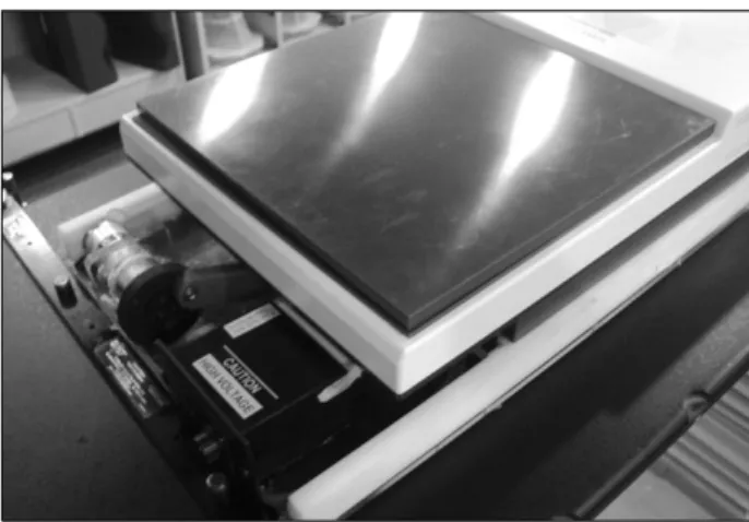
Fig. 1. To simulate the patients' respiratory motion, the moving platform was fabricated in laboratory. The solid water phantom with thickness of 1.3 cm and the MatriXX were placed on the simulating platform as shown figure. The SSD was 100 cm and the depth of measurement layer became 1.6 cm, which D
maxfor 6 MV photon beams. We let the platform move with range of 0, 1, 2, and 3 cm and with cycle of 3.6 s.
다.
22,23)따라서 환자 내부 장기 움직임을 고려한 호흡연동
방사선치료(respiratory-gated radiation therapy), 호흡조절방사 선치료(active breathing control radiation therapy), 및 동적병 소추적방사선치료(dynamic tumor tracking radiation therapy) 와 같이 호흡을 고려한 4차원방사선치료방법에 대한 치료 가 활발히 이루어지고 있다.
24-26)이러한 호흡연동방사선치 료를 시행함에도 흉부나 복부의 IMRT 시행 전 정도관리시 호흡에 의한 움직임은 고려되고 있지 않고 있다.
본 연구의 목적은 호흡연동방사선치료법을 적용한 IMRT 시 환자의 호흡에 따른 장기운동에 따른 선량분포의 차이 를 감마지표로 분석하여 장기운동에 따른 최적의 연동창 (gating window)의 범위와 ITV (internal target volume) 여유 분(margin)을 알아보고자 한다.
재료 및 방법
실제 환자의 호흡운동을 모사하기 위하여 호흡연동방사 선치료법을 적용하여 IMRT를 완료한 폐암환자 25명과 간 암 환자 23명의 사차원컴퓨터단층촬영(four-dimensional co- mputed tomography, 4DCT) 영상을 후향적(retrospective)으로 분석하였고 모든 환자의 평균 호흡주기 및 호흡에 의한 횡 경막의 움직임을 4DCT 영상으로 측정하였다. 4DCT는 RPM (Real-time Position Management
TM, Varian Medical Sy- stems, Inc., Palo Alto, CA, USA) 시스템과 연동하여 4DCT 영상을 획득하였다. 호흡에 의한 장기의 움직임은 비교적 분석이 용이하고 움직임이 두드러진 횡격막을 기준으로
27,28)두미축(craniocaudal) 방향으로 분석하여 환자들의 평균 호 흡패턴을 자체 제작한 호흡운동모사 플랫폼(respiration sim- ulating platform) 운동에 적용하였다.
본 연구에서는 호흡운동에 따른 IMRT의 계산된 선량분 포와 실제 전달된 선량분포의 차이를 분석하기 위해 치료 를 완료한 폐암환자 2명과 간암환자 2명의 IMRT 치료계획 을 선택하였다. 한 환자 당 조사면(field)은 5개씩 이었으며 치료계획시스템(Eclipse 8.1, Varian Medical Systems, Inc., Palo Alto, CA, USA)에서 가상의 팬텀을 만들어 각각의 갠 트리(gantry)를 수직방향(0
o)으로 변경하여 재계산된 선량분 포를 얻었다.
환자의 다양한 호흡운동 패턴을 모사하기위해 자체 제 작한 호흡모사 플랫폼은 전동기의 회전운동을 크랭크축 (crankshaft)을 통해 직선왕복운동하도록 설계하여 다양한 진폭과 회전속도로 사인파(sinusoidal)형태로 움직이도록 고 안하였다. 플랫폼 위에는 고체팬텀과 필름 또는 2차원선량
계 등을 올려놓아 움직이는 병소의 2차원적 선량분포를 측 정할 수 있도록 고안하였다. 본 실험에서는 환자의 횡경막 움직인 거리를 분석하여 가능성 있는 횡경막의 3가지 움직 임을 모사시켰고 주기는 환자의 평균호흡주기를 사용하였 다.
각 조사면의 실제 전달 선량을 측정하기 위해 2차원 이
온전리함 배열(I’mRT MatriXX, IBA Dosimetry, Germany)을
호흡모사 플랫폼 위에 위치시켰다. 2차원 이온전리함 배열
은 2차원 선량분포를 측정하는 장비로 내부에 모서리 부분
의 4개를 제외한 총 1020개의 이온전리함이 배열되어 있
다. 유효면적(active area)은 24.4×24.4 cm
2이고, 유효층(ac-
tive layer)은 2차원 이온전리함배열의 표면으로부터 0.3 cm
아래에 위치하고 있다.
29)이를 고려하여 계산된 선량의 조
건과 동일한 기하학적 구조를 재현하기 위해 Fig. 1과 같이
움직이는 받침대 위에 2차원 이온전이람 배열을 설치하고,
30.0×30.0×1.3 cm
3의 고체팬텀(Virtual water
◯R, Civco Medical
Solutions, Orange City, IA, USA)을 2차원 이온전리함 배열
위에 위치시켰다. 그리고 선원표면간거리(source-to-surface
distance, SSD)를 100 cm로 설치하여 최대선량 깊이(D
max)에
서 움직이는 표적의 이차원적 선량분포를 측정할 수 있도
록 하였다. 치료용선형가속기(Clinac iX, Varian Medical Sy-
stems, Inc., Palo Alto, CA, USA)에서 각각의 치료 계획과
동일하게 6 MV의 광자선, 400 MU/min 선량율을 사용하였
고, 호흡연동방사선치료시 본원에서 선택하는 최대 연동창
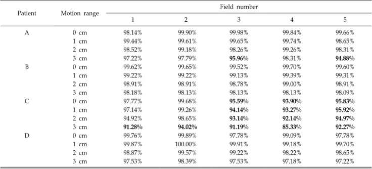
Table 2. The pass rate according to the motion ranges: The pass rate for the calculated and the measured dose distribution for the gated IMRT according to the target motion ranges; The pass rates tend to decrease with the motion range.
Patient Motion range Field number
1 2 3 4 5
A 0 cm 98.14% 99.90% 99.98% 99.84% 99.66%
1 cm 99.44% 99.61% 99.65% 99.74% 98.65%
2 cm 98.52% 99.18% 98.26% 99.26% 98.31%
3 cm 97.22% 97.79% 95.96% 98.31% 94.88%
B 0 cm 99.62% 99.65% 99.52% 99.70% 99.60%
1 cm 99.22% 99.22% 99.13% 99.39% 99.31%
2 cm 98.91% 98.91% 98.78% 99.00% 98.91%
3 cm 98.18% 98.13% 98.13% 98.13% 98.09%
C 0 cm 97.77% 99.68% 95.59% 93.90% 95.83%
1 cm 97.14% 99.26% 94.14% 93.27% 95.92%
2 cm 94.92% 98.65% 93.14% 92.14% 94.97%
3 cm 91.28% 94.02% 91.19% 85.33% 92.27%
D 0 cm 99.76% 99.89% 97.78% 99.09% 97.78%
1 cm 99.87% 100.00% 99.91% 99.18% 99.70%
2 cm 98.87% 99.57% 99.22% 98.22% 98.65%
3 cm 97.53% 98.39% 97.53% 97.18% 97.22%
Table 1. Respiratory patterns for 48 lung and liver cancer patients (SD: Standard deviation).
Treatment site Number of patients Breathing period (s) Motion range (cm)
Mean±SD Range Mean±SD Range
Lung 25 3.4±0.5 2.6∼4.9 1.3±0.6 0.2∼2.4
Liver 23 4.0±0.8 2.6∼6.0 1.2±0.5 0.2∼2.3
Total 48 3.6±0.7 2.6∼6.0 1.3±0.6 0.2∼2.4
크기인 30∼70%의 연동창(gating windows)으로 호흡연동방 사선치료법을 적용하여 움직임이 0, 1, 2, 및 3 cm의 경우 각각 방사선을 조사하였다.
2차원 이온전리함배열로 획득한 각각의 조사면의 선량 분포는 계산된 선량분포와 비교하기 위해 Omni-Pro IMRT (IBA Dosimetry, Germany) 프로그램의 감마지표(gamma in- dex) 분석을 사용하여 계산된 선량분포와 비교 분석하였다.
호흡연동 진폭에 의한 각 조사문의 합격률은 감마지표 3 mm/3% 수준으로 설정하여 합격률을 계산하였고, 0.0 cm, 1.0 cm, 2.0 cm 및 3.0 cm 의 크기로 움직이는 표적에 대한 합격률을 비교하였다.
결과 및 고찰
자체 제작한 호흡운동모사 플랫폼의 움직임은 각각 1.0, 2.0, 3.0 cm으로 사인파 형태의 1차원 왕복운동을 하였다.
주기는 환자의 평균호흡주기인 3.2초로 호흡운동을 모사하 였다. 이 플랫폼은 고체팬텀과 이온함배열 등을 올려놓고 움직일 수 있도록 고안하였기 때문에 호흡연동방사선치료 등의 이차원 선량분포 분석이 가능하였다. 임상욱 등
31)의 연구에서는 컴퓨터와 연결된 동팬텀이 표적 내의 한 점의 움직임을 실제환자의 불규칙한 호흡운동을 모사하기 위한 것과는 달리 본 연구에서 개발한 플랫폼은 무거운 계측기 등을 움직일 수 있도록 고안하였다. 상용화된 QUASAR
TM와 같은 호흡동조 구동 팬톰은 3차원 왕복운동을 하나 실 제 환자의 불규칙한 운동을 모사하지는 못하며, 무거운 계 측기는 적용이 불가하여 작은 필름 등을 이용한다.
32)Table 1은 호흡모사 플랫폼에 적용하기위해 48명의 폐암
과 간암 환자의 4차원 컴퓨터단층영상에서 횡격막의 두미
축 방향의 움직임 크기와 평균 호흡주기를 측정한 결과를
보여준다. 모든 환자의 평균 호흡주기는 3.6 s, 횡격막의 평
균 움직임 크기는 1.3 cm으로 측정되었다.
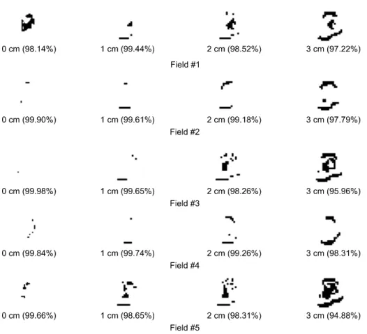
Fig. 2. Example of five respiratory gated IMRT fields for case of A patient; the phantom simulated re- spiratory motion with four scenario (0, 1, 2, and 3 cm motion range and 3.6 s cycle). Gamma index were used with 3 mm/3% tolerance to compare calculated and measured dose distribution. White and black area in the figures represent pass and fail respectively.
Table 2는 30∼70% 범위의 연동창을 설정하여 호흡연동 방사선치료법을 적용한 IMRT의 20개 조사면을 계산된 선 량분포를 기준으로 3 mm/3% 허용범위에서 감마지표 합격 률을 보여준다. 환자 C를 제외하고는 움직임의 크기가 2 cm 이하일 경우 합격률이 98% 이상으로 호흡연동방사선치 료법이 효과적이었음을 알 수 있다. 움직임이 없을 때 평균 합격률은 98.63%이고, 총 20개 조사면 중 17개의 조사면이 97% 이상의 합격률을 보였다. 움직임이 없을 때 감마 지표 의 평균 합격률이 98.63%였으며, 움직임을 1.0, 2.0, 3.0 cm 으로 모사할 경우 평균 합격률이 각각 98.59%, 97.82%, 95.84%으로 낮아졌다.
Fig. 2는 환자 A의 5개 조사면에 대한 호흡에 의한 움직 임 크기별로 계산된 선량분포와 비교한 감마 지도(gamma map) 형태로 보여준다. 감마 지도에서 흰색은 합격을 나타 내며, 검은색은 불합격을 나타낸다. 우측으로 갈수록 움직 임을 크게 모사한 것인데 불합격 영역이 많아짐을 볼 수 있다. 5번 조사면의 경우 움직임을 모사하지 않았을 경우 합격률이 99.66%였고 2 cm으로 움직임을 모사할 경우도 98.31% 였으나 3 cm으로 움직임을 모사할 경우 94.88%로
합격률이 급격히 떨어짐을 알 수 있다.
결 론
호흡연동방사선치료 시 30∼70%의 연동창에서 움직임 크기가 커질수록 합격률이 낮아지는 것을 알 수 있었다. 환 자 C의 경우 3, 4, 5번 조사면은 다른 조사면에 비하여 크 기가 커서 정지상태에서도 합격률이 94∼96% 정도로 낮았 으나 움직임이 커질수록 합격률은 85% 정도까지 낮아짐을 알 수 있었다. 따라서 합격률이 낮은 조사면의 경우 호흡에 의한 움직임은 감마지표에 더 큰 영향을 주는 것으로 보인 다. 이 경우 계획된 선량을 충분히 전달하기 위해서는 마선 영 등
30)의 연구와 같이 움직이는 표적에 대하여 호흡연동 방사선치료 시 연동창을 좁게 선택하거나 치료계획 시 ITV (internal target volume)의 여유분을 크게 설정하여야 한다.
A환자와 같은 20×20 cm
2이하의 일반적인 크기의 IMRT
조사면의 경우 움직임 거리가 2 cm 까지는 30∼70% 연동
창 범위는 충분해 보인다. 이는 마선영 등
27)의 연구결과에
서도 30∼70% 연동창 범위에서도 3 cm의 움직임은 감마지
표 합격률이 98% 이상이었다. 단, 조사면의 크기가 커질 경우 감마지표 합격률은 낮아질 것으로 생각된다.
호흡연동방사선치료법을 적용한 세기조절방사선치료 시 움직임을 고려하지 않았을 때 3 mm/3% 기준에 대해 99%
이상 합격률을 보이더라도 병소의 움직임이 2 cm 이상이 될 경우 합격률이 97% 이하로 급격히 떨어질 수 있기 때문 에 ITV 여유분을 크게 설정하거나 연동창을 좁게 선택할 것을 권장한다.
References
1. Ezzell GA, Burmeister JW, Dogan N, et al: IMRT com- missioning: Multiple institution planning and dosimetry compar- isons, a report from AAPM Task Group 119, Med Phys 36:5359-5373 (2009)
2. Ahn WS, Cho BC: Intensity modulated radiation therapy com- missioning and quality assurance: Implementation of AAPM TG119. Progress in Medical Physics 22:99-105 (2011) 3. Hussein M, Rowshanfarzad P, Ebert MA, Nisbet A,
Clark CH: A comparison of the gamma index analysis in vari- ous commercial IMRT/VMAT QA systems. Radiother Oncol 109:370-376 (2013)
4. Arumugam S, Xing A, Goozee G, Holloway L:
Independent calculation-based verification of IMRT plans using a 3D dose-calculation engine. Med Dosim 38:376-384 (2013) 5. Anjum MN, Parker W, Ruo R, Aldahlawi I, Afzal M:
IMRT quality assurance using a second treatment planning system. Med Dosim 35:274-279 (2010)
6. Li G, Zhang Y, Jiang X, et al: Evaluation of the ArcCHECK QA system for IMRT and VMAT verification. Physica Medica 29:295-303 (2013)
7. Guillot M, Gingras L, Archambault L, Beddar S, Beaulieu L: Performance assessment of a 2D array of plastic scintillation detectors for IMRT quality assurance. Phys Med Biol 58:4439–4454 (2013)
8. Sabet M, Rowshanfarzad P, Vial P, Menk FW, Greer PB: Transit dosimetry in IMRT with an a-Si EPID in direct de- tection configuration. Phys Med Biol 57:N295-N306 (2012) 9. Vial P, Gustafsson H, Oliver L, Baldock C, Greer PB:
Direct-detection EPID dosimetry: investigation of a potential clinical configuration for IMRT verification. Phys Med Biol 54:7151-7169 (2009)
10. Carlone M, Cruje C, Rangel A, et al: ROC analysis in pa- tient specific quality assurance. Med Phys 40:042103-1- 042103-7 (2013)
11. Intensity Modulated Radiation Therapy Collaborative Working Group: Intensity modulated radiotherapy: current status and issues of interest. Int J Radiat Oncol Biol Phys 51:880-917 (2001)
12. Wilcox EE, Daskalov GM, Pavlonnis III, et al: Dosimetric verification of intensity modulated radiation therapy of 172 pa- tients treated for various disease sites: comparison of EBT film
dosimetry, ion chamber measurements, and independent MU Calculations. Med Dosim 33:303-309 (2008)
13. Anjum MN, Parker W, Ruo R, Afzal M: Evaluation criteria for film based intensity modulated radiation therapy quality assurance. Physica Medica 26:38-43 (2010)
14. Roesink JM, Moerland MA, Battermann JJ, Hordijk GJ, Terhaard CHJ: Qantitative dose-volume response analysis of changes in parotid gland function after radiotheraphy in the head-and-neck region. Int J Radiat Oncol Biol Phys 51:938-946 (2001)
15. Sasaki R, Soejima T, Matsumoto A, et al: Clinical sig- nificance of serum pulmonary surfactant proteins A and D for the early detection of radiation pneumonitis. Int J Radiat Oncol Biol Phys 50:301-307 (2001)
16. Lee N, Xia P, Quivey JM, et al: Intensity-modulated radio- therapy in the treatment of nasopharyngeal carcinoma: an up- date of the UCSF experience. Int J Radiat Oncol Biol Phys 53:12-22 (2002)
17. Nutting CM, Convery JD, Cosgrove PV, et al: Reduction of small and large bowel irradiation using an optimized in- tensity-modulated pelvic radiotherapy technique in patients with prostate cancer. Int J Radiat Oncol Biol Phys 48:649-656 (2000) 18. Sanguineti G, Cavey ML, Endres EJ, et al: Is IMRT needed to spare the rectum when pelvic lymph nodes are part of the initial treatment volume for prostate cancer? Int J Radiat Oncol Biol Phys 64:151-160 (2006)
19. Luxton G, Hancock SL, Boyer AL: Dosimetry and radio- biologic model comparison of IMRT and 3D conformal radio- therapy in treatment of carcinoma of the prostate. Int J Radiat Oncol Biol Phys 59:267-284 (2004)
20. Wang-Chesebro A, Xia P, Coleman J, Akazawa C, Roach III: Intensity-modulated radiotherapy improves lymph node coverage and dose to critical structures compared with three-dimensional conformal radiation therapy in clinically lo- calized prostate cancer. Int J Radiat Oncol Biol Phys 66:654-662 (2006)
21. Wang L, Hoban P, Paskalev K, et al: Dosimetric advant- age and clinical implication of a micro-multileaf collimator in the treatment of prostate with intensity-modulated radiotherapy. Med Dosim 30:97-103 (2005)
22. ICRU REPORT 62: Prescribing, Recording and Reporting Photon Beam Therapy (Supplement to ICRU Report 50). Inte- rnational Commission in Radiation Units and Measurements (1999)
23. Chu SS, Cho KH, Lee CG, Suh CO: Development of con- formal radiotherapy with respiratory gate device. Radiat Oncol J 20:41-52 (2002)
24. Kini VR, Vedam SS, Keall PJ, et al: Patient training in res- piratory-gated radiotherapy. Med Dosim 28:7-11 (2003) 25. Lim S, Park SH, Ahn SD, et al: Guiding curve based on
the normal breathing as monitored by thermocouple for regular breathing. Med Phys 34:4514-4518 (2007)
26. Lim S: Research on the respiratory synchronization system for
dynamic tumor tracking radiation therapy. Gyeonggi, Kyonggi
University doctorate thesis (2008)
호흡연동방사선치료시 폐암과 간암환자의 병소 움직임 크기에 따른 선량분포 차이 분석