Received: July 1, 2020 Revised: September 18, 2020 Accepted: November 6, 2020
OPEN ACCESS
HORTICULTURAL SCIENCE and TECHNOLOGY 39(1):122-131, 2021
URL: http://www.hst-j.org pISSN : 1226-8763 eISSN : 2465-8588
This is an Open Access article distributed under the terms of the Creative Commons Attribution Non-Commercial License which permits unrestricted non-commercial use, distribution, and reproduction in any medium, provided the original work is properly cited.
Copyrightⓒ2021 Korean Society for Horticultural Science.
본 논문은 농촌진흥청 연구과제(과제번호: PJ01272801) 지원에 의해 이루어진 것임.
Genomic DNA의 FT-IR 스펙트럼을 이용한 복숭아
‘유명’ 관련 품종의 유연관계 분석
송승엽 · 이주현 · 황기동 · 정남희 · 최인명 · 권정현*
농촌진흥청 국립원예특작과학원 과수과
Genetic Relationship Analysis by using FT-IR
Spectrum of Genomic DNA from Peach Cultivars Related to ‘Yumyeong’
Seung-Yeob Song, Ju-Hyun Lee, Kidong Hwang, Namhee Jeong, In Myung Choi, and Jung Hyun Kwon*
Fruit Research Division, National Institute of Horticultural & Herbal Science, Rural Development Administration, Wanju 55365, Korea
*Corresponding author: [email protected]
Abstract
The genetic relationships of offspring cultivars from ‘Yumeyong’ peach were evaluated by using Fourier transform infrared (FT-IR) spectroscopy. Four offspring cultivars were bred by controlled hybridization between ‘Yumyeong’ and ‘Chiyomaru’, 2 from open pollination, and 1 from bud sport mutation. The FT-IR spectrum of genomic DNA were analyzed by using principal component analysis (PCA), partial least square discriminant analysis (PLS-DA), and hierarchical clustering analysis (HCA). FT-IR spectra showed that typical spectral differences were existed in the frequency regions of guanine ring (C6 = C6), thymine ring (C4 = O4), cytosine, N-H stretching (amide I), C = O stretching vibrations (amide II), and PO2- ionized asymmetric and symmetric stretching, respectively. Among the offspring cultivars between ‘Yumyeong’ and ‘Chiyomaru’, ‘Mihong’ and
‘Misshong’ showed higher genetic similarity to ‘Yumyeong’ than ‘Chiyomaru’. On the other hand,
‘Yumi’ and ‘Soomee’ showed higher genetic similarity to ‘Chiyomaru’ than ‘Yumyeong’. ‘Wolmi’, a mutant cultivar from ‘Yumyeong’, showed close relationship with ‘Yumyeong’, however, the genetic similarity was lower compared to other crossbreeding cultivars, ‘Misshong’, and ‘Mihong’.
Additional key words: discrimination system, hierarchical clustering analysis, multivariate analysis, partial least square discriminant analysis (PLS-DA), principal component analysis (PCA)
서 언
장미과(Rosaceae), 자두나무속(Prunus)에 속하는 복숭아(Prunus persica (L.) Batsch)는 낙엽성 온 대과수로 중국이 원산지인 세계적인 과일이다(Ahmad et al., 2004). 우리나라에서는 사과와 감귤 다음
라 복숭아(유모종)와 천도(무모종)로 분류된다. 복숭아는 실생선발과 변이선발이 쉽고, 영양번식으로 쉽게 증식이 되기 때문 에 품종 식별을 위한 분자표지 개발과 유연관계 분석이 필요하다(Hong et al., 2013).
장미과 작물 중 복숭아의 genome size는 226.6Mb로 가장 작다(Verde et al., 2013). 복숭아에 대한 유전적 다양성 및 유연관 계를 파악하기 위하여 분자생물학적 분석법인 randomly amplified polymorphic DNA (RAPD), amplified fragment length polymorphism (AFLP), simple sequence repeats, sequence-related amplified polymorphism (SRAP)와 같은 다양한 DNA 마커들이 이용되고 있다(Warburton and Bliss, 1996; Casas et al., 1999; Ahmad et al., 2004; Hu et al., 2005; Xu et al., 2006;
Yoon et al., 2006; Bouhadida et al., 2009; Cheng and Huang, 2009). 복숭아 품종의 친자관계를 확인하기 위해 simple sequence repeat (SSR) 마커를 이용하여 분석하였고(Yamamoto et al., 2003), 그리고 sequence characterized amplified region (SCAR) 마커를 이용하여 복숭아 품종 판별 연구가 진행되었다(Han et al., 2010). 이러한 방법들은 정확한 유전적 분 석을 통해 품종에 대한 정밀한 분석이 가능하지만, 분석에 대한 복잡성 그리고 많은 시간과 비용이 소요되는 한계점을 가지고 있다. 이러한 한계점을 FT-IR fingerprinting 기술을 이용하여 사전 식별 분석을 통해 효과적으로 보완 가능하다. FT-IR spectroscopy는 화학, 농업, 제약, 식품 산업 등 다양한 연구 분야에서 널리 사용되고 있고, FT-IR 스펙트럼은 DNA의 특성을 나타낸다. 각각의 영역에서 나타내고 있는 스펙트럼은 화학적 작용기의 진동을 나타내고, DNA의 전체 화학 구조와 서로 다른 작용기작 또는 화학적 구조가 스펙트럼에 반영된다. 또한, cell의 전체 화학구조에 대한 lipids, proteins, nucleic acids 그리고 polysaccharides을 포함하고 있는 전체 화학구조를 매우 구체적으로 나타낸다(Lee and Chapman, 1986; Naumann et al., 1991; Mungur et al., 2005; Lei et al., 2011). IR 스펙트럼의 변화는 nucleic acid의 분자 모양에 의해 결정되며, 광범위한 수소 결합과 대칭구조의 phosphate 증가, 그리고 단백질의 C-OH 결합 감소를 나타낸다(Dukor, 2002). FT-IR을 이용한 분석은 미 생물, 곰팡이 그리고 박테리아 식별에 사용되었고(Ngo-Thi et al., 2003; Naumann et al., 2005; Fischer et al., 2006; Preisner et al., 2007; Sundaram et al., 2012), 그리고 생식 세포질의 대사 평가(Yesudas et al., 2013), DNA를 이용한 질병과 암 식별에 사용되었다(Malins et al., 1997, 2003, 2005). 또한 바이오 마커로 이용하여 암 진단에 활용되었고(Argov et al., 2002; Sahu et al., 2008), 유전자 변형 DNA 판별에 적용 되었으며(Emura et al., 2006), indica와 japonica의 DNA 수준에서 유전적 차이 가 높음을 확인하였다(Emura et al., 2006). 또한, FT-IR을 이용하여 국내에서 수집된 야생종 복숭아의 유연관계에 대해 분석 된 바 있으나(Song et al., 2020), 육종내력이 있는 품종들 즉, 양친의 정보가 알려져 있거나 기존 품종의 돌연변이로 탄생한 계 통들에 대한 분석은 미미하다.
복숭아 ‘유명(Yumyeong)’은 1977년 국내에서 육성된 품종으로 에틸렌이 합성되지 않아 단단한 과육이 장기간 유지되는 경육종(stony hard) 이다(Haji et al., 2001). 복숭아 품종군에서 경육종이 차지하는 비율은 매우 낮기 때문에 ‘유명’은 경육종 연구에서 자주 다루어지고 있으며 (Pan et al., 2015), 육종적으로는 저장성이 우수한 품종 육성을 위해 모본으로 활용되고 있 다 (Jun et al., 2013). 현재까지 ‘유명’에서 다수의 품종들이 선발되었으나, 이들 품종 사이의 유연관계나 유전적 특성에 대한 연구는 부족한 실정이다. 따라서 본 연구는 복숭아 ‘유명(Yumyeong)’에서 파생된 인공교배품종, 자연교잡실생, 그리고 돌연 변이 품종을 대상으로 genomic DNA의 FT-IR 스펙트럼 데이터를 이용하여 근연관계를 분석하고, 다양한 유전자 분석의 사 전 분석 기법의 가능성을 확인하고자 하였다.
재료 및 방법 식물재료
본 연구에서는 복숭아 ‘유명’(Yumyeong, UM)과 유명(UM)을 모본으로 사용하여 육성된 4개의 육성품종 ‘유미’(Yumi, YM), ‘수미’(Soomee, SM), ‘미홍’(Mihong, MH) 그리고 ‘미스홍’(Misshong, MS), 부본으로 사용된 ‘치요마루’(Chiyomaru,
Table 1. Accession, origin, pubescence of fruit skin, flesh color, and pedigree characteristics of peach varieties released by the National Institute of Horticultural and Herbal Science
Accession Label Origin Pubescence of fruit skin
Flesh
color Pedigree
Chiyomaru JU Japan Present Yellow (Nakatsu Hakuto × Nunomewase) × Nunomewase Yumyeong Seedling (Daewol) DW Korea Present White Open pollination of Yumyeong
Mihong MH Korea Present White Yumyeong × Chiyomaru
Misshong MS Korea Present White Yumyeong × Chiyomaru
Soomee SM Korea Present White Yumyeong × Chiyomaru
Yumyeong Mutation (Wolmi) WM Korea Present White Bud mutation of Yumyeong
Yumi YM Korea Present White Yumyeong × Chiyomaru
Yumyeong UM Korea Present White (Yamatowase × Nunomewase) or (Hakuto × Okayama 3) Yumyeong Seedling (Eumseong) YS Korea Present White Open pollination of Yumyeong
JU), 유명의 자연교잡실생인 ‘유명실생-음성(Eumseong, YS)’과 ‘대월(Daewol, DW)’, 유명의 돌연변이 품종인 ‘월미 (Wolmi, WM)’를 분석하였다(Table 1). 국립원예특작과학원 과수과 시험포장에 재식된 시험수를 품종별로 3주씩 3반복으로 잎 시료를 채취하였다. 각각의 잎 시료는 DNA 추출을 위해 동결건조하고 막자사발을 이용하여 분말 형태로 분쇄하였다. 분쇄 된 잎 시료는 –70°C 초저온냉동고에 보관하여 분석에 사용하였다.
복숭아 DNA 분리
수집된 식물 잎 시료를 genomic DNA 추출에 사용하였다. 동결 건조된 잎 시료 10mg을 사용하여 DNeasy Plant Mini Kit (QIAGEN, Dusseldorf, Germany) 방법에 따라 DNA를 추출하였다. 추출된 DNA는 AE buffer 를 이용하여 희석하였다.
DNA 농도와 순도는 NanoDrop 2000을 이용하여 280, 260와 230nm에서 측정하였고, 추출된 DNA 순도를 확인하기 위해서 흡광비(OD260/OD280)를 계산하였다. 이 비율은 polyphenol, carbohydrate contamination 과 함께 protein을 측정하였다 (Sambrook et al., 1989). AE buffer를 이용하여 DNA 최종 농도를 20ng/㎕으로 조정하였다. 또한, 효과적인 FT-IR 스펙트럼 측정을 위해 genomic DNA를 체크하여 DNA 오염도를 최소화하고, Genomic DNA 농도는 아가로스 겔 전기영동을 통해 확 인하였다. 확인된 genomic DNA는 –20°C에 보관하여 실험에 사용하였다(Song et al., 2014).
FT-IR 스펙트럼과 데이터 전처리 및 다변량 통계분석
FT-IR (Fourier transform infrared) 스펙트럼 조사는 Tensor 27 (Bruker Optics GmbH, Ettlingen, Germany)과 DTGS (Deuterated triglycine sulfate) 검출기를 사용하였다(Song et al., 2014). 추출된 각각의 시료는 5µL씩 5반복으로 384-well ZnSe plate에 분주하여, 37°C hot plate에서 약 20–30분간 건조한 후 HTS-XT (Bruker Optics GmbH) 고효율 자동화 장치 를 이용하여 스펙트럼을 측정하였다. 스펙트럼은 4000–400cm-1 범위에서 4cm-1 간격으로 128회 반복 측정되었고, 측정된 스펙트럼의 평균 스펙트럼을 분석에 사용하였다. Bruker 에서 제공하는 OPUS Lab (ver. 7.0, Bruker Optics Inc.)를 사용하여 FT-IR 스펙트럼 조사 및 데이터 변환을 하였고, baseline 교정을 위해 FT-IR 스펙트럼 분석 영역의 양 끝점(800–1,800cm-1) 의 흡광도를 0으로 조정하였다. 실험상의 오차를 최소화하기 위하여 각 스펙트럼을 동일 면적으로 normalization을 실시하였 으며, mean centering 과정을 거쳐 2차 미분하여 스펙트럼의 전처리 과정을 실시하였다.
가공된 FT-IR 스펙트럼 데이터는 NIPALS 알고리즘(Wold, 1966)을 이용하여 PCA (Principal component analysis)와 PLS-DA (Partial least squares discriminant)분석을 R 프로그램(version 3.1.2)에서 수행하였다(Fiehn et al., 2000; Trygg et
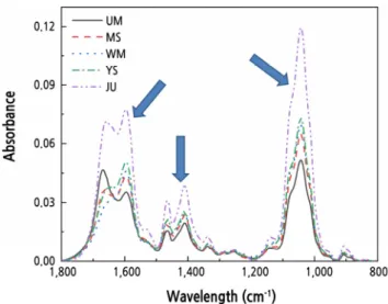
Fig. 1. Representative FT-IR spectral from genomic DNA of peach leaves. FT-IR spectral ranges showed quantitative information of 1,750–1,600 cm-1 (amide I (N-H stretching) and amide II (C = O stretching vibrations)), 1,550–1,400 cm-1 (purine and pyrimidine ring), and 110–950 cm-1 (PO2- ionized asymmetric and symmetric stretching). UM : Yumyeong, MS : Misshong, WM : Yumyeong Mutation (Wolmi), YS : Yumyeong Seedling (Eumseong), JU : Chiyomaru. *n = 5.
al., 2007). PCA 및 PLS-DA 분석을 통해 얻어진 score를 이용하여 HCA (hierarchical clustering analysis) 분석하였으며 유 사도 지수로 UPGMA (unweighted pair group method with arithmetic mean analysis)를 사용한 Euclidean distance를 측정 하여 각 시료의 유연관계를 덴드로그램으로 나타냈다.
FT-IR 스펙트럼 데이터를 이용한 Classification, prediction 그리고 cross-validation
FT-IR 스펙트럼 데이터를 기반으로 DNA를 통한 품종간 예측을 위해서 PLS regression 분석을 적용하여 확립하였다. 품종 간 예측을 위해 2개의 변수 X와 Y를 나누고 X 변수에는 FT-IR 스펙트럼 데이터를 Y 변수에는 품종간 DNA의 번호를 그룹화 시키고 각각의 정보를 연결시켜 PLS regression을 실시하였다. 예측 모델의 예측 능력을 테스트하기 위해 트레이닝 세트를 만 들어 모델 구축에 사용하였고, 트레이닝 세트는 테스트 세트에 사용되지 않았다. X 변수와 Y 변수간의 측정 모델을 만들기 위 하여 상관관계를 예측한 후 회귀계수를 사용하여 상관관계를 계산하였다. 전체의 데이터는 임의의 두 부분으로 나눴고, 먼저 트레이닝 세트를 선발 후 트레이닝 세트에 사용되지 않은 데이터를 테스트 세트로 사용하여 예측 모델을 구축하는데 사용되었 다. PLS 모델의 예측 능력은 실측 값과 PLS 예측 값 사이의 정확도를 10 bootstrap 테스트하여 나타냈고, RMSEP (Root Mean Square Error of Prediction)와 MSEP (Mean Square Error of Prediction)를 측정하여 예측의 정확도를 확인하였다 (Liu et al., 2006; Terhoeven-Urselmans et al., 2010).
결과 및 고찰
복숭아의 FT-IR 스펙트럼 비교 분석
복숭아 genomic DNA의 FT-IR 스펙트럼 데이터로부터 다변량통계분석을 통해 복숭아 품종간의 유연관계를 분석하였다.
각각의 복숭아 품종들은 FT-IR 스펙트럼 1,750–1,600, 1,550–1,400, 1,150–950cm-1 파장에서 특징적인 스펙트럼 패턴 변화가 나타났다(Fig. 1). FT-IR 스펙트럼 패턴의 차이는 DNA의 구조적 특성과 상호작용을 나타내는 것으로 보고되고 있다 (Falk et al., 1963; Liquier et al., 1991; Zhou-Sun et al., 1997; Brewer et al., 2002; Banyay et al., 2003; Gonzalez-Ruiz et
A B
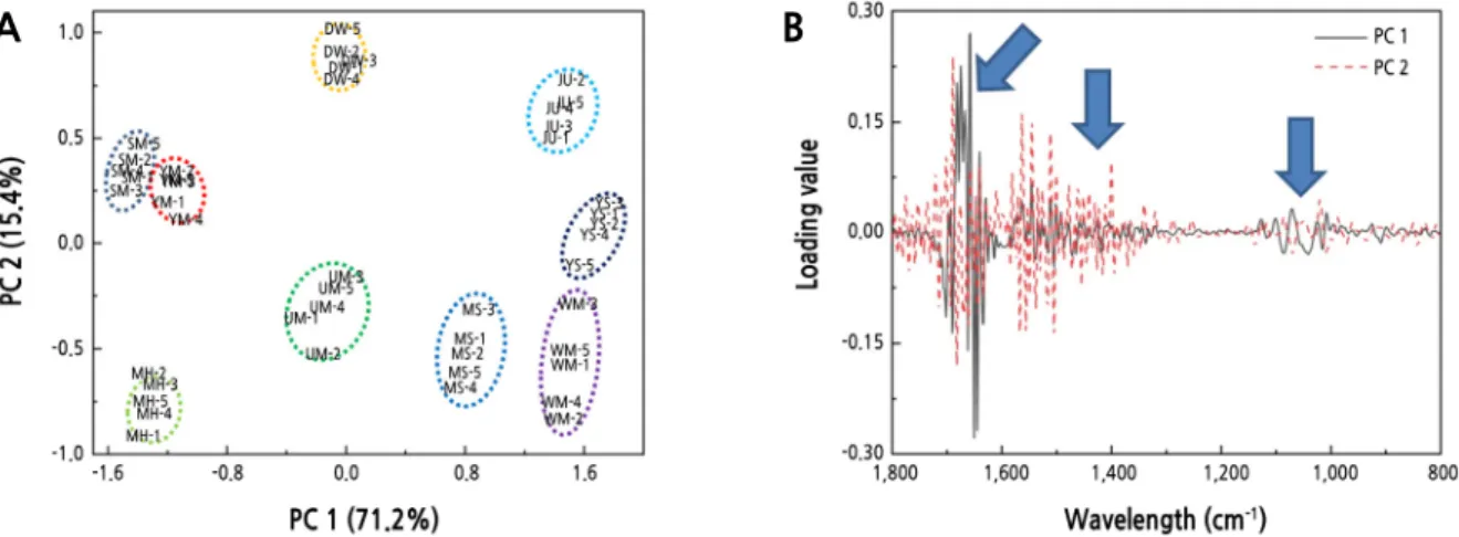
Fig. 2. PCA scores (A) and loading values plot (B) of PCA scores from FT-IR data of peach leaves. UM : Yumyeong, JU : Chiyomaru, WM : Yumyeong mutation (Wolmi), DW : Yumyeong seedling (Daewol), YS : Yumyeong seedling (Eumseong), MH : Mihong, MS : Misshong, SM : Soomee, YM : Yumi.
al., 2011). 1,750–1,600cm-1 부위는 전체적인 DNA의 염기구조인 C = O, C = N 그리고 C = C 형태와 –NH2결합 형태를 나타낸다(Brewer et al., 2002; Banyay et al., 2003). purine과 pyrimidine ring 구조는 1,550–1,400cm-1 부위에서 나타나며 (Falk et al., 1963; Liquier et al., 1991; Zhou-Sun et al., 1997), phosphodiester-deoxyribose backbone내의 PO2- 그룹의 대 칭적 구조와 비대칭적 구조는 1,100–950cm-1 부위에서 나타난다(Falk et al., 1963; Zhou-Sun et al., 1997). 이 세가지 스펙 트럼 영역은 backbone과 sugar pucker 사이의 base pairing, base stacking 그리고 glycosidic bond 회전과 밀접하게 연관되어 있다(Brewer et al., 2002; Gonzalez-Ruiz et al., 2011). 이처럼 FT-IR 스펙트럼의 차이가 복숭아 품종 고유의 DNA 염기구조 가 서로 다름을 의미하며, FT-IR 스펙트럼 분석을 통해 복숭아 품종별 DNA 구조적 차이와 이로 인한 유전적 차이를 확인할 수 있 을 것으로 판단된다.
FT-IR 스펙트럼 다변량 통계분석 및 대사체 수준 유연관계 비교
9품종 복숭아의 FT-IR 스펙트럼 데이터를 PCA 분석한 결과 PC 1과 2의 설명력이 71.2%와 15.4%로 각각 나타났다(Fig.
2A). ‘유명(UM)’을 중심으로 교잡 품종과 돌연변이 품종이 분포하는 것을 확인할 수 있었다(Fig. 2A). 또한, ‘치요마루(JU)’
는 우측 상단에 위치하였고, ‘치요마루(JU)’ 품종이 교잡 품종과 돌연변이 품종에 비해 ‘유명(UM)’ 품종과의 유전적 유사성이 낮은 것으로 판단된다. ‘유명(UM)’의 자연 교잡 품종인 ‘음성(YS)’와 ‘대월(DW)’ 품종은 중앙 상단과 우측 중간에 위치하며
‘유명(UM)’ 품종과의 유전적 유사성이 낮게 나타났고, ‘유명(UM)’의 돌연변이 품종인 ‘월미(WM)’ 품종은 우측 하단에 위치 하면서 교배 품종인 ‘수미(SM)’에 비해 유전적 유사성이 낮게 나타났다. ‘유명 x 치요마루’ 교배종인 ‘유미(YM)’, ‘수미 (SM)’, ‘미홍(MH)’ 그리고 ‘미스홍(MS)’ 품종은 ‘유명(UM)’ 품종의 좌, 우에 위치하였다. 이들 4품종 중 ‘유명(UM)’과 유전 적 유사성이 가장 높은 것은 ‘미스홍(MS)’으로 나타났으며, ‘유미(YM)’와 ‘수미(SM)’ 품종이 유전적 유사성이 ‘미스홍 (MS)’에 비해 낮게 나타났다.
복숭아 9품종의 genomic DNA 식별에 중요하게 작용한 FT-IR 스펙트럼 부위를 확인한 결과, PC 1과 2를 분류하는 중요한 기준은 1,700–1,500, 1,500–1,300 그리고 1,100–950cm-1 부위임을 알 수 있었다(Fig. 2B). 1,700–1,500, 1,500–1,300 그리고 1,100–950cm-1 부위는 FT-IR 스펙트럼상에서 차이를 나타냈던 부위(Fig. 1)와 일치하였으며, 이 부위가 나타내는 DNA 염기구조 C = O, C = N 그리고 C = C 형태와 –NH2결합 형태, guanine ring (C6 = C6), thymine ring (C4 = O4), uracil (C4 = O4), purine과 pyrimidine ring 구조, phosphodiester-deoxyribose backbone의 PO2- 그룹에 대한 대칭적 구조와 비대
A B
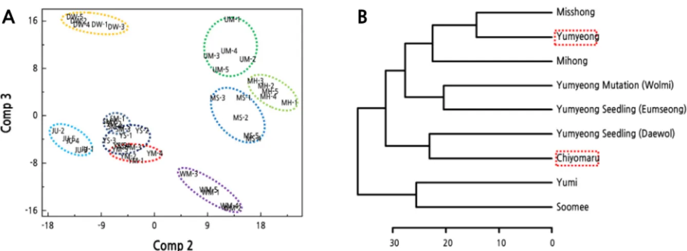
Fig. 3. PLS-DA score plot (A) and the HCA dendrogram from PLS-DA (B) of FT-IR data from peach leaves. UM : Yumyeong, JU : Chiyomaru, WM : Yumyeong mutation (Wolmi), DW : Yumyeong seedling (Daewol), YS : Yumyeong seedling (Eumseong), MH : Mihong, MS : Misshong, SM : Soomee, YM : Yumi.
칭적 구조의 차이가 genomic DNA 식별에 중요한 역할을 하는 것으로 알 수 있었다(Schulz and Baranska, 2007)(Fig. 2B). PLS-DA 분석의 경우 PCA 분석 보다 복숭아 품종간 차이가 뚜렷하게 나타났다(Fig. 3A). PLS-DA score plot을 보면 PCA 와 동일하게 ‘치요마루(JU)’와 ‘유명(UM)’는 서로 좌우에 위치하면서 유전적 유사성이 낮은 것으로 나타났다. ‘치요마루 (JU)’와 ‘유명(UM)’의 교배를 통해 얻어진 4품종 중 ‘미홍(MH)’, ‘미스홍(MS)’ 품종이 ‘유명(UM)’ 품종과 ‘유미(YM)’, ‘수 미(SM)’ 품종이 ‘치요마루(JU)’과 유전적 유사성이 높은 것으로 각각 나타났으며 이 결과는 HCA dendrogram을 통해서도 확 인할 수 있었다(Fig. 3B). 또한, ‘유명(UM)’의 돌연변이 품종인 ‘월미(WM)’ 품종은 교잡 품종에 비해 유전적 유사성이 낮은 것으로 나타났다(Fig. 3A). ‘유명(UM)’의 자연교잡실생인 ‘음성(YS)’은 ‘유미(YM)’, ‘수미(SM)’ 품종과 유연관계가 매우 높 은 것으로 나타났으며, ‘대월(DW)’ 품종은 다른 품종에 비해 ‘유명(UM)’과의 유전적 유사성이 낮은 것으로 판단된다(Fig. 3A).
일반적으로 돌연변이 품종은 일부 염기서열에 변이가 일어나는 것이기 때문에 본래 품종과 유전적 정보가 매우 유사하며, Yamamoto(2003)에 따르면 복숭아 ‘아카츠키(Akatsuki)’와 그 돌연변이 품종 ‘효성(Gyousei)’을 17개의 SSR마커를 통해 분석하였을 때 다형성을 찾을 수 없었다. 한편 사과의 경우에는 분석법에 따라 아조변이를 구분하기도 하였는데 SSR로는 아 조변이의 차이를 보이지 않았던 반면, Sequence-Specific Amplified Polymorphism(S-SAP) 분석으로는 그 차이를 확인할 수 있었다(Kuras et al., 2013). 하지만, FT-IR 스펙트럼을 이용한 분석은 SSR, S-SAP 등의 방법과는 다르게 특정 염기서열을 사용하여 분석하는 것이 아니라 파장별 흡광도를 비교하여 genomic DNA의 전체적인 구조를 분석하고 측정된 염기서열 중 달라지는 염기를 비교 분석하는 방법이다. 이번 실험에서 ‘유명’과 ‘월미’가 가장 큰 차이를 보인 파장대는 1,750–1,500cm-1 로 조사되었으며(Fig. 1), 이 파장대의 흡광도는 guanine (G), adenine (A), cytosine (C), uracil (U) 그리고 thymine (T)의 단 일, 이중, 삼중 결합에 의해 결정된다. 특히 U와 T (C2 = O2 stretch of U, T)의 단일과 이중 결합이 나타나는 1,698–
1,691cm-1 파장대에서 가장 극명한 차이를 보여 피리미딘 염기에 관련된 변화가 있을 것으로 추정되며 결합 값 등의 특정을 통 해 복숭아 변이에 대한 분석이 가능할 것으로 예상된다(Banyay et al., 2003).
복숭아 품종간 Classification, prediction 그리고 cross-validation
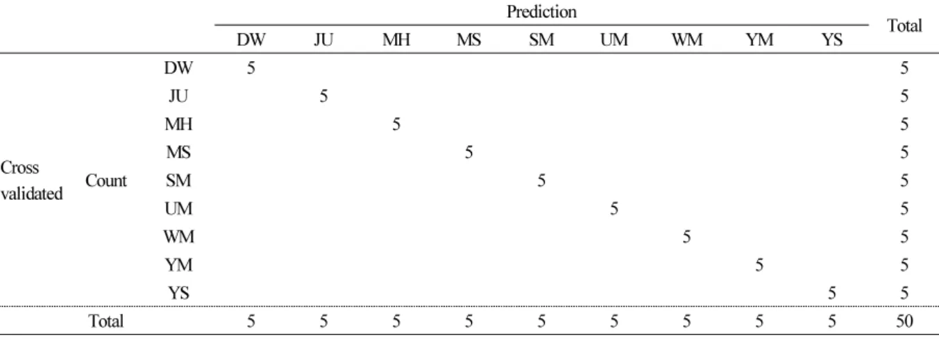
각각의 복숭아로부터 품종간 PLS-DA 식별이 bootstrap test를 통해 실제 적용 가능성을 확인하였다(Table 2). Bootstrap test의 정확도는 99%로 통계적으로 유의하였으며(p < 0.01), RMSEP (Root Mean Square Error of Prediction)은 0.23, MSEP (Mean Square Error of Prediction)는 0.005로 예측값에 대한 정확도를 나타냈다(Table 2). 이러한 결과는 복숭아 품종
Table 2. Summary of the PLS-DA classification results from FT-IR spectral data of genomic DNA. In bootstrapping, each case was classified by the functions derived from all other cases. DW : Yumyeong seedling (Daewol), JU : Chiyomaru, MH : Mihong, MS : Misshong, SM : Soomee, UM : Yumyeong, WM : Yumyeong mutation (Wolmi), YM : Yumi, YS : Yumyeong seedling (Eumseong)
Prediction
Total
DW JU MH MS SM UM WM YM YS
Cross
validated Count
DW 5 5
JU 5 5
MH 5 5
MS 5 5
SM 5 5
UM 5 5
WM 5 5
YM 5 5
YS 5 5
Total 5 5 5 5 5 5 5 5 5 50
MSEP (Mean Square Error of Prediction) = 0.005.
RMSEP (Root Mean Square Error of Prediction) = 0.23.
R2 = 0.99.
p < 0.01.
간의 차이를 PLS 모델링을 통해 높은 정확도로 명확히 구별 할 수 있다는 것을 나타냈다. 본 연구에서는 9개의 복숭아 품종을 테스트하였고, 테스트를 통해 정확하게 예측하고 자신의 그룹으로 분류 되었다. Genomic DNA의 특정한 nucleoside base 그 룹은 물리적인 진동으로 인해 변하기 쉬운 구조의 DNA이지만 서로 다른 FT-IR 스펙트럼의 특징적인 polypeptide-DNA 복 합체 정보를 나타낼 수 있다고 보고되었다(Mello and Vidal, 2012). 따라서 품종간 예측 정확도를 통해 새로운 샘플이 사용되 면 다양한 통계 연산을 사용하여 FT-IR 스펙트럼의 재현성을 나타낼 수 있다.
복숭아 품종에는 매우 많은 종류가 있지만 유전적 다양성은 낮은 것으로 알려져 있다(Kwon et al., 2015). 복숭아는 Prunus persica라는 단일 종으로 구성 되어 있으며, 자가결실이 가능하다는 생물학적인 특징 이외에 육종 과정에서 반복적으로 유사 한 품종을 모수로 활용되는 경우가 많기 때문에 유사한 유전자가 집적되고 있으며, SSR 마커 등 분자유전학적인 분석을 통해 서도 유전적 다양성이 낮은 것으로 보고되고 있다(Xie et al., 2010). 현재까지 복숭아 genomic DNA의 FT-IR 스펙트럼 데이 터를 이용한 복숭아에서 근연관계 분석에 대한 보고는 거의 없지만, 다른 작물에서는 genomic DNA의 FT-IR 스펙트럼 데이 터 분석을 통해 품종 식별을 한 사례가 보고되어 있다. 특히, 적배추에서는 서로 다른 부모에서 F1 자손 식별이 가능하였고 (Song et al., 2014), genomic DNA의 스펙트럼 유사성으로 고등 식물의 분류학적 관계를 나타냈으며(Muntean et al., 2009), FT-IR 스펙트럼의 유사성을 기준으로 차나무의 품종을 식별하였다(Song et al., 2017). 또한, 대목용 복숭아와 야생 복숭아의 유전적 유사도를 확인하고, 수집 지역에 따른 유전적 다양성이 낮은 것으로 보고된 바 있다(Cho et al., 2012). 이상의 연구 결과 로 판단할 때 복숭아 genomic DNA의 FT-IR 스펙트럼 데이터를 이용한 근연관계 식별이 가능함을 알 수 있었다.
본 연구에서 확립된 genomic DNA를 이용한 복숭아의 품종 식별 기술은 품종, 계통의 신속한 선발 수단으로 활용이 가능할 것으로 기대되며 육종을 통한 품종개발 가속화에 기여 할 수 있을 것으로 예상된다.
초 록
본 연구는 FT-IR 스펙트럼 데이터를 기반으로 다변량통계 기법을 이용하여 ‘유명’ 복숭아 자손 품종의 유전적 관계를 분석
하였다. ‘유명’과 ‘치요마루’의 교배를 통해 육성된 4품종, ‘유명’ 품종의 자연교잡실생 2품종 그리고 아조변이 1품종을 사용 하였다. Genomic DNA의 FT-IR 스펙트럼 데이터로부터 principal component analysis(PCA), partial least square discriminant analysis (PLS-DA) 그리고 hierarchical clustering analysis(HCA) 분석을 실시하였다. IR 스펙트럼은 구아닌 고리(C6 = C6), 티민 고리(C4 = O4), 사이토신, N-H 스트레칭(amide I), C = O 스트레칭 진동(amide II) 및 PO2- 이온화 대칭 과 비대칭 스트레칭의 주파수 영역에서 전형적인 스펙트럼 차이가 존재함을 나타낸다. ‘유명’과 ‘치요마루’의 교배 품종 중 ‘미 홍’과 ‘미스홍’은 ‘치요마루’보다 ‘유명’에 유전적 유사성이 더 높았다. 반면 ‘유미’와 ‘수미’는 ‘유명’보다 ‘치요마루’와 유전 적 유사성이 더 높았다. ‘유명’의 변이 품종 ‘월미’는 ‘유명’과 밀접한 관계를 보였지만, 다른 교배 품종인 ‘미홍’과 ‘미스홍’에 비해 유전적 유사성이 낮았다.
추가 주요어 : 식별시스템, 위계적군집분석, 다변량통계분석, 부분최소제곱회귀, 주성분분석
Literature Cited
Ahmad R, Potter D, Southwick SM (2004) Genotyping of peach and nectarine cultivars with SSR and SRAP molecular markers. J Am Soc Hortic Sci 129:204-210. doi:10.21273/JASHS.129.2.0204
Argov S, Ramesh J, Salman A, Sinelnikov I, Goldstein J, Guterman H, Mordechai S (2002) Diagnostic potential of Fourier-transform infrared microspectroscopy and advanced computational methods in colon cancer patients. J Biomed Opt 7:248-254. doi:10.1117/1.1463051 Banyay M, Sarkar M, Graslund A (2003) A library of IR bands of nucleic acids in solution. Biophys Chem 104:477-488. doi:10.1016/S0301-
4622(03)00035-8
Bouhadida M, Casas AM, Gonzalo MJ, Arús P, Moreno MA, Gogorcena Y (2009) Molecular characterization and genetic diversity of Prunus rootstocks. Sci Hortic 120:237-245. doi:10.1016/j.scienta.2008.11.015
Brewer SH, Anthireya SJ, Lappi SE, Drapcho DL, Franzen S (2002) Detection of DNA hybridization on gold surfaces by polarization modulation Infrared Reflection Absorption Spectroscopy. Langmuir 18:4460-4464. doi:10.1021/la0492815
Casas AM, Igartua E, Balaguer G, Moreno MA (1999) Genetic diversity of Prunus rootstocks analyzed by RAPD markers. Euphytica 110:139-149. doi:10.1023/A:1003745311408
Cheng Z, Huang H (2009) SSR fingerprinting Chinese peach cultivars and landraces (Prunus persica) and analysis of their genetic relationships. Sci Hortic 120:188-193. doi:10.1016/j.scienta.2008.10.008
Cho KH, Nam EY, Bae KM, Shin IS, Kim SH, Kim HR, Chung KH (2012) Analysis of genetic diversity among Wild Peach germplasm using DNA markers. Korean J Breed Sci 44:588-596. doi:10.9787/KJBS.2012.44.4.588
Dukor RK (2002) Vibrational spectroscopy in the detection of cancer. In: Chalmers JM, Griffiths PR (eds) Handbook of vibrational spectroscopy 5. Wiley London pp 3335-3361. doi:10.1002/0470027320.s8107
Emura K, Yamanaka S, Isoda H, Watanabe KN (2006) Estimation for different genotypes of plants based on DNA analysis using Near-infrared (NIR) and Fourier-transform Infrared (FT-IR) Spectroscopy. Breed Sci 56:399-403. doi:10.1270/jsbbs.56.399 Falk M, Hartman KA, Lord RC (1963) Hydration of deoxyribonucleic acid. III. A spectroscopic study of the effect of hydration on the
structure of deoxyribonucleic acid. J Am Chem Soc 85:391-394. doi:10.1021/ja00887a005
Fiehn O, Kopka J, Drmann P, Altmann T, Trethewey R, Willmitzer L (2000) Metabolite profiling for plant functional genomics. Nat Biotechnol 18:1157-1161. doi:10.1038/81137
Fischer G, Braun S, Thissen R, Dott W (2006) FT-IR spectroscopy as a tool for rapid identification and intra-species characterization of airborne filamentous fungi. J Microbiol Methods 64:63-77. doi:10.1016/j.mimet.2005.04.005
Gonzalez-Ruiz V, Olives AI, Martin MA, Ribelles P, Ramos MT, Menendez JC (2011) An overview of analytical techniques employed to evidence drug-DNA Interactions. Applications to the Design of Genosensors. In: Biomedical engineering, trends, research and technologies, M. A. Komorowska and S. Olsztynska-Janus (eds.). In Tech pp 65-90. doi:10.5772/13586
Haji T, Yaegaki H, Yamaguchi M (2001) Changes in ethylene production and flesh firmness of melting, nonmelting and stony hard peaches after harvest. J Jpn Soc Hortic Sci 70:458-458. doi:10.2503/jjshs.70.458
Han SE, Cho KH, Nam EY, Shin IS, Kim CH, Kim HR, Kim DH (2010) Identification of new breeding lines by Prunus Persica cultivar-specific SCAR primers. Korean J Breed Sci 42:495-501. uci:G704-000329.2010.42.5.009
Hong JH, Yi SI, Kwon YS, Kim Y, Choi KJ (2013) Genetic diversity analysis of peach [Prunus persica (L.) Batsch] varieties using SSR markers.
Korean J Breed Sci 45:201-211. doi:10.9787/KJBS.2013.45.3.201
Hu D, Zhang Z, Zhang D, Zhang Q, Li J (2005) Genetic relationship of ornamental peach determined using AFLP markers. HortScience 40:1782-1786. doi:10.21273/HORTSCI.40.6.1782
Jun JH, Kwon JH, Nam EY, Chung KH, Yun IK, Yun SK, Kwack YB, Kim SJ, Kang SJ (2013) ‘Yumi’ peach. HortScience 48:1416-1417.
doi:10.21273/HORTSCI.48.11.1416
Kuras A, Antonius K, Kalendar R, Kruczyn´ska D, Korbin M (2013) Application of five DNA marker techniques to distinguish between five apple (Malus x domestica Borkh.) cultivars and their sports. J Hortic Sci Biotechnol 88:790-794. doi:10.1080/14620316.2013.11513040 Kwon JH, Jun JH, Nam EY, Chung KH, Hong SS, Yoon IK, Yun SK, Kwack YB (2015) Profiling diversity and comparison of Eastern and
Western cultivars of Prunus persica based on phenotypic traits. Euphytica 206:401-415. doi:10.1007/s10681-015-1494-0 Lee D, Chapman D (1986) Infrared spectroscopic studies of biomembranes and model membranes. Biosci Rep 6:235-256. doi:10.1007/B
F01115153
Lei Z, Huhman DV, Sumner LW (2011) Mass spectrometry strategies in metabolomics. J Biol Chem 286:25435-25442. doi:10.1074/jbc.
R111.238691
Liquier J, Coffinier P, Firon M, Taillandier E (1991) Triple helical polynucleotidic structures: sugar conformations determined by FTIR spectroscopy. J Biomol Struct Dyn 9:437-445. doi:10.1080/07391102.1991.10507927
Liu Y, Ying Y, Yu H, Fu X (2006) Comparison of the HPLC method and FT-NIR analysis for quantification of glucose, fructose, and sucrose in intact apple fruits. J Agric Food Chem 54:2810-2815. doi:10.1021/jf052889e
Malins DC, Gilman NK, Green VM, Wheeler TM, Barker EA, Anderson KM (2005) A cancer DNA phenotype in healthy prostates, conserved in tumors and adjacent normal cells, implies a relationship to carcinogenesis. Proc Natl Acad Sci 102:19093-19096.
doi:10.1073/pnas.0509630102
Malins DC, Johnson PM, Barker EA, Polissar NL, Wheeler TM, Anderson KM (2003) Cancer-related changes in prostate DNA as men age and early identification of metastasis in primary prostate tumors. Proc. Natl Acad Sci 100:5401-5406. doi:10.1073/pnas.0931396100 Malins DC, Polissar NL, Gunselman SJ (1997) Models of DNA structure achieve almost perfect discrimination between normal prostate, benign prostatic hyperplasia (BPH), and adenocarcinoma and have a high potential for predicting BPH and prostate cancer. Proc Natl Acad Sci 94:259-264. doi:10.1073/pnas.94.1.259
Mello MLS, Vidal BC (2012) Changes in the infrared microspectroscopic characteristics of DNA caused by cationic elements, different base richness and single-Stranded form. PLoS ONE 7:e43169. doi:10.1371/journal.pone.0043169
Mungur R, Glass ADM, Goodenow DB, Lightfoot DA (2005) Metabolite fingerprinting in transgenic Nicotiana tabacum altered by the Escherichia coli glutamate dehydrogenase gene. J Biomed Biotechnol 2005:198-214. doi:10.1155/JBB.2005.198
Muntean CM, Halmagyi A, Puia MD, Pavel I (2009) FT-Raman of genomic DNA from plant tissues. Spectroscopy 23:59–70. doi:10.3233/
SPE-2009-0375
Naumann A, Navarro-Gonzalez M, Peddireddi S, Kues U, Polle A (2005) Fourier transform infrared microscopy and imaging: detection of fungi in wood. Fungal Genet Biol 42:829-835. doi:10.1016/j.fgb.2005.06.003
Naumann D, Helm D, Labischinski H (1991) Microbiological characterizations by FT-IR spectroscopy. Nature 351:81-82. doi:10.1038/351 081a0
Ngo-Thi NA, Kirschner C, Naumann D (2003) Characterization and identification of microorganisms by FT-IR microspectrometry. J Mol Struct 661-662:371-380. doi:10.1016/j.molstruc.2003.08.012
Pan L, Zeng W, Niu L, Lu Z, Liu H, Gui G, Zhu Y, Chu J, Li W, et al. (2015) PpYUC11, a strong candidate gene for the stony hard phenotype in peach (Prunus persica L. Batsch), participates in IAA biosynthesis during fruit ripening. J Exp Bot 66:7031-7044.
doi:10.1093/jxb/erv400
Preisner O, Lopes JA, Guiomar R, MacHado J, Menezes JC (2007) Fourier transform infrared (FT-IR) spectroscopy in bacteriology: towards a reference method for bacteria discrimination. Anal Bioanal Chem 387:1739-1748. doi:10.1007/s00216-006-0851-1
Sahu RK, Mordechai S, Manor E (2008) Nucleic acids absorbance in mid IR and its effect on diagnostic variates during cell division: a case study with lymphoblastic cells. Biopolymers 89:993-1001. doi:10.1002/bip.21048
Sambrook J, Fritsch EF, Maniatis T (1989) Molecular cloning: a laboratory manual, 2nd ed., Cold Spring Harbor Laboratory Press, Cold Spring Harbor, New York
Schulz H, Baranska M (2007) Identification and quantification of valuable plant substances by IR and Raman spectroscopy. Vib Spectrosc 43:13-25. doi:10.1016/j.vibspec.2006.06.001
Song SY, Jie EY, Ahn MS, Lee IH, Nou IS, Min BW, Kim SW (2014) Fourier transform infrared (FT-IR) spectroscopy of genomic DNA to discriminate F1 progenies from their paternal lineage of Chinese cabbage (Brassica rapa subsp. pekinensis). Mol Breed 33:453-464.
doi:10.1007/s11032-013-9963-4
Song SY, Kim CH, Joung YB, Lim CK, Seong KC, Song KJ, Moon DG (2017) Genomic DNA data of FT-IR spectroscopy using discriminate from different species in tea trees. J Korean Tea Soc 23:45-51. doi:10.29225/jkts.2017.23.1.45
Song SY, Nam EY, Yun SK, Kim SJ, Kwon JH (2020) Multivariate analysis of wild peach accessions for rootstock breeding using FT-IR spectroscopy of genomic DNA data. Korean J Breed Sci 52:25-31. doi:10.9787/KJBS.2020.52.1.25
Sundaram J, Park B, Hinton A, Yoon SC, Lawrence KC (2012) Identification and characterization of Salmonella serotypes using DNA spectral characteristics by Fourier transform infrared (FT-IR) spectroscopy. Proc SPIE pp 8369. doi:10.1117/12.918585
Terhoeven-Urselmans T, Vagen TG, Spaargaren O, Shepherd KD (2010) Prediction of soil fertility properties from a globally distributed soil mid-infrared spectral library. Soil Sci Soc Am J 74:1792-1799. doi:10.2136/sssaj2009.0218
Trygg J, Holmes E, Londstedt T (2007) Chemometrics in metabonomics. J Proteomes Res 6:467-479. doi:10.1021/pr060594q Verde I, Abbott AG, Scalabrin S, Jung S, Shu S, Marroni F, Zhebentyayeva T, Dettori1 MT, Grimwood J, et al. (2013) The high-quality draft
genome of peach (Prunus persica) identifies unique patterns of genetic diversity, domestication and genome evolution. Nat Genet
45:487-496. doi:10.1038/ng.2586
Warburton ML, Bliss FA (1996) Genetic diversity in peach (Prunus persica L. Batch) revealed by randomly amplified polymorphic DNA (RAPD) markers and compared to inbreeding coefficients. J Am Soc Hortic Sci 121:1012-1019. doi:10.21273/JASHS.121.6.1012 Wold H (1966) Estimation of principal components and related models by iterative least squares, pp 391-420. In: K. R. Krishnaiah (ed.).
Multivariate Analysis. Academic Press, New York.
Xie R, Li X, Chai M, Song L, Jia H, Wu D, Chen M, Chen K, Aranaza MJ, et al. (2010) Evaluation of the genetic diversity of Asia peach accessions using a selected set of SSR markers. Sci Hortic 125:622-629. doi:10.1016/j.scienta.2010.05.015
Xu DH, Wahyuni S, Sato Y, Yamaguchi M, Tsunematsu H, Ban T (2006) Genetic diversity and genetic relationships of Japanese peach (Pr unus persica L.) cultivars reveales by AFLP and pedigree tracing. Genet. Resour. Crop Evol 53:883-889. doi:10.1007/s10722-004-05 75-z
Yamamoto T, Mochida K, Hayashi T (2003) Shanhai Suimitsuto, one of the origins of Japanese peach cultivars. J Jpn Soc Hortic Sci 72:116-121. doi:10.2503/jjshs.72.116
Yesudas CR, Bashir R, Geisler MB, Lightfoot DA (2013) Identification of germplasm with stacked QTL underlying seed traits in an inbred soybean population from cultivars Essex and Forrest. Mol Breed 31:693-703. doi:10.1007/s11032-012-9827-3
Yoon JH, Liu DC, Song WS, Liu WS, Zhang AM, Li SH (2006) Genetic diversity and ecogeographical phylogenetic relationships among pe ach and nectarine cultivars based on simple sequence repeat (SSR) markers. J Am Soc Hortic Sci 131:513-521. doi:10.21273/JASHS.1 31.4.513
Zhou-Sun B, Sun J, Gryaznov SM, Liquier J, Garestier T, Helene C, Taillandier E (1997) A physico-chemical study of triple helix formation by an oligodeoxythymidylate with N34’-P5’ phosphoramidate linkages. Nucleic Acids Res 25:1782-1787. doi:10.1093/nar/25.9.1782