Asian Spine Journal Vol. 4, No. 1, pp 44~47, 2010 doi:10.4184/asj.2010.4.1.44
Copyright � 2010 by Korean Society of Spine Surgery
This is an Open Access article distributed under the terms of the Creative Commons Attribution Non-Commercial License (http://creativecommons.org/licenses/by-nc/3.0) which permits unrestricted non-commercial use, distribution, and reproduction in any medium, provided the original work is properly cited.
Asian Spine Journal�pISSN 1976-1902 eISSN 1976-7846
Received Jun 18, 2009; 1st Revised Jul 29, 2009; 2nd Revised Aug 12, 2009; Accepted Aug 17, 2009 Corresponding author:Chang Bum Lee, MD
Department of Orthopaedic Surgery, Maryknoll Hospital, 12 Daecheong-dong 4-ga, Jung-gu, Busan 600-730, Korea
Tel: +82-51-465-8801, Fax: +82-51-463-1194, E-mail: [email protected]
Copyright � 2010 by Korean Society of Spine Surgery
This is an Open Access article distributed under the terms of the Creative Commons Attribution Non-Commercial License (http://creativecommons.org/licenses/by-nc/3.0) which permits unrestricted non-commercial use, distribution, and reproduction in any medium, provided the original work is properly cited.
Asian Spine Journal�pISSN 1976-1902 eISSN 1976-7846
Treatment for the Lumbosacral Soft Tissue Defect after Spine Surgery
Sun Jin Choi1, Chang Bum Lee1, Hyung Taek Park1, Jong Hoon Park1, Hyeong Seok Lee1, Yong Jin Kim2
1Department of Orthopaedic Surgery, Maryknoll Hospital, Busan, Korea,
2Department of Orthopaedic Surgery, West Busan Centum Hospital, Busan, Korea
The lumbosacral area is one of the most frequently operated spine regions because of the prevalence of disease in that area.
Although a lumbosacral soft tissue defect after surgery due to inflammation and other causes is rare, such soft tissue defects are difficult to treat. Therefore, suitable methods for treating lumbosacral soft tissue defects are necessary. There- fore, this study introduces a case-treated with a transverse lumbosacral rotational flap.
Key WWords: Lumbar spine, Lumbosacral flap, Soft tissue defect
Introduction
Although lumbosacral soft tissue defects after back surgery are rare, this complication can be disastrous and dif- ficult to treat. A few treatment methods have been intro- duced, but they are technically difficult to perform. We report a case treated with a transverse lumbosacral rotation- al flap, which is a simple and useful method for treating soft tissue defects after back surgery.
Case Report
A 55-year-old female visited our hospital complaining of gait disturbance and weakness on her right leg for 3 years.
She had a history of back surgery at L4-5 herniated nucleus pulposus (HNP) and radiation therapy for uterine cervical cancer after a total hysterectomy. Pain and light touch sens- es were decreased on the right L3 to L4 dermatomes and deep tendon reflexes were not evoked in the legs. Contrac-
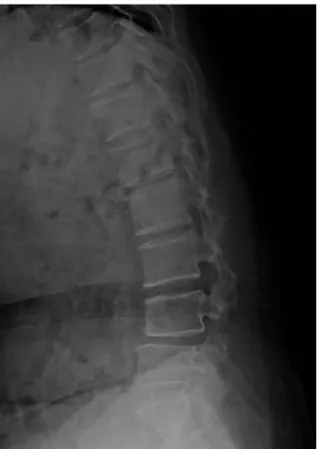
tion of the right tibialis anterior and peroneus longus was not detected. A stoppage circumduction gait was noted dur- ing walking. The skin and soft tissue of the lower abdomen and back were stiff, hard and non-elastic because of radio- therapy. A dynamic radiographic study showed retro- spondylolisthesis, L4 on L5 (Fig. 1). Magnetic resonance imaging revealed HNP with caudal migration, L4-L5, and a right posterolateral type. In addition, there was soft tissue adhesion around the nerve root. Posterior decompression and lumbar interbody fusion with cages were performed using the posterior approach (Fig. 2). It was difficult to close the skin, muscle and fascia directly because of the soft tissue defects, which were characterized by stiffness and hardness of the tissue. Therefore, the wound was left open using an interrupted wiring suture. The postoperative wound did not heal well and a serious discharge was drained consecutively. Debridement and a temporary suture were performed twice after surgery to control the surgical wound. However, healing of the soft tissue had not improved due to the sclerotic changes to the tissue and the impairment of circulation (Fig. 3). As a result, wide
debridement and a transverse lumbosacral rotational flap were performed with good results and without complica- tions (Fig. 4).
Discussion
When we encountered this case, we could not find useful methods or references despite an extensive search of the lit- erature on plastic and microsurgery for the management of lumbosacral defects. However, most of these articles report- ed treatments for bed sores. In addition, they mainly used a gluteus maximus flap to treat soft tissue defects. Free or local flaps based on the perforating arteries of the gluteal resion are among the most commonly used procedures due to the excellent blood supply and adequate bulkiness [1].
On the other hand, limited shifting capacity that sacrifices the muscle in ambulatory patients is a major drawback of this procedure. In addition, they take a great deal of operat- ing time, and there is excessive blood loss [2,3]. Occasion- ally, we found a suitable method by reviewing the articles.
The lumbosacral flap offers a reliable and reasonable
Treatment for the Lumbosacral Soft Tissue Defect after Spine Surgery/ 45
Fig. 3. The soft tissue had not healed well because of the sclerotic changes in the tissue and the impairment of cir- culation.
Fig. 2. Posterior decompression and lumbar interbody fusion with cages were performed using the posterior approach.
Fig. 1. Pre-operative dynamic radiographic study show- ing instability and retrospondylolisthesis, L4 on L5.
46 / ASJ: Vol. 4, No. 1, 2010
method for the initial coverage of lumbosacral defects. Its design is anatomically sound when the lumbar perforators, which serve as the vascular pedicle, are preserved. A trans- verse lumbosacral flap has the following advantages: it is smaller and simple in execution; minimal loss of blood occurs; donor site defects can be closed directly; and the gluteal areas are intact for possible use later to cover recur- rent or previous failed flapa [4]. The transverse lumbosacral flap is a good method for covering lumbosacral defects. Its
reliability appears to be due to the axial pattern of its proxi- mal portion, and to an uninterrupted subdermal vascular plexus (Fig. 5) across the midline of the back [5,6].
Considerable preparation is needed if a post-operative wound problem can be predicted. If there is some possibili- ty of wound problem due to repeated surgery, immunologic diseases and radiotherapy before surgery, then operator should consider other methods, such as the anterior approach, minimal invasive surgery, and posterolateral approach. In our case, the patient had received radiotherapy 20 times for uterine cervical cancer and patient had wide previous operative scar on her abdomen as a result of a total hysterectomy. Secondarily posterior adhesiolysis was required for the nerve root entrapped by the previous opera- tive scar tissue. Therefore, there was no choice but to per- form the previous posterior approach.
A local flap is very useful for small sacral sores or skin defects. The average size of the sores for which unilateral flaps were used was 10×12 cm, while the size of the sores for which two flaps had to be raised was 22×12 cm and 22
×10 cm [4]. In our case, the skin defect was 7×8 cm, this unilateral flap was used easily and safely without excessive skin tension. Proper flap design is most important for achieving better results. Sufficient skin cover is essential for avoiding skin tension. The debridement of unhealthy skin and soft tissue must be performed. Overall, it is believed that a transverse lumbosacral flap is a simple and useful method for covering soft tissue defects after back surgery.
Fig. 4. Wide debridement and a transverse lumbosacral dorsal rotational flap were performed.
Fig. 5. Our interpretation of the subdermal plexus across the midline (regarding the contributions of the lumbar per- forators).
A B C
Treatment for the Lumbosacral Soft Tissue Defect after Spine Surgery/ 47 REFERENCES
01. Kroll SS, Rosenfield L. Perforator-based flaps for low pos- terior midline defects. Plast Reconstr Surg 1988;81:561-6.
02. Minami RT, Mills R, Pardoe R. Gluteus maximus myocu- taneous flaps for repair of pressure sores. Plast Reconstr Surg 1977;60:242-9.
03. Ramirez OM, Orlando JC, Hurwitz DJ. The sliding gluteus
maximus myocutaneous flap: its relevance in ambulatory patients. Plast Reconstr Surg 1984;74:68-75.
04. Rawat SS, Mathur BS. Transverse lumbar flap for sacral bed sores. Plast Reconstr Surg 1991;88:154-8.
05. Hill HL, Brown RG, Jurkiewicz MJ. The transverse lum- bosacral back flap. Plast Reconstr Surg 1978;62:177-84.
06. Smith PJ. The vascular basis of axial pattern flaps. Br J Plast Surg 1973;26:150-7.