관절경적 회전근 개 봉합술 이후 발생한 상완골두의 급성 골수염
이화여자대학교 의학전문대학원 정형외과학교실 신상진∙정병진∙국성환∙신승준
Acute Osteomyelitis of the Humeral Head after Arthroscopic Rotator Cuff Repair
Sang-Jin Shin, M.D., Byoung-Jin Jeong, M.D., Seung Hwan Kook, M.D., Sung-Joon Shin, M.D.
Department of Orthopaedic Surgery, Ewha Womans University School of Medicine, Seoul, Korea
A 57-year-old man who simultaneously underwent an operation for repair of rotator cuff and a revision operation for nonunion of a Pilon fracture presented with osteomyelitis of the humeral head on the 19thday after surgery due to MRSA (Methicillin-resistant Staphylococcus aureus) infection. Infection was controlled after administra- tion of appropriate intravenous antibiotic therapy and performance of several surgical procedures. However, dev- astating defects at the humeral head and the rotator cuff remained. No case of short term MRSA induced osteomyelitis has been reported.
Volume 16, Number 2, December, 2013 doi:http://dx.doi.org/10.5397/CiSE.2013.16.2.141
Arthroscopic rotator cuff repair showed satis- factory clinical outcomes with less postoperative complications compared to those of open repair.
Complication rate after arthroscopic rotator cuff repair is reported 4.8~10.6% which includes shoulder stiffness, infection, failure of the repair, reflex sympathetic dystrophy and deep venous thrombosis.1-3) Among them, postoperative infec- tion leaves a potentially devastating complication when once occurred. Serious functional disorder may result from fibrosis and destructive carti- lagenous impairment after postoperative septic arthritis, eventually making an early diagnosis
and treatment very important. The most common organisms which cause postoperative shoulder infection are Staphylococcus epidermidis, Staphy- lococcus aureus and Propionibacterium acne.2-4) Although Methicillin-resistant Staphylococcus aureus (MRSA) is the most common pathogen of community onset of sepsis, however, there has been no documents regarding MRSA induced septic arthritis after arthroscopic rotator cuff repair.5)We present an MRSA induced septic arthritis after arthroscopic rotator cuff repair which lead to acute osteomyelitis in a short period of less than 3 weeks.
※통신저자: 신 상 진
서울시 양천구 안양천로 1071
이화여자대학교 의학전문대학원 정형외과학교실
Tel: 02) 250-5010, Fax: 02) 2642-0349, E-mail: [email protected]
접수일: 2013년 4월 24일, 1차 심사완료일: 2013년 7월 17일, 2차 심사완료일: 2013년 10월 24일, 게재 확정일: 2013일 11월 14일
Case Report
A 57-year-old male visited emergency room due to ankle injury after a fall from a 1.5 m height. He was diagnosed as a Pilon fracture of his left ankle and underwent open reduction and internal fixation. There were no operative wound complications while routine follow up visits. Two months after open reduction and internal fixa- tion, nonunion of left medial malleolus was found and the patient was scheduled for revision operation. However, he also complained persis- tent left shoulder pain with limited range of motion which started a few days after the previ- ous accident. Despite aggravation of his shoulder pain, the patient was treated conservatively with oral NSAIDs. He had no history of shoulder joint injection. Physical examination of left shoulder revealed positive Jobe test and Hawkins test.
Range of motion of left shoulder was 100。for- ward flexion, 30。external rotation and L4-5 vertebral level of internal rotation. The shoulder MRI showed a full-thickness tear near the insertion site of the supraspinatus tendon.
Arthroscopic rotator cuff repair was also planned at the same day after the revision oper-
ation of the Pilon fracture. Preoperative evalua- tion showed no recent history of any febrile event, erythema or heating around the previous operation site except mild tenderness on the medial malleolus of the left ankle was present, which was thought to be an inflammatory reac- tion resulting from non-union. Medical history of the patients was nonspecific, except a 10 year-history of hypertension. Blood count, ery- throcyte sedimentation rate (ESR), C-reactive protein (CRP) values and other laboratory tests including electrolytes, liver and renal function tests were within normal limits. Open reduction and internal fixation with allogenous bone graft was done for the non-union of the medial malle- olus without any intraoperative complications.
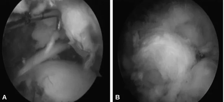
No cultures or biopsy were performed for there were no suspicious findings for infectious nonunion on the previous surgical site. After aseptic dressing and short leg cast application, another proper preparation including aseptic setting with lateral position for shoulder opera- tion was done. Arthroscopic findings of left shoulder revealed a medium sized full thickness rotator cuff tear (20×15 mm) (Fig. 1A). Arthro- scopic rotator cuff repair was performed using
Fig. 1. (A) Arthroscopic images showing medium sized (20×15 mm) full thickness tear at the tendinous portion of the supraspinatus muscle, (B) Arthroscopic image after rotator cuff repair using transosseous equivalent technique.
A B
transosseous equivalent technique with 2 bio- suture anchors (Spiralok 5.0 mm suture anchor;
Mitek, Raynham, MA, USA) and 2 knotless anchors (Bio-PushLock: Arthrex, Naples, FL, USA) without any complications (Fig. 1B). The patient managed well until the postoperative third day, however, showed erythematous swelling only around the shoulder joint with spiking fever over 38。C that lasted for 3 days in spite of application of intravenous second gener- ation cephalosporins and antipyretics. On labo- ratory tests of the 7th day after surgery, white blood cell count was elevated to 15,500/mm3(nor- mal range, 4,000 to 10,000/mm3), ESR showed 61 mm/h (normal range, 0 to 28 mm/h) with a high- ly elevated CRP level of 34.5 mg/dL (normal range, 0 to 0.3 mg/dL). Under guidance of an infectious disease specialist, antibiotics regimen was changed to Tabaxin (4.5 g: tazobactam 0.5 g
+ piperacillin 4 g), on the 6th day for 3 days.
Fluid collection in the subacromial space and glenohumeral joint was confirmed by ultra- sonography on the 8th day (Fig. 2). Arthroscopic debridement with irrigation was carried out on the same day after ultrasonographic examina- tion, under suspicion of septic arthritis (Fig. 3).
Arthroscopic findings revealed that increased turbid synovial fluid and synovitis, but repaired tendon was well preserved and no bony or carti- laginous destruction was found. Intraoperative fluid culture and biopsy of the tissue was done through posterior portal. After the arthroscopic surgery, empirical Vancomycin was planned to additionally apply, however, patients had reacted allergic response to Vancomycin. Teicoplanin (400 mg) was additionally applied with Tabaxin for 3 days and later on Tabaxin was discontinued due to culture results of MRSA with proper guidance of an infectious disease specialist.
Surgical drainage was removed and tip culture was done on the 6th day after arthroscopic surgery. However, fever recurred at the 11th day
Fig. 2. Ultrasonography after 7thday of the arthroscopic rota- tor cuff repair showing fluid collection in the subdeltoid bursa extending to the joint space.
Fig. 3. Arthroscopic image at 7thday of the arthroscopic rota- tor cuff repair showing turbid fluid collection in the subacro- mial space which was found in septic arthritis.
after the second surgery and the CRP was re- elevated from 2.56 mg/dL to 4.03 mg/dL. Cul- ture result of the drainage tip also showed remaining MRSA. Shoulder MRI was performed to find out the cause of relapse of symptoms.
T1-weighted coronal and axial MRI of the shoul- der demonstrated a joint effusion and contrast enhancement in the humeral head, especially around the greater tuberosity and adjacent mus- cle, and it was correspond to osteomyelitis on the greater tuberosity of the humerus with remaining myositis of the rotator cuff and the deltoid muscle (Fig. 4). At 12thday after the sec- ond surgery, open debridement of greater tuberosity and humeral head were done without arthroscopic evaluation. Turbid fluid collection with granulation change of the tissue around the great tuberosity was found. Repaired supraspinatus tendon was not noticeable due to dissolution, and loosening of the suture anchors was found. Removal of the suture anchors and massive irrigation with debridement was done.
And Vancomycin (2 g) cement bead inserted in the glenohumeral joint, subacromial space and proximal humerus to prevent recurrence. There
was no change in antibiotics regimen (total peri- od of 6 weeks and 5 days of intravenous Teicoplanin, 400 mg). On the meantime, daily dressing and closed observation for the left ankle was done showing no sign for infection, stitch out and short leg cast change was done on routine schedule. Surgical drainage was removed and drainage tip culture was done on the 10thday after antibiotic cement bead insertion. On the basis of afebrile post-operative condition and negative culture results, bead removal was done at 3 weeks after open debridement with a 2 cm incision under regional anesthesia without any additional debridement or manipulation on rup- ture site. ESR and CRP value normalized to 35 mm/h and 0.25 mg/dL, respectively at the 3rd week after the last operation. During a 3-year- follow up period, there was no infectious sign clinically and laboratory tests showed normal range. However, there remains a defect of humeral head and a large rotator cuff tear with a limited range of motion (Fig. 5). His active for- ward flexion is 100。, external rotation 30。, and internal rotation L3 at last follow-up visit.
Although there remains limitation in range of
Fig. 4. Shoulder MRI showing osteomyelitis of the greater tuberosity of the humerus and myositis of the rotator cuff and the deltoid muscle on (A) oblique-coronal view and (B) axial view.
A B
motion of the shoulder joint, the patient has lit- tle discomfort in daily living. Reverse total shoulder arthroplasty is planned to be done later as the patient is not yet in an age indicative for reverse total shoulder arthroplasty.
Discussion
Infectious complication is uncommon after arthroscopic rotator cuff repair with a lower rate in arthroscopic procedures reported 0.04~0.23%, compared to those of open and mini-open pro- cedures reported 0.27~1.9%.2,6) There are some preoperative predisposing factors for shoulder septic arthritis including acupuncture which is popular in Asian culture, corticosteroid injection and medical disease such as diabetes mellitus or immune suppression state.7) However, this patient had no history of acupuncture on the shoulder, medical history susceptible to infection such as diabetes. Preoperative positive infectious
sign or infection-suspicious preoperative blood tests were not found in this patient. Additionally for postoperative pain management he had not undergone indwelling intra-articular catheteri- zation with infusion pump of bupivacaine which is suspected to induce chondrolysis following arthroscopic shoulder operation.8)
However unlike other septic arthritis of the shoulder, this patient underwent combined sur- gical procedure for the non-union fracture of medial malleolus, before arthroscopic rotator cuff repair at the same day. Although there were no suspicious infection sign at the ankle on pre- operative physical exams and laboratory tests, we presume that there might have been an undetected low grade infection which might had contributed both to the non-union of the frac- ture site and the postoperative shoulder infection in a hematogenous manner, in spite of appropri- ate preoperative and intraoperative antimicrobial prophylaxis following the Centers for Disease Control and Prevention (CDC) guidelines.9) Therefore, leading us to consider an optimal arthroscopic procedure to be delayed after a previous complicated operation, especially when there is a possibility of underlying undetected infection such as fracture-nonunion as in this case. Additionally there are possibilities of the long operation time in spite of intraoperative antibiotics according to CDC guidelines that contributed to the infection of our patient.
As for postoperative infectious symptoms after arthroscopic rotator cuff repair, patients present symptoms such as increased shoulder pain, wound drainage, erythema around the wound at approximately 4 to 6 weeks after surgery.6) Patients showed less constitutional symptoms like fever and chills and laboratory test showed less sensitivity in ESR elevated in 60% and CRP level elevated in 50%. However, this patient pre- sented pain and erythema around the shoulder associated with high fever, leukocytosis and ele- vated ESR, CRP within a week and eventually Fig. 5. A plain radiograph of left shoulder at last follow-up
visit showing bone destruction at the lateral portion of the humeral head.
abruptly progressed into osteomyelitis within 3 weeks after arthroscopic surgery. This difference in clinical presentation as for fever and labora- tory tests seemed to be a consequence from dif- ferent pathogen. A similar presentation is found in a case of a MRSA septic arthritis after 5 days of anterior cruciate ligament reconstruction with febrile presentation and elevated ESR and CRP levels.10) We consider an early bacterial culture from joint aspirations is mandatory when patients present early infectious presentations such as fever and elevated ESR and CRP level, because early initiation of MRSA sensitive antibiotics is important for preventing devastat- ing results from septic arthritis.
According to Kwon et al.4) retention of the suture anchors had not interfere clinical results in the success of bacterial eradication. However, the remained suture anchor with suture strands inside the humeral head after the first arthro- scopic debridement possibly might accelerate osteomyelitis of the humeral head. As in this case, osteomyelitis and bone defect at the posterior aspect of the humeral head around the previous bio-suture anchor site, may support this possibil- ity. Although due to lack in data related to the relationship of infection and bone anchor reten- tion, anything that could be a reservoir for the pathogen should be removed for efficient infection control and improved postoperative outcomes.
In conclusion, combined operation can be one of predisposing factor for septic shoulder arthri- tis in patients who underwent recent operation with complications. Septic arthritis at the shoul- der joint due to MRSA is possible when a patient demonstrated early infectious symptoms and elevated laboratory tests postoperatively. Prompt diagnosis and early antibiotic administration fol- lowing a removal of devices such as suture anchors are considered to prevent both postop- erative septic arthritis and osteomyelitis.
Conflict of Interest
No potential conflict of interest relevant to this article was reported.
REFERENCES
01) Brislin KJ, Field LD, Savoie FH. Complications after arthroscopic rotator cuff repair. Arthroscopy.
2007;23:124-8.
02) Osti L, Papalia R, Del Buono A, Denaro V, Maf- fulli N. Understanding and preventing complications in repairing rotator cuff tears. Med Sport Sci. 2011;
57:178-83.
03) Settecerri JJ, Pitner MA, Rock MG, Hanssen AD, Cofield RH. Infection after rotator cuff repair. J Shoulder Elbow Surg. 1999;8:1-5.
04) Kwon YW, Kalainov DM, Rose HA, Bisson LJ, Weiland AJ. Management of early deep infection after rotator cuff repair surgery. J Shoulder Elbow Surg. 14:1-5, 2005.
05) Frazee BW, Fee C, Lambert L. How common is MRSA in adult septic arthritis? Ann Emerg Med.
2009;54:695-700.
06) Athwal GS, Sperling JW, Rispoli DM, Cofield RH. Deep infection after rotator cuff repair. J Shoul- der Elbow Surg. 2007;16:306-11.
07) Zachary C, Landman BA, C Benjamin M. Acute multifocal osteomyelitis following acupuncture: A case report. J Bone Joint Surg Case Connector.
2012;2:e1-4.
08) Anderson SL, Buchko JZ, Taillon MR, Ernst MA.
Chondrolysis of the glenohumeral joint after infusion of bupivacaine through an intra-articular pain pump catheter: a report of 18 cases. Arthroscopy. 2010;26:
451-61.
09) Mangram AJ, Horan TC, Pearson ML, Silver LC, Jarvis WR. Guideline for Prevention of Surgical Site Infection, 1999. Centers for Disease Control and Prevention (CDC) Hospital Infection Control Prac- tices Advisory Committee. Am J Infect Control. 1999;
27:97-132.
10) Kurokouchi K, Takahashi S, Yamada T, Yamamo- to H. Methicillin-resistant Staphylococcus aureus- induced septic arthritis after anterior cruciate liga- ment reconstruction. Arthroscopy. 2008;24:615-7.
초 록
회전근개 전층 파열과 원위 경골 관절면 골절 불유합을 동시에 시행한 57세 남자가 수술 19일만에 MRSA (Methicillin-resistant Staphylococcus aureus)에 의한 상완골두 급성 골수염이 발생하였다. 골수염은 3 차례의 수술과 정맥 항생제 요법으로 치료되었으나 상완골두와 회전근개 결손은 남아있었다. MRSA에 의한 상완골두 골수염은 보고된 바가 없기에 문헌고찰과 함께 보고하는 바이다.