560
Clinical Significance of PTEN and Ki-67 Expression in Prostate Cancer
Il Kang, Yoon Seup Kum1, Kwan Kyu Park1, Duk Yoon Kim, Jae Shin Park
From the Departments of Urology and 1Pathology, College of Medicine, Daegu Catholic University, Daegu, Korea
Purpose: Phosphatase and tensin homolog (PTEN) is a novel tumor sup- pressor gene located at chromosome 10q23. Ki-67 antigen is a human nuclear protein that is expressed in all active parts of the cell cycle. We evaluated the significance of PTEN and Ki-67 expression in prostate cancer and investigated the relation of this expression with clinico- pathological factors in prostate cancer.
Materials and Methods: Initially, we did two kinds of immunohistoche- mical staining for PTEN and Ki-67. Immunohistochemical staining was performed on 75 formalin-fixed paraffin-embedded cancer specimens.
Staining on paraffin blocks from prostate carcinomas was compared with that for adjacent normal prostate. Stainings were considered positive if nuclear staining was seen. Positive stainings were analyzed with the patient's clinico-pathological findings. Statistical analysis was performed by using chi-square test with p<0.05 considered significant.
Results: PTEN was expressed in 65 (86.6%) of 75 specimens. Ki-67 was expressed in 63 (84.0%) of 75 specimens. The staining scores of the tumor cells for PTEN and Ki-67 were higher than those of the adjacent normal cells (p<0.05). The staining scores for PTEN were negatively correlated with the serum prostate-specific antigen (PSA) level and Gleason score, but this was not statistically significant (p>0.05). PTEN expression was negatively correlated with lymph node or distant metastases (p<0.05).
Ki-67 was positively correlated with the serum PSA level, the Gleason score, and metastases (p<0.05).
Conclusions: PTEN and Ki-67 staining correlated well with Gleason score and PSA level in prostate cancer. These could be a possible predictor of prostatic neoplasms. (Korean J Urol 2009;50:560-566)
Key Words: Prostatic neoplasms, Human PTEN protein, Ki-67 antigen
Korean Journal of Urology Vol. 50 No. 6: 560-566, June 2009 DOI: 10.4111/kju.2009.50.6.560
대구가톨릭대학교 의과대학
비뇨기과학교실, 1병리학교실
강 일ㆍ금윤섭1ㆍ박관규1
김덕윤ㆍ박재신
Received:February 6, 2009 Accepted:May 22, 2009
Correspondence to: Jae Shin Park Department of Urology, College of Medicine, Daegu Catholic University, 3056-6, Daemyeong 4-dong, Nam-gu, Daegu 705-718, Korea
TEL: 053-650-4662 FAX: 053-623-4660 E-mail: jspark@cu.ac.kr
Ⓒ The Korean Urological Association, 2009
서 론
전립선암은 서구에서 가장 빈발하며, 악성종양 중 세 번 째 높은 사망률을 보이고 있다.1-3 전립선암의 형질변환과 진행에는 여러 생물학적 인자가 작용할 것으로 추정된다.
아직까지 이에 대한 정확한 기전은 알려지지 않았지만, 여 러 종양유전자 및 종양억제유전자 등이 세포증식과 세포고 사에 관여한다는 보고가 있다. 이 중에서 phosphatase and
tensin homolog (PTEN)는 대표적인 종양억제 유전자로 잘 알려져 있고, 염색체 10q23.3에 위치해 있다.4 PTEN 유전자 는 방광암, 유방암, 폐암 등의 암에서 돌연변이 혹은 결손이 밝혀졌고, 세포주기 정지 및 세포사멸을 유도할 뿐만 아니 라 세포이동과 분화를 조절하는 것으로 알려졌다.5-7 PTEN 의 결손은 PIP3의 축적을 유발하고, PI3-kinase/Akt pathway 를 활성화시켜서, 이를 통해 세포주기의 진행과 세포의 생 존과 이동을 자극시킨다.8 최근의 연구는 PTEN이 저산소 상태에서 전립선암의 angiogenesis에 관여한다는 것을 밝혀
Table 1. Comparison of clinicopathological characteristics accord- ing to phosphatase and tensin homolog (PTEN) expression
Variables PTEN expression
p-value Negative Positive
Age (years)
<70 (%) 4 (12.1) 29 (87.9)
0.784
≥70 (%) 6 (14.3) 36 (85.7)
Prostate volume (cc)
<37 (%) 5 (10.6) 42 (89.4)
0.374
≥37 (%) 5 (17.9) 23 (82.1)
냈고, PTEN의 결손이 PI-3 kinase를 불활성화 시킴으로써 전립선암의 혈관신생을 증가시킨다고 보고했다.9,10 한편, 종양의 증식능력 평가를 통해 종양의 성장 정도와 악성도 를 간접적으로 알아보는 연구가 시도되고 있다. 이러한 세 포의 증식은 세포의 분열률, S기 분획, 분열 중인 세포의 핵 항원에 대한 면역조직화학적 염색 등을 통해 평가할 수 있다.11 이 중 Ki-67은 휴지기 세포를 제외한 증식하는 세포 들에서 발현되는 항원으로 면역조직화학 염색법을 통해 여 러 종류의 종양에 세포증식정도의 척도로 사용되고 있다.12 이에, 저자들은 인체 전립선암의 조직에서 면역화학적 염 색을 통하여 종양세포에서 결손이나 돌연변이를 일으키는 종양억제유전자인 PTEN과 세포증식에 관한 표지자로 알려 진 Ki-67의 역할과 임상적 지표와의 상관관계에 대해 알아 보고자 하였다. 또한, 이 두가지의 표지자들이 PSA와 같이 전립선암의 진행정도와 예후에 영향을 미치는지 알아보고 자 하였다.
대상 및 방법 1. 대상
2005년 6월부터 2008년 5월까지 본원에서 경직장 초음파 전립선생검 결과 전립선암으로 진단받은 환자 75명 중 근 치적 전립선절제술을 시행받은 25명과 호르몬 치료를 받은 환자 50명에서 수집된 파라핀 블록 75개를 본 실험에 사용 하였다. 대상 환자들의 평균 나이는 70.64±8.311세 (54-89) 였다. 평균 전립선 용적은 37.73±16.950 cc였다. 진단 당시 prostate-specific antigen (PSA)는 36.40±60.637 ng/ml였다.
2. 방법
1) 조직학적 검사: 75개의 파라핀 블록을 5μm 두께로 연 속절편을 제작하여 통상적인 H&E 염색을 실시하여 광학현 미경으로 관찰하였다. 그리고 일부는 면역조직화학 염색을 위해 준비해 두었다.
2) 면역조직화학적 염색: 미리 준비된 파라핀 블록 절편 으로 면역조직화학 염색을 실시하였다. 일차 항체로 PTEN (Novovastra, Newcastle, U.K), Ki-67 (Zymed, Sanfrancisco, USA)을 사용하였고, 면역조직화학 염색은 labelled strepta- vidin biotin (LASB) 방법으로 시행하였다. 조직 절편을 자일 렌으로 탈파라핀한 후 알코올로 함수시키고, 0.3% 과산화 수소로 5분간 반응시켜서 내재성 과산화효소와 비오틴을 억제시킨 다음 phosphate buffered saline (PBS) 완충액으로 10분간 세척하고 정상 면양 혈청을 가하여 20분간 반응시 켰다. 정상 면양 혈청을 제거한 다음 희석한 일차항체들 (α -SMA, 1:200, CK7, 1:200, CK19, 1:200, Hep, 1:200)을 첨가하
여 4oC 항온기에 하룻밤 두었다가 다음날 PBS 완충액으로 10분간 세척하고 비오틴과 결합된 항 생쥐 면양 혈청인 이 차 항체와 20분간 반응시켰다. 세척 후 streptavidin과 결합 한 과산화효소 복합체를 가하여 20분간 반응시킨 후 amino-ethylcarbazole (AEC)로 발색하고 Mayer's hematoxylin 으로 대조 염색한 후 광학현미경으로 관찰하였다.
3) 면역조직화학적 염색의 판정: PTEN에 대한 염색의 판 정은 암세포와 인근에 분포한 정상세포의 핵에 적갈색으로 염색되었을 때를 양성으로 판단하였다. 편도조직의 세포핵 양성을 비교군으로 하고, PTEN의 발현정도는 이전의 타연 구 논문에 근거하여 평가되었다.13 염색의 정도는 4가지로 분류되었다. 종양세포의 30% 미만이 염색되는 경우를 grade I, 30% 이상에서 60% 미만으로 염색되는 경우를 grade II, 60% 이상에서 염색되는 경우를 grade III로 정의하였다. Ki- 67의 경우도 편도 조직을 대조군으로 하여 세포의 핵에 적 갈색의 과립이 보이는 세포를 양성으로 판정하였다. 전체 종양세포의 5% 미만이 염색되는 경우를 grade I, 5-15%가 염색되는 경우를 grade II, 15% 이상이 염색되는 경우를 grade III로 하였다.
3. 결과분석 및 통계처리
H&E 염색에서 나타난 암세포의 Gleason score, PSA 수치 와 림프절 침범 및 원격전이 여부와 PTEN과 Ki-67 염색발 현 정도와의 상관관계에 대하여 알아보았다. 통계는 chi- square test를 사용했고, 유의수준 알파는 0.05로 설정하였 다. 분석은 SPSS software, Windows 13.0 version (SPSS, Chicago, USA)을 사용했다.
결 과 1. 임상 및 병리학적 소견
대상의 나이 및 전립선 용적과 PTEN, Ki-67 염색정도 간 에는 통계학적인 관련성이 없었다 (Table 1, 2). 조직검사에
Table 2. Comparison of clinicopathological characteristics accord- ing to Ki-67 expression
Variables Ki-67 expression
p-value Negative Positive
Age (years)
<70 (%) 5 (15.2) 28 (84.8)
0.859
≥70 (%) 7 (16.7) 35 (83.3)
Prostate volume (cc)
<37 (%) 6 (12.8) 41 (87.2)
0.322
≥37 (%) 6 (21.4) 22 (78.6)
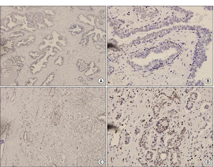
Fig. 1. (A, B) Absence of immunoreactivity for phosphatase and tensin homolog (PTEN) in normal prostate gland, (C, D) Numerous strongly positive nucleoli staining among cancer cells for PTEN (A, C: H&E x40, B, D: H&E x400).
서 나타난 Gleason score는 저등급 (6 미만)이 9명, 중등급 (6-8)이 46명, 고등급 (9-10)이 20명이었다.
2. PTEN 염색양상
전립선암 조직 75례의 PTEN 단백에 대한 면역조직화학염
색 결과 86.6% (65/75명)에서 양성반응을 보였고, 정상조직 과 전립선비대 조직에는 염색이 되지 않았다 (Fig. 1, 2).
PTEN 염색 정도와 Gleason score (p=0.595), PSA (p=0.216)와 는 음의 상관관계에 있었으나 통계학적으로 유의하지 않았 다. PSA 수치와 Gleason score 등급에 따라 3가지 집단으로 분류하고 분석한 결과 PSA와 Gleason score가 낮은 등급에 서 PTEN의 염색정도가 높았으나 통계학적인 의미는 없었 다 (Table 3). 또한, 추적 관찰 중 림프절 전이나 골전이가 발생한 경우에는 발생하지 않은 경우보다 유의하게 낮은 Grade로 염색이 되었다 (p=0.041) (Fig. 3).
3. Ki-67 염색양상
Ki-67 염색에 대해서는 84% (63/75명)가 양성을 보였다.
염색된 핵을 가진 세포는 전립선암의 소견을 보였고, 정상 조직과 전립선비대 조직에는 염색되지 않았다 (Fig. 1, 2).
Ki-67 염색 정도와 Gleason score, PSA와는 양의 상관관계에
Fig. 2. (A, B) Absence of immunoreactivity for Ki-67 in normal prostate gland, (C, D) Discrete nuclear staining among cancer cells for Ki-67 (A, C: H&E x40, B, D: H&E x400).
Table 3. The correlation of PTEN expression with PSA and Gleason score in prostate cancer
Variables PTEN
Grade 0 Grade 1 Grade 2 Grade 3 p-value PSA
<10 10-20 ≥20 G score <6 6-8 >8
2 2 6 3 0 7
5 19 7 10 6 15
1 16 3 6 5 9
1 9 4 3 5 6
0.077
0.527
PTEN: phosphatase and tensin homolog, PSA: prostate-specific
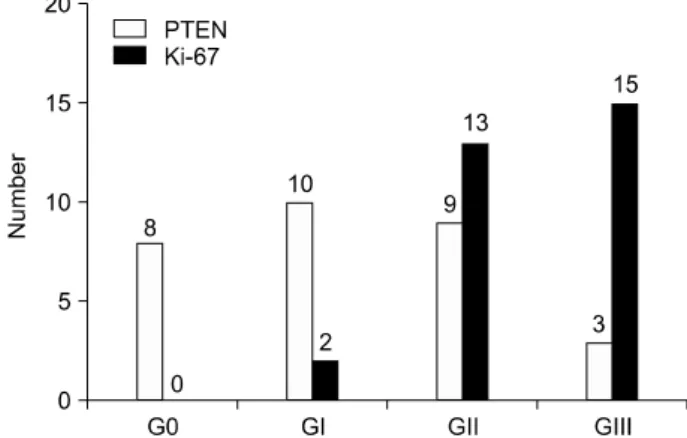
antigen Fig. 3. The correlation between staining scores of phosphatase and
tensin homolog (PTEN) and Ki-67 and the number of patients with metastases.
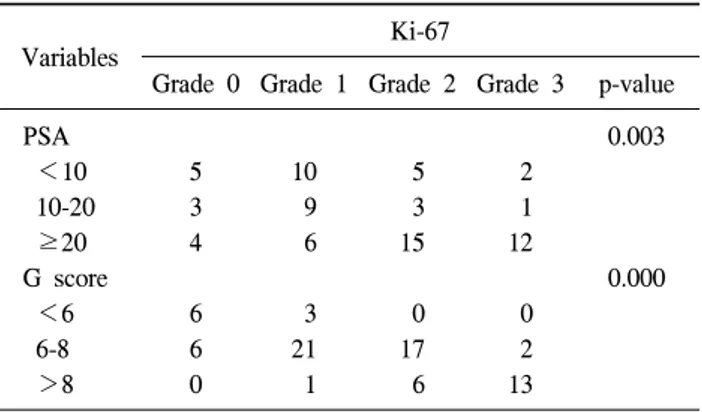
있었다 (p<0.05). PSA 수치와 Gleason score 등급에 따라 3 가지 집단으로 분류하고 분석한 결과 PSA와 Gleason score 가 높은 등급에서 Ki-67의 염색정도가 높았고 통계학적으
로 유의하였다 (PSA: p=0.003, Gleason score: p=0.000) (Table 4). 또한, 추적 관찰 중 림프절이나 골전이가 발생한 경우
Table 4. The correlation of Ki-67 expression with PSA and Gleason score in prostate cancer
Variables Ki-67
Grade 0 Grade 1 Grade 2 Grade 3 p-value
PSA 0.003
<10 5 10 5 2
10-20 3 9 3 1
≥20 4 6 15 12
G score 0.000
<6 6 3 0 0
6-8 6 21 17 2
>8 0 1 6 13
PSA: prostate-specific antigen
발생하지 않은 경우보다 유의하게 높은 Grade로 염색이 되 었다 (p<0.05) (Fig. 3).
고 찰
전립선암 환자의 진단과 예후추정을 위하여 발암인자, 성장인자, 종양억제유전자 등에 대한 연구가 활발하게 진 행되고 있다. 대표적인 종양억제유전자로 PTEN에 관한 연 구들이 보고되었다. PTEN은 10번 염색체에 위치하는 종양 억제유전자로서 mutated in multiple advanced cancers (MMAC1) 혹은 TGF-β-regulated and epithelial cell-enriched phosphatase (TEP1)로 명명되기도 한다. PTEN은 phosphatidylinositol 3'-kinase/protein kinase B/Akt 신호경로를 통하여 세포주기 의 정지와 세포사멸을 유도할 뿐만 아니라 세포생리학면에 서 세포의 유착, 이동과 분화를 조절하는 것으로 알려져 있 다.4 신경교아세포종이나 유방암 같은 고형종양에서 PTEN 의 돌연변이가 있음이 밝혀졌으며,14 국내에서도 Park 등6에 의해 PTEN이 유방암의 혈관 신생과 관련 있다는 것이 밝혀 졌다. 1984년 Gibas 등15이 방광암에서 염색체 10q의 결손을 보고한 이후 Cappellen 등16은 방광암에서 염색체 10q의 LOH (loss of heterozygosity)를 밝혀냈고 침윤성 방광암에서 빈도가 높음을 알아냈다. 그 이후 Cairns 등17에 의해 방광암 에서 PTEN의 돌연변이가 있음이 밝혀졌고, 국내에서도 Shin 등18은 침윤성 방광암에서 PTEN의 발현소실이 나쁜 예후와 연관성이 있다고 주장했다. McMenamin 등19은 전립 선암 환자의 암조직에서 PTEN 발현을 관찰하였는데 20.2%
에서 PTEN이 음성이었으며, 종양의 병기가 높아지거나 Gleason score가 올라갈수록 PTEN 발현율이 낮아 PTEN의 발현소실은 전립선암에서 나쁜 예후를 의미한다고 보았다.
Rubin 등20은 전립선암 환자에서 림프절 전이와 염색체 10q23의 LOH 관계를 연구하였는데, 10q23이 전이성 진행
의 표지자로 이용될 수 있다고 제시하였다.
이와 같이 대부분의 논문에서 PTEN의 발현소실이 전립 선암의 진행과 연관된다고 보았다. 하지만, Fenic 등21은 prostatic intraepithelial neoplasia (PIN)를 대상으로 한 PTEN 에 관한 연구에서, PIN에서는 오히려 PTEN의 발현이 증가 하는 상반된 결과를 보였지만 통계적으로는 유의하지 않다 고 보고했다. 본 연구에서는 전립선암 환자의 암세포가 인 근한 정상세포보다 PTEN에 염색이 더 잘 되었다. 또한, PSA 수치와 Gleason score 등급에 따라 3가지 집단으로 분 류하고 분석한 결과 PSA와 Gleason score가 낮은 등급에서 PTEN의 염색정도가 높았으나 통계학적인 의미는 없었다.
다만, 추적관찰에서 전이가 확인된 환자들이 그렇지 않은 환자들에 비해 PTEN의 소실이 유의하게 높은 것으로 나와 서, PTEN의 소실이 전립선암의 예후에 영향을 준다는 것을 확인했다.
Ki-67 항체는 휴지기 세포를 제외한 증식하는 세포들에 서만 발현되는 핵항원을 인식하는 것으로 Gerdes 등22에 의 해 세포증식에 연관된 핵 내 항원과 반응한다고 보고된 이 후 종양세포의 증식활성도를 나타내는 척도로 사용되어 왔 다. 과거에는 Ki-67에 대한 면역조직화학적 염색은 동결절 편을 이용해야 가능했으나, 최근에는 Ki-67 유전자 산물에 대한 MIB-1 단클론 항체를 이용하여 파라핀 포매 조직에도 염색이 가능해졌다.23 Ki-67의 증가는 S-phase 세포의 분획 과 이수체 개체수의 증가를 반영하는 것으로 종양의 병기, 분화도, 재발, 진행, 생존율의 감소와 연관이 있으며, Ki-67 발현 정도가 높은 경우에는 재발률이 높다.24 Ki-67 발현과 비뇨기 종양과의 관련성은 이미 국내외 여러 연구에서 확 인되었다. Papadopoulos 등25과 국내의 Kim 등26은 신세포암 에서 Ki-67의 발현증가가 TNM 병기와 핵분화도의 증가와 연관된다고 하였고, 다른 많은 연구에서도 Ki-67의 발현 증 가는 신세포암에서의 핵분화도의 증가, 생존율, 혹은 무병 생존율의 감소와 연관되는 것으로 알려졌다.27 Chon 등28은 Ki-67과 방광이행상피세포암의 병기 및 조직학적 분화도에 대하여 연구하였고, Ki-67 표지지수는 질병의 재발과 유의 한 상관관계가 있다고 발표하였다. Bubendorf 등12은 전립선 침생검에서 획득한 111례의 암조직을 이용한 연구에서 Ki-67 표지지수가 생존율과 연관성이 있다고 하였고, Stattin 등11도 125례의 전립선암 조직을 이용하여 Ki-67에 대한 면 역조직화학염색을 시행한 결과 Ki-67 표지지수가 조직학적 분화도 및 병기와 통계학적으로 유의한 연관성을 나타냈다 고 발표하였다. 하지만, Uzoaru 등29과 국내의 Kim 등30은 생 존율이 Ki-67 표지지수, 나이, ploidy 등과 통계학적 연관성 이 없다는 상반된 결과를 나타냈다. 본 연구에서도 PSA 수 치와 Gleason score 등급에 따라 3가지 집단으로 분류하고
분석한 결과 PSA와 Gleason score가 높은 등급에서 Ki-67의 염색정도가 높은 결과가 나와서 Ki-67이 종양 활성도와 관 련이 있다는 것을 확인할 수 있었다. 본 연구에서는 상대적 으로 대상수가 적어 앞으로 대량연구와 더불어 전립선암의 발생기전, 암의 진행, 치료 및 예후판정에 도움이 되는 분자 생물학적인 연구가 이루어져야 할 것이다. 또한 PTEN, Ki-67 외에도 암세포에 관여하는 다양한 유전자에 대한 연 구가 필요하며, 각 유전자들의 상호 연관성에 대한 조사 및 평가도 이루어져야 한다고 생각한다.
결 론
전립선암에서 종양억제유전자인 PTEN과 세포 증식능력 에 관여한다고 알려진 Ki-67 단백발현을 면역조직화학법으 로 관찰했고, 그 결과 전립선암에서 PTEN과 Ki-67은 모두 높은 발현율을 나타냈다. Ki-67은 Gleason score, PSA, 골전 이 및 림프절 전이와의 통계학적인 유의성을 나타냈지만, PTEN은 전이가 발생한 경우에만 유의한 결과를 나타냈다.
REFERENCES
1. Jemal A, Siegel R, Ward E, Murray T, Xu J, Smigal C, et al. Cancer statistics, 2006. CA Cancer J Clin 2006;56:106-30 2. Cancer Registration and Biostatistics Branch, National Cancer
Center. Cancer Statistics in Korea, 2006
3. Javidan J, Deitch AD, Shi XB, de Vere White RW. The androgen receptor and mechanisms for androgen independence in prostate cancer. Cancer Invest 2005;23:520-8
4. Steck PA, Pershouse MA, Jasser SA, Yung WK, Lin H, Ligon AH, et al. Identification of a candidate tumor suppressor gene, MMAC1, at chromosome 10q23.3 that is mutated in multiple advanced cancers. Nat Genet 1997;15:356-62
5. Gray IC, Phillips SM, Lee SJ, Neoptolemos JP, Weissenbach J, Spurr NK. Loss of chromosomal region 10q23-25 in prostate cancer. Cancer Res 1995;55:4800-3
6. Park JK, Jung MJ, Chun BK, Hur B. The relationship between PTEN tumor suppressor gene and vascular endothelial growth factor-mediated angiogenesis in breast cancer. Korean J Pathol 2004;38:100-5
7. Simpson L, Parsons R. PTEN: life as a tumor suppressor. Exp Cell Res 2001;264:29-41
8. Liliental J, Moon SY, Lesche R, Mamillapalli R, Li D, Zheng Y, et al. Genetic deletion of the PTEN tumor suppressor gene promotes cell motility by activation of Rac1 and Cdc42 GTPases. Curr Biol 2000;10:401-4
9. Koul D, Shen R, Garyali A, Ke LD, Liu TJ, Yung WK.
MMAC/PTEN tumor suppressor gene regulates vascular endo- thelial growth factor-mediated angiogenesis in prostate cancer.
Int J Oncol 2002;21:469-75
10. Mazure NM, Chen EY, Laderoute KR, Giaccia AJ. Induction of vascular endothelial growth factor by hypoxia is modulated by a phosphatidylinositol 3-kinase/Akt signaling pathway in Ha-ras-transformed cells through a hypoxia inducible factor-1 transcriptional element. Blood 1997;90:3322-31
11. Stattin P, Damber JE, Karlberg L, Bergh A. Cell proliferation assessed by Ki-67 immunoreactivity on formalin fixed tissues is a predictive factor for survival in prostate cancer. J Urol 1997;157:219-22
12. Bubendorf L, Tapia C, Gasser TC, Casella R, Grunder B, Moch H, et al. Ki67 labeling index in core needle biopsies independently predicts tumor-specific survival in prostate cancer. Hum Pathol 1998;29:949-54
13. Depowski PL, Rosenthal SI, Ross JS. Loss of expression of the PTEN gene protein product is associated with poor outcome in breast cancer. Mod Pathol 2001;14:672-6 14. Rhei E, Kang L, Bogomolniy F, Federici MG, Borgen PI,
Boyd J. Mutation analysis of the putative tumor suppressor gene PTEN/MMAC1 in primary breast carcinomas. Cancer Res 1997;57:3657-9
15. Gibas P, Prout GR Jr, Connolly JG, Pontes JE, Sandberg AA.
Nonrandom chromosomal changes in transitional cell carci- noma of the bladder. Cancer Res 1984;44:1257-64
16. Cappellen D, Gil Diez de Medina S, Chopin D, Thiery JP, Radvanyi F. Frequent loss of heterozygosity on chromosome 10q in muscle-invasive transitional cell carcinomas of the blaader. Oncogene 1997;14:3059-66
17. Cairns P, Evron E, Okami K, Halachmi N, Esteller M, Herman JG, et al. Point mutation and homozygous deletion of PTEN/
MMAC1 in primary bladder cancers. Oncogene 1998;16:3215-8 18. Shin DJ, Chung MJ, Kim HJ. Clinical significance of PTEN
expression in bladder cancer. Korean J Urol 2001;42:594-7 19. McMenamin ME, Soung P, Perera S, Kaplan I, Loda M,
Sellers WR. Loss of PTEN expression in paraffin-embedded primary prostate cancer correlates with high Gleason score and advanced stage. Cancer Res 1999;59:4291-6
20. Rubin MA, Gerstein A, Reid K, Bostwick DG, Cheng L, Parsons R, et al. 10q23.3 loss of heterozygosity is higher in lymph node-positive (pT2-3, N+) versus lymph node-negative (pT2-3, N0) prostate cancer. Hum Pathol 2000;31:504-8 21. Fenic I, Franke F, Failing K, Steger K, Woenckhaus J. Ex-
pression of PTEN in malignant and non-malignant human prostate tissues: comparison with p27 protein expression. J Pathol 2004;203:559-66
22. Gerdes J, Schwab U, Lemke H, Stein H. Production of a mouse monoclonal antibody reactive with a human nuclear antigen associated with cell proliferation. Int J Cancer 1983;
31:13-20
23. Burger PC, Shibata T, Kleihues P. The use of the monoclonal antibody Ki-67 in the identification of proliferating cells:
application to surgical neuropathology. Am J Surg Pathol
1986;10:611-7
24. Cohen MB, Waldman FM, Carroll PR, Kerschmann R, Chew K, Mayall BH. Comparison of five histopathologic methods to assess cellular proliferation in transitional cell carcinoma of the urinary bladder. Hum Pathol 1993;24:772-8
25. Papadopoulos I, Weichert-Jacobsen K, Wacker HH, Sprenger E. Correlation between DNA ploidy, proliferation marker Ki-67 and early tumor progression in renal cell carcinoma. A prospective study. Eur Urol 1997;31:49-53
26. Kim BH, Kim CI, Park CH. Caveolin-1 and Ki-67 expression as prognostic factors in clear cell carcinoma of the kidney.
Korean J Urol 2008;49:99-106
27. Delahunt B, Bethwaite PB, Thornton A, Ribas JL. Proliferation of renal cell carcinoma assessed by fixation-resistant poly-
clonal Ki-67 antibody labeling. Correlation with clinical outcome. Cancer 1995;75:2714-9
28. Chon WH, Lee SD, Lee JZ, Choi KW. The relationship of Clusterin expression and Ki-67 labeling index with clinicopa- thologic factors in human transitional cell carcinoma. Korean J Urol 2008;49:688-95
29. Uzoaru I, Rubenstein M, Mirochnik Y, Slobodskoy L, Shaw M, Guinan P. An evaluation of the markers p53 and Ki-67 for their predictive value in prostate cancer. J Surg Oncol 1998;67:33-7
30. Kim DG, Kim KK, Lee KS. Expressions of p53, bcl-2 protein and Ki-67 labelling index and their relationships with pro- gnostic factors in prostate cancer. Korean J Urol 2001;42:
828-33