REPO RT
Int J Thyroidol 2017 May 10(1): 56-60 https://doi.org/10.11106/ijt.2017.10.1.56Received February 20, 2017 / Revised April 25, 2017 / Accepted April 27, 2017
Correspondence: Kyung Ae Lee, MD, PhD, Division of Endocrinology and Metabolism, Department of Internal Medicine, Chonbuk National University Medical School, 20 Geonji-ro, Deokjin-gu, Jeonju 54907, Korea
Tel: 82-63-250-2749, Fax: 82-63-254-1609, E-mail: [email protected]
Copyright ⓒ 2017, the Korean Thyroid Association. All rights reserved.
This is an open-access article distributed under the terms of the Creative Commons Attribution Non-Commercial License (http://creative- commons.org/licenses/by-nc/4.0/), which permits unrestricted non-commercial use, distribution, and reproduction in any medium, provided the original work is properly cited.
A Case of Acute Cerebral Infarction and Thyroid Storm Associated with Moyamoya Disease
Seol A Jang, Young Ha Baek, Tae Sun Park and Kyung Ae Lee
Division of Endocrinology and Metabolism, Department of Internal Medicine, Chonbuk National University Hospital, Chonbuk National University Medical School, Jeonju, Korea
Coexistence of moyamoya disease and Graves’ disease is rare. A 41-year-old woman presented with symptoms of left-sided hemiparesis and dysarthria. Magnetic resonance imaging and angiography revealed acute infarction of the right thalamus and occipital lobe with complete obstruction of the distal internal carotid arteries and obstruction of the right P2. Free thyroxine, thyroid-stimulating hormone (TSH), and TSH receptor antibody levels were 79.33 pmol/L, 0.007 uIU/mL, and 151.5 u/L, respectively. She received antiplatelet therapy and standard antithyroid drug dose. After admission, seizure and unexplained fever occurred. The thyroid storm score (Burch and Wartofsky scale) was 90 points. After intensive treatment, mental status and thyrotoxicosis- related symptoms ameliorated and vital signs stabilized. We describe a case of thyroid storm following cerebrovascular ischemic events in a Korean woman with moyamoya disease and Graves’ disease. Thyroid storm combined with cerebrovascular events can lead to severe morbidity and mortality. Prompt recognition and strict management are crucial.
Key Words: Moyamoya disease, Graves’ disease, Thyroid storm
Introduction
Moyamoya disease (MMD) is a cerebrovascular condition that is characterized by progressive occlu- sion of the terminal portion of the internal carotid artery, resulting in the formation of collateral abnormal vessels at the base of the brain. MMD has been reported to be associated with various disease entities.1) Coexis- tence of MMD and Graves’ disease is rare; however, various cases of concurrent MMD and Graves’ dis- ease have been reported recently. Thyroid storm is a life-threatening endocrine condition associated with uncontrolled hyperthyroidism and a high mortality rate.
Thyroid storm in a patient with underlying MMD can lead to increased morbidity and mortality rates. We
herein describe a case of thyroid storm following cer- ebrovascular ischemic events in a Korean patient with MMD and Graves’ disease.
Case Report
A 41-year-old Korean woman was referred to our emergency department owing to symptoms of left- sided hemiparesis and dysarthria. The patient had been diagnosed with Graves’ disease about 10 years ago and had been taking antithyroid drug irregularly.
There was no history of treatment with other drugs or alcohol abuse. The patient showed an alert mental state, slurred speech, and left-sided weakness corre- sponding to the MRC (Medical Research Council) grade II. A neurological examination demonstrated
Fig. 2. Cerebral angiograms showing complete obstruc- tion of both distal internal car- otid arteries.
Fig. 1. Brain magnetic resonance of T2 FLAIR axial image, diffusion-weighted image and brain MR angiography maximum intensity projection showing acute infarction of the right thalamus and occipital lobe.
left-sided hemiparesis. A physical examination re- vealed bilateral exophthalmos and a diffuse goiter (Grade 2, according to the WHO differentiation). In the emergency room, the patient’s blood pressure was 125/75 mmHg, heart rate was 109 bpm, respiratory rate was 18 breaths per minute, and body temper- ature was 36.2oC. Electrocardiography revealed sinus tachycardia and magnetic resonance imaging (MRI) of the brain revealed acute infarction of the right thala- mus and occipital lobe (Fig. 1). Cerebral angiography revealed complete obstruction of both distal internal carotid arteries and obstruction of the right P2 (Fig. 2).
Thyroid function test revealed the following: thy-
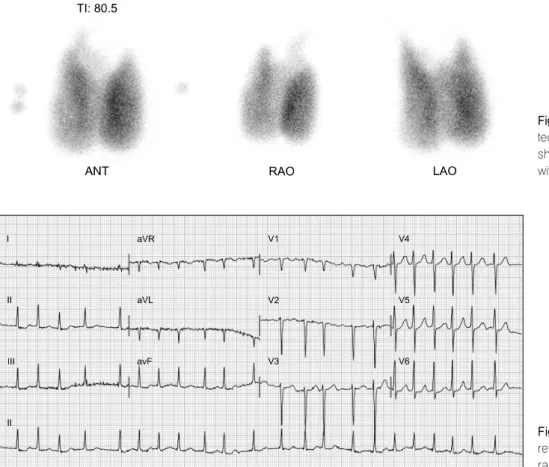
roid-stimulating hormone (TSH), 0.007 microunits/mL (reference range, 0.55-4.78 microunits/mL); triiodo- thyronine, 3.60 ng/mL (reference range, 0.78-1.82 ng/mL); free thyroxine (T4), 79.33 pmol/L (reference range, 11.5-22.7 pmol/L); and TSH receptor anti- body, 151.5 U/L (reference range, 0-10 U/L). A thy- roid ultrasonography revealed a diffusely enlarged thyroid gland without any nodule or mass. Thyroid scintigraphy revealed homogeneously increased Tc- 99m uptake (thyroid uptake: 50.5%) (Fig. 3).
The patient was admitted to the neurology depart- ment and diagnosed with MMD combined with acute cerebral infarction and Graves’ disease. The patient
Fig. 3. 99m Technetium per- technetate scintigraphy scan showing an enlarged thyroid with diffusely increased uptake.
Fig. 4. Electrocardiography reveal atrial fibrillation with a rapid ventricular response.
Fig. 5. Plain chest radiograph showing cardiomegaly.
received antiplatelet therapy and propylthiouracil (200 mg/day). As the patient exhibited an alert mental state and stable vital signs, she was prescribed a standard dose of antithyroid drug. However, after admission, the patient suddenly developed an unexplained fever and had a seizure. The mental status of patient was checked as stupor and seizure was repeated. The vi- tal signs were as follows: blood pressure, 170/113 mmHg; heart rate, 140 bpm (tachycardia); and fever, 38.8oC. Physical examination revealed bibasilar crackles and an irregular rhythm. Electrocardiography revealed atrial fibrillation with a rapid ventricular re- sponse (Fig. 4) and chest radiography showed car- diomegaly (Fig. 5). To exclude additional infarction or cerebral hemorrhage due to aggravated neurologic symptoms, brain MRI was performed again, which did not reveal any evidence of newly progressed in- farction or hemorrhagic lesion and there was no inter- val change compared with the prior image. The thy- roid storm score (Burch and Wartofsky score) was 90 points.2) For thyroid storm management, we initiated high-dose propylthiouracil, Lugol’s solution, and hy-
drocortisone. An echocardiogram revealed global hy- pokinetic wall movement with mild left ventricular sys- tolic dysfunction (ejection fraction, 48%). Rate control and management for heart failure were conducted simultaneously. After intensive treatment, the patient’s mental status improved and thyrotoxicosis-related
symptoms ameliorated. Moreover, her vital signs sta- bilized and she was discharged in an improved state.
After 6 weeks of treatment, hyperthyroidism had stabilized (free T4, 18.89 pmol/L; TSH, 0.010 uIU/mL) and neurologic symptoms had ameliorated significantly.
The patient maintained regular antithyroid medication and had no additional cerebral complications.
Discussion
To date, about 50 cases of concurrent MMD and Graves’ disease have been described in the literature.3) In Korea, interesting cases associated with MMD and Graves’ disease were reported.4,5) Although the rela- tionship between Graves’ disease and cerebrova- scular accidents in MMD is unclear, the coexistence of both diseases is rare and noteworthy.6)
MMD is symptomatic whenever the collateral ves- sels fail to compensate for the cerebral blood flow def- icit produced by the progressive stenotic lesion in the major cerebral arteries.7) In the thyrotoxicosis state, excessive thyroid hormones can alter vascular re- activity and cause cerebral perfusion impairment.
Therefore, cerebrovascular ischemic symptoms in MMD can be precipitated by activities associated with thyrotoxicosis.1,8) Moreover, previous reports have suggested that altered cerebral hemodynamics and hypercoagulability in thyrotoxicosis may trigger vas- cular attacks in patients with MMD.9,10)
Some physicians have argued that an autoimmune mechanism, which contributes to the development of Graves’ disease, may also play an important role in the onset and progression of MMD-related cere- brovascular events.7) Lei et al.11) demonstrated that in- creased thyroid function and autoantibodies are asso- ciated with MMD. A Korean study observed that pa- tients with MMD were more likely to have elevated thyroid autoantibody levels than those with non-MMD strokes, despite being in a euthyroid state.12) Similarly, the patient in our study had high levels of TSH re- ceptor antibody when symptoms developed. Another possible link between these disorders is atheroscle- rosis. Colleran et al.13) demonstrated a positive corre- lation between free T4 levels and both homocysteine
and methylmalonic acid levels, suggesting that thyro- toxicosis may induce hyperhomocysteinemia. As hy- perhomocysteinemia has been implicated in athero- sclerotic and embolic disorders,14,15) it could be anoth- er key to the pathophysiology of MMD.16) In our case, due to the patient had a history of uncontrolled long- standing Graves’ disease, aggravation of MMD can be affected by chronic hyperthyroidism.
Although a thyroid storm associated with cere- brovascular ischemic events is very rare in patients with both MMD and Graves’ disease, it is still a life-threatening condition that can cause severe mor- bidity and mortality. Hsu et al.17) reported a case of Graves’ disease with thyroid storm associated with MMD in 2006, which unlike previously reported cases, progressed rapidly and was ultimately fatal.
In our case, the thyroid storm occurred during the management of cerebral infarction. Thyroid storm is typically triggered by stressful medical events or conditions. The cerebrovascular accidents may con- tribute to the occurrence of thyroid storm. Although the order of the incidents is not clear, if transient ischemic attacks or cerebral infarction occurs in a patient with both Graves’ disease and MMD, it can also induce a thyroid storm. Prompt recognition and treatment are crucial for improving prognosis. Furthermore, main- tenance of a long-term euthyroid state is essential and it is important to prevent a relapse of hyperthyroidism after normal thyroid function is restored in MMD patients.1)
Here, we report a case of thyroid storm and acute cerebral infarction associated with MMD in a patient with Graves’ disease. The case has enhanced our understanding of a few aspects. First, we should keep in mind the possibility of combined problems with GD, such as MMD, especially patients that have neuro- logical signs and symptoms. In such cases, further studies are warranted. Second, the cerebrovascular ischemic events can be a triggering factor (or risk factor) for thyroid storm, like in our case. Therefore, if transient ischemic attacks or cerebral infarction occurs in a patient with Graves’ disease combined with MMD, emergent treatment should be initiated.
References
1) Im SH, Oh CW, Kwon OK, Kim JE, Han DH. Moyamoya disease associated with Graves disease: special considerations regarding clinical significance and management. J Neurosurg 2005;102(6):1013-7.
2) Burch HB, Wartofsky L. Life-threatening thyrotoxicosis.
Thyroid storm. Endocrinol Metab Clin North Am 1993;22(2):
263-77.
3) Ren SC, Gao BQ, Yang WL, Feng WX, Xu J, Li SW, et al. Von Willebrand factor and coagulation factor VIII in Moyamoya disease associated with Graves' disease: a case report.
Exp Ther Med 2016;12(5):3195-200.
4) Back JH, Kang HM, Min BD, Gil SH, Kim SJ, Oh BC, et al. A case of Graves' disease associated with myasthenia gravis and complicated with Moyamoya disease. Korean J Med 2010;79(2):195-200.
5) Shin DW, Seo JY, Lee JG, Kim JS, Lee KB, Roh H, et al.
Acute ischemic stroke in Moyamoya disease associated with thyrotoxic crisis. J Korean Neurol Assoc 2014;32(2):95-7.
6) Cheon CK, Kim SY, Yoo JH. Two adolescent patients with coexistent Graves' disease and Moyamoya disease in Korea.
Korean J Pediatr 2014;57(6):287-91.
7) Shen AL, Ryu SJ, Lin SK. Concurrent moyamoya disease and Graves' thyrotoxicosis: case report and literature review. Acta Neurol Taiwan 2006;15(2):114-9.
8) Sasaki T, Nogawa S, Amano T. Co-morbidity of moyamoya disease with Graves' disease. report of three cases and a review of the literature. Intern Med 2006;45(9):649-53.
9) Siegert CE, Smelt AH, de Bruin TW. Superior sagittal sinus
thrombosis and thyrotoxicosis. Possible association in two cases.
Stroke 1995;26(3):496-7.
10) Ni J, Zhou LX, Wei YP, Li ML, Xu WH, Gao S, et al.
Moyamoya syndrome associated with Graves' disease: a case series study. Ann Transl Med 2014;2(8):77.
11) Lei C, Wu B, Ma Z, Zhang S, Liu M. Association of moyamoya disease with thyroid autoantibodies and thyroid function: a case-control study and meta-analysis. Eur J Neurol 2014;21(7):996-1001.
12) Kim SJ, Heo KG, Shin HY, Bang OY, Kim GM, Chung CS, et al. Association of thyroid autoantibodies with moyamoya-type cerebrovascular disease: a prospective study. Stroke 2010;41(1):
173-6.
13) Colleran KM, Ratliff DM, Burge MR. Potential association of thyrotoxicosis with vitamin B and folate deficiencies, resulting in risk for hyperhomocysteinemia and subsequent thromboem- bolic events. Endocr Pract 2003;9(4):290-5.
14) Iso H, Moriyama Y, Sato S, Kitamura A, Tanigawa T, Yamagishi K, et al. Serum total homocysteine concentrations and risk of stroke and its subtypes in Japanese. Circulation 2004;109(22):2766-72.
15) van Diemen-Steenvoorde R, van Nieuwenhuizen O, de Klerk JB, Duran M. Quasi-moyamoya disease and heterozygosity for homocystinuria in a five-year-old girl. Neuropediatrics 1990;
21(2):110-2.
16) Lee R, Sung K, Park YM, Yu JJ, Koh YC, Chung S. A case of Moyamoya disease in a girl with thyrotoxicosis. Yonsei Med J 2009;50(4):594-8.
17) Hsu SW, Chaloupka JC, Fattal D. Rapidly progressive fatal bihemispheric infarction secondary to Moyamoya syndrome in association with Graves thyrotoxicosis. AJNR Am J Neuroradiol 2006;27(3):643-7.