< 목 차 >
Ⅰ. 서 론
Ⅱ. 실험재료 및 방법
Ⅲ. 실험성적
Ⅳ. 총괄 및 고찰
Ⅴ. 결 론 참고문헌 사진부도 영문초록
I. 서 론
치조제의 과도한 흡수는 의치의 유지 ∙ 안정 및 기 능의 장애 그로인한 심리적 위축감을 초래하기 때문에 (1)예로부터 고정성 보철물의 지지를 위한 매식체의 개발 및 연구가 있어왔다(2, 3, 4, 5).
초기의 매식체 재료로는 금, 은, 납, 고무, 금속 등 이 사용되어 왔으나 부식문제등으로 1930년경 stainless steel과 vitallium이 개발, 사용되어 오면 서 1950년경 순 타이타늄(com-mercially pure titanium)과 골과의 유착 가능성이 보고된 이래, 순 타이타늄과 그 합금이 매식체 재료의 주종을 이루고 있다.
그 외 형태적 변형이 용이한 plastic polymers(8, 9,
10)와 조직적합성이 우수한 alumina, carbon등의 ceramics(11-16)이 사용되어 왔으며, 최근에는 골조직 과의 친화성 및 조직 적합성이 우수하며 골조직과 화 학결합을 통한 생유착이 이루어진다는 calcium phosphate ceramics을 프라스마 분사법(plasma spray) 등으로 금속표면에 입힘으로써 금속과 ceramics의 장점을 이용한 매식체가 연구개발 되어 오고 있다(17-33).
또한 형태적으로 1940년 helix wire spiral(34, 35)로 부터 tripodial pin(36), bonespiral(37, 38), vent- plant(37), bladevent(46)에 이르기까지 다양한 형태의 변화가 있어왔으며 특히 조직의 내방성장을 유도하여 생기계적 유지를 얻기 위하여 여러 크기의 기공형태를 부여한 매식체(41,42)와 매식체의 초기 움직임을 최소화 하고 조직과의 최대접촉을 얻을 수 있는 원통형의 나 사형태의 매식체가 주로 이용되고 있다(43, 44).
또한 매식체 주위조직의 양상은 매식체의 성공과 실 패에 밀접한 관계가 있으며(45)Babbush(46), Weiss(47) 등은 매식체 주위의 섬유성 결체조직은 충격흡수 및 주위골조직 형성을 촉진한다고 하여 섬유유착이론을 주장하고 있으며 Cllins(48), Southham(49) 등은 금속 매식체 주위에는 필연적으로 섬유성 결체조직이 형성 된다고 하였으며 Cranin(49)은 매식체 주위조직 형성 에는 매식체의 재료 및 표면 상태가 영향을 미친다고 하였으며 Brunski(50)는 생역학적 요인이 중요한 요소
- 대한 치과 보철학회지 Vol. 28 No. 1, 1990 -
수종의 치근형 골내매식체와 조직간의 반응에 관한 조직학적 연구
서울대학교 치과대학 치과보철학교실
이 호 영ㆍ김 영 수
�본 논문은 1988년도 서울대학교병원 임상연구비 보조로 이루어진 것임.
�본 논문의 요지는 1989년도 제38회 미국치과임플랜트학회에서 발표하였음.
로 작용한다고 언급하고 있다.
반면에 매식체가 장기간 안정된 고정을 위하여는 신 생골과의 유착이 필요하다고 하였으며(52), 이는 1980 년 Bränemark(53-66) 등이 골유착 매식체의 성공율, 기 전 및 방법 등을 소개한 이후 최근에는 골유착 개념이 지배적이론으로 보편화되어가고 있는 현황이며 매식 술식을 비롯한 기존의 매식학 개념이 변화하고 있다.
이러한 신학들의 보고를 토대로 본인은 임상적으로 사용되고 있는 수종의 매식체와 서울대 학교에서 개발 중인 시험용 타이타늄 매식체(kimplant) 및 한국산 Nephrite 매식체를 성견 치조졸 내에 매식하여 이들 과 골조직과의 반응을 육안적 소견, 방사선검사, 광학 현미경, 주사전자현미경, 투과전자현미경적 검경을 통 하여 비교 관찰한 결과 다소의 지견을 얻었기에 보고 하는 바이다.
Ⅱ. 실험재료 및 방법
가) 실험재료
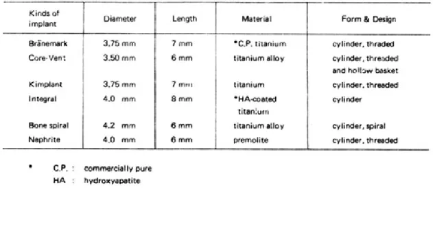
체중 15Kg 정되의 성견 2마리가 암수 구별 없이 사 용되었으며, 치조골내 매식한 것은(Table 1)(Fig 2, 3) 과 같다.
나) 실험방법
1) 발치
전신 마취제인 pentotal sodium을 성견 앞발의 cephalic vein에 kg당 20mg을 정맥내 주사하여 전 신 마취시키고 하악우측 견치후방의 소구치, 대구치들 을 발치하고 3-0봉합사로 봉합하였다.
2) 매식
발치후 5개월이 지난후 발치부위를 방사선 촬영하 여 하치조관과 치조골 정상과의 거리를 측정하여 Bränemark, Core-Vent, Kim-plant, Intergral, Bone spiral, Nephrite를 각각 adelle 등(56), Niznick(67), 김(68), Calciteck社(69), Oratronic社(70),
김(68) 등이 제시한 술식에 의해 매식하였다(Fig. 3, 4).
절개한 부위는 3-0봉합사로 봉합한 후 1일 4mg의 gentamycine을 7일간 근육주사 하였다. 수술후 1개 월마다 방사선 촬영을 실시하였다.
3) 실험동물의 희생 및 조직표본 제작
매식 4개월후 실험동물을 희생시켜 하악을 적출하 여 골막을 박리한 후 부검용톱을 이용하여 매식체 주 변의 골을 절단하여 한 개의 매식체가 포함된 골 블록 을 제작하였다. 그림 1에서 보듯이 각각의 골 블록은 chisel을 이용하여 매식체 중앙부위 골을 쪼개어 광학 현미경, 주사전자현미경시편을 제작하고 매식체가 붙 어있는 시편은 투과전자현미경용으로 이용하였다.
① 광학 현미경용 표본제작
매식체와 접하고 있는 골에서 치아장축방향으로 절 단한 완전한 면을 포함하는 시편을 재료로 사용하였 다. 제작된 시편은 10% neutral buffered formalin 용액에서 24시간 고정한 후 10% EDTA 용액을 이용 하여 탈회하였으며 2일에 1번씩 탈회 용액을 교환하 였다. 방사선 사진과 탐침을 이용하여 탈회의 정도를 확인한 후 흐르는 물에 12시간 세척하고 통법에 따라 파라핀 블록을 제작하고 5μ두께의 박편을 제작한 후 Hematoxlin과 Eosin 중염색을 하였다.
② 주사전자 현미경용 표본제작
매식체와 접한 면을 포함한 골절편을 5×5×1mm 크 기 의 납 작 한 시 편 으 로 제 작 하 여 2.5%
glutaraldehyde(pH.7.2)에 전고정하였다. 전고정된 골절편을 다시 1% osmium teroxide용액(pH7.2)에 후고정한 후 탈수과정을 거쳐 임계점 건조기(Hitachi, ACP-2)로 건조시켰고 ion coater(Eiko, IB-3)로 표 면에 gold-platinum을 입힌 후 주사전자 현미경 (Hitachi s-520)으로 관찰하였다.
③ 투과전자 현미경용 표본제작
2.5% glutaraldehyde(pH 7.2)로 48시간 전고정하 고 전고정한 시편을 동일한 phosphate buffer로 24 시간 세척한 후 전자현미경용 10% EDTA 용액으로 탈회하였다. 1개월간 탈회한 후 조심스럽게 매식체를
분리하였다.
매식체와 접한 면을 포함한 골절면을 2×2×2mm 의 크기로 잘라 다시 10% EDTA용액으로 계속 탈회 하였다. 탈회과정 중에는 2일마다 10% EDTA 용액을 교환하여 주었으며 방사선 사진을 이용하여 탈회 정도 를 조사하였다.
탈회가 완전히 된 저리편은 다시 1×1×1mm의 크 기로 제작한 후 2% osomimn teroxide용액(pH 7.2) 에 후고정하고 각급 alcohol과 acetone으로 탈수시킨 다 음 Epon에 포 매 하 였 다 . 포 매 된 조 직 은 Ultramicrotome으로 먼저 1μ두께의 조직소절편을
제작하여 1% toluidine blue로 염색한 후 관찰할 부 위를 선정하여 500-800A 두께의 초박절편을 제작하 여 2% uranyl acetate와 lead citrate에 염색하고 Hitachi H-600 투과전자현미경으로 관찰하였다.
Ⅲ. 실험결과
1. 육안적 소견
적출된 하악에서 각 매식체 주위는 정상적인 치은조 직으로 피개되어 있으며 골편 제작시 Nephrite는 자 연 적 으 로 탈 락 되 었 다 . 골 유 착 성 매 식 체 인 Bränemark, Core-Vent, Kim-Plant, Integral 매 식체에서는 그 주위로 골조직이 증식되어 있음이 관찰 되었으며 Nephrite를 제외한 나머지 매식체들은 전혀 동요가 없었다(Fig. 3).
2. 방사선상의 소견
Nephrite매식체 주위에는 인정할 만한 정도의 치 조백선이 형성된 것을 관찰할 수 있었으며 Bone spiral과 함께 근단부위에 방사선 투과성을 나타내고 있었으며 그 주위에 골경화 현상이 보였다(Fig. 4).
Table 1. Experimental materials used in this study
Fig. 1. Schematic drawing illustrating of sectioning for light microscopic and electron microscopic examination.
3. 조직학적 소견
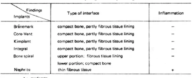
가) 광학 현미경적 소견(Table 2참조)
(1) Bränemark
층판상의 치밀골로 이루어져 있으며 골조직이 매식 체의 외형에 준해 형성되어있으며 계면(interface)은 골조직과 직접 접촉되어있는 상을 보이며 골면에서 섬 유막이나 세포막은 볼 수 없다. 부분적으로 계면의 골 표면은 무구조층이 골면을 덮고 있기도 하였다.
골수강이 노출되어 있는 경우는 섬유아세포 및 파 골 세포가 출현하고 있었다(Fig. 5).
(2) Core-Vent
총판상의 치밀골로 이루어져 있는 매식체의 상층부 는 부분적으로 엷은 섬유조직이 피개되어 있었으며 골 면은 매식체 외형에 준하고 있었으며 계면 부위는 치 밀골로 이루어져 있었다. 함입부위에는 단일층의 세포 층이 부분적으로 인지되었다(Fig. 6).
(3) Kimplant
층판구조의 치밀골이 매식체 외형에 준하여 형성되 어 있었으며 매식체와 접하는 계면는 완전한 치밀골로 피개되었다. 부분적으로 계면부위에 단세포층이 관찰 되었으나 조골세포 또는 조섬유세표 인지의 여부는 식 별하기 곤란하였다(Fig. 7).
(4) Integral
치밀골로 이루어져 있으며 계면 부분은 직접골로 이 루어져 있었고 매식체 외형에 준해서 층판상으로 골이 형성되어 있었으며 간혹 골수강이 매식체 주행에 평행 하게 존재하고 있었다(Fig. 8).
(5) Bone spiral
매식체의 상층부는 섬유막으로 피개되어 있었으나 근단부위에는 매식체 외형에 준하는 골형태를 보였으 며 계면 부분의 섬유막은 소실되었고 골이 직접 노출 되거나 또는 계면 부근에 비교적 화골이 덜된 무구조 물이 골면을 피개하고 있었다(Fig. 9).
(6) Nephrite
매식체 외형에 준해서 골의 형성이 이루어져 있고 계면은 엷은 섬유막으로 피게되었다.
골실질을 뚫고 골수까지 도달한 예에서는 섬유결합 조직막으로 덮혀 있는 계면 부분은 매식체 외형을 나 타냈으며 매식체에 노출된 고리수강은 지방골수의상 을 보이며 매식체와의 경계 부위는 섬유결합조직막이 골수강을 피개하여 매식체와 골수를 격리시키고 있다.
섬유막에는 다쉬의 임파세포와 대식세포의 침윤상을 보였다(Fig. 10).
Table 2. Summary of light microscopic findings
나) 주사전자 현미경적 소견
(1) Bränemark
불규칙한 배열의 교원질 사상체를 덮고 있었다(Fig.
11, 12).
(2) Core-Vent
교원질 섬유가 불규칙하게 배열되어있으며 교원질 속을 형성하고 있으며, 과립형 기질이 부착되어 있었 다(Fig. 13).
(3) Kimplant
불규칙하게 배열된 교원질 사상체와 과립형 기질이 나타났다(Fig. 14).
(4) Integral
불규칙적으로 배열된 교원질 사상체위로 과립형기 질이 보였다(Fig. 16).
(5) Bone spiral
하부구조물을 덮고 있는 과립형 기질이 계면에 재개 되어 있었다(Fig. 16).
(6) Nephrite
매식체의 계면에서 교원질이 보이지 않으며 골편으 로 여겨지는 것으로 덮혀있다(Fig. 17).
다) 투과전자 현미경적 소견
(1) Bränemark
계면 부위는 거칠고 불규칙하게 교원질 사상체가 배 열되어 있었다(Fig. 18).
(2) Core-Vent
매식체는 electron dakr zone과 접하고 있었으며 교원질 섬유가 불규칙하게 배열된 측과 수직으로 배열 된 층의 3가지 층이 나타났다(Fig. 19).
(3) Kimplant
교원질 사상체가 불규칙하게 배열되어있었다(Fig.
20).
(4) Integra
표면은 무구조한 층으로 되어있었으며 교원질 사상 체가 불규칙하게 배열되어있었다(Fig. 21).
(5) Bone spiral
교원질 사상체가 매식체 외형에 불규칙하게 배열되 어 있었으며 계면에는 조섬유 세포층으로 피개되어 있 었다(Fig. 22).
(6) Nephrite
교원질 사상체가 매식체의 외형에 불규칙하게 배열 되어 있었다(Fig. 23).
Ⅳ. 총괄 및 고찰
매식체의 성공과 실패는 매식체 표면에서의 세포활 동과 관계있으며 매식후 주위에 형성되는 경계부위는 결체성 섬유조직으로 변하거나 사골의 형태로 남아 있 거나 또는 신생골로 고체되는 양상을 나타내게 되는데 Atwood(71)는 골내 매식체가 이상 감각, 불편감, 동통, 감염, 골의 과도한 흡수, 그리고 심리적인 문제점을 야 기하지 않고 구강내에서 장기간 제기능을 수행할 수 있을 때 임상적으로 성공했다고 판단된다고 언급하고 있으나 NIH-Harvard 토론회와 Toronto 토론회에 서 동요도, 매식체주위의 바이사선상, 골의 흐비수, 염 증을 포함한 제반증상 및 5년, 10년 후의 성공율의 관 점에서 섬유유착성 매식체와 골유착성 매식체의 성공 에 대한 기준을 제시하였다(72).
Anderson(73, 74)과 Salthouse(75)는 대식세포 및 조 섬유세포가 초기에 출연하다가 그후에 결체조직의 증 식으로 결체성 섬유조직낭을 형성하게 된다고 언급하 면서 그동안 섬유유착성 매식체에서는 이것이 필요하 고 바람직한 것으로 받아들여 졌으나 Bränemark(55) 등은 섬유조직과 같은 연조직은 구강내에서 발생하는
기계적 화학적 자극에 저항이 약한 뷔위로 노출되어 상피조직의 하방이행을 초래하게 되어 결국 매식체의 동요 및 매식체의 탈락을 초래하게 된다고 언급하였 으며, Albrektsson 등(58)은 성공적인 골유착의 요구조 건으로 매식체의 재료, 매식체의 형태, 매식체 표면상 태, 골조직의 상태, 외과적 술식 및 매식체에 가해지 는 외력과 보철물설계, 구강위생의 유지를 말하고 있 으며 특히 소독개념을 포함한 외과적 술식과 외력등의 생역학적 요소를 강조하고 있다.
Lundskog(76)는 수술시 골조직에서 발생하는 온도 가 50℃에서 30초간 지속될때는 조골세포의 형태학 적 변형을 가져온다고 하였으며 Mattews(77)등은 56
℃에서는 알칼리성 분해효소가 파괴됨을 보고 하였으 며 Eriktsson(78)은 47℃를 골조직 손상을 가져오는 경 계온도로 언급하고 있다.
Satomi(79) 등은 매식시 tapping과 non-tapping 의 차이를 비교∙관찰하여 tapping insertion을 하여 야만 골유착현상이 일어남을 보고하고 있으며, 2000rpm이하의 drill속도 및 내부와 외부에서 충분 한 냉각제를 공급해 주는 것을 필요로 한다고 하였다.
또한 Bränemark(54, 55, 56)등은 매식초기에 외부압 력이 가해지거나 동요가 일어난 경우에는 결체조직낭 이 형성되기 때문에 3~6개월간 골내에서의 치유기간 이 필요하다고 언급하고 있으며 김(80) 등은 임상에서는 1~2개월의 치유기간을 연장하는 것을 추천하고 있다.
Brunski(51)는 저작기능을 하는 매식체에서는 결체조 직에 의해 저작기능을 하지 않는 매식체에서는 골조직 에 의해 둘러싸인다는 것을 보고하여 매식체 주위 조 직형성에는 재료보다는 생역학적인 요소가 더욱 중요 하다고 언급하고 있다.
매식체 주위의 골조직 재생을 위하여는 조골세포, 골세포, 파골세포의 활동이 활발하여야 하며 이러한 세포들을 위한 영양공급이 원할히 이루어져야 하며 또 한 적당한 자극이 있어야 하며 이러한 자극에는 전기 적자극, 기질유도작용, 세포간유도작용 등이 있다(54).
Roberts(81)는 매식체 주위의 신경골은 가골상태 및 층판구조를 갖는 골조직의 형태를 갖추면서 안정기, 강화기를 거쳐 내구력이 있는 골조직으로 완성된다고 하였으며 매식체와 골조직간의 직접적인 접촉은
30~90%정도에서 일어난다고 하였으며 그양은 매식 체의 종류와 골조직에 힘이 가해지느냐의 여부에 좌우 된다고 말하고 있으며, Albrektsson(57)은 매식체 표면 의 90%이상에서 직접적인 골과의 접촉이 이루어져야 골 유착이 되었다고 할 수 있다고 언급하고 있다. 본 논문의 골유착 매식체에서는 양은 알수 없으나 대부분 직접적인 골접촉을 볼 수 있었으며 부분적으로 세포막 이 매식체 표면에 피개되어 있는 것을 관찰할 수 있었 다. 본 실험에서 사용된 Nephrite 경우 보석절삭 기계 를 이용한 수제품으로서 정교성이 떨어져 정확하게 dill과 일치되지 않았기 때문에 임상적으로 골조직이 자라서 접촉할 수 있는 1~2mm보다 많은 양의 간격 이 형성되어 섬유조직이 형성된 것으로 사료된다.
Strock(82)과 김(83) 등에 의해 섬유유착성 매식체로 확인된 Bone spiral 매식체에서도 중상충 부위에서는 섬유조직이 관찰되었지만 중하부에서는 골과의 직접 적인 접촉을 나타내고 있는데 그 상태가 골유착성과 유사하게 치밀골에 의해 피개되어 있었던 것은 비기능 상태에서는 모든 매식체에서 골 유착이 일어날 수 있 다는 Brunski(51)의 연구결과와 일치되는 것으로 사료 된다.
임상적으로 골 유착여부를 평가하는 것으로 동요도 검사, 방사선학적 평가, tapping에 전달되는 sound diagram 등이 있으나 위의 방법으로는 확실한 골유 착을 입증할 수 없고 특히 방사선 사진상의 해상능력 은 0.1mm 범위에 있고 세포의 크기는 0.01mm 범위 에 있기 때문에 단 세포층은 방사선 사진상에 나타나 지 않는다고 하였으며 동물실험과 생검을 통한 조직학 적 증거의 필요성을 강조하였다(57). 본 연구에서도 골 유착 매식체들은 전혀 동요를 나타내지 않았지만 조직 학적으로 부분적인 결체성 섬유조직이나 세포층과 같 은 연조직층이 관찰됨은 임상적 소견과 조직학적 소견 이 반드시 일치하는 것은 아니지만 동요가 있는 매식 체에서는 항상 조직학적 소견이 그에 상응되는 것을 알수 있었다(45).
매식체 재료로서 주로 사용되는 타이타늄이 다른 금 속과 특이한 것은 조직과의 접촉이 금속자체가 아닌 표면산화물이 접촉하기 때문에 생화화적 측면에서 일 종의 ceramic이 골조직과 접촉하게 된다고 하며(59),
kasemo(34)는 oxide의 두께는 50~100Å이며 다른 금 속에 비하여 높은 dielectric constant 갖고 있어 생 체 조직내의 분자들과 1~10 kcal/mol정도의 약하지 만 긴 범위의 Van der Waals interaction을 갖는다 고 하며, 또한 산화현상, 수산화물형성, 무기이온의 성 장 등으로 10~100 kcal/mol의 강한 화학적 결합을 하게 된다고 언급하고 있다.
타이타늄 합금 매식체(7)는 90% titanium, 6%
aluminium, 4% vanadium으로 되어 있으며 순 타이 타늄에 비해 기계적 강도가 약 60% 높은 것으로 되어 있는 반면 Albrektsson(57)은 순 타이타늄에서는 proteoglycans과 glucoseaminoglycans이 형성하 는 무구조성 기질층의 두께가 200~400Å을 나타내 는 반면 타이타늄 합금은 5000Å의 두께를 나타내어 조직 친화성이 순 타이타늄이 우수한 것으로 보고하고 있다.
본 실험의 골유착성 타이타늄 매식체에서의 투과전 자현미경적 소견에서 이러한 두께는 확인할 수 없었으 며 교원질 섬유속으로부터 나온 교원질 사상체가 매식 체에 접근하는 것을 확인할 수 있으며 주사 전자현미 경에서는 교원질 사상체를 덮고 있는 많은 양의 기질 이 과립형태로 존재하는 것을 관찰할 수 있엇다. 이러 한 기질이 매식체를 골조직에 접착시키는 매개체로서 작용한다고 Albrektsson(57, 58, 59)은 언급하고 있다.
Linder(61) 등은 12주 동안 이식된 타이타늄 매식체 주위에서 3가지 layer 즉 50~100Å의 oxide layer, 100~200Å의 proteoglycan layer, 200~500Å의 교원질 사상체층이 관찰되며 부분적으로 조섬유세포, 포식세포 및 골세포 등의 돌기들이 매식체에 접근하 고 있으며 이러한 세포돌기들은 항상 proteoglycan layer에 의해 매식체와 분리되어 있음을 보고하였으 며 본 실험에서의 골유착성 타이타늄 매식체에서는 3 가지 층은 구분할 수 없었으며 교원질사상체와 세포돌 기의 접근 및 매식체와 그들간에 무구조 층의 기질이 확인되었다.
이러한 전자 현미경적 소견은 수산화 인회석을 입힌 Intergral에서도 유사하게 나타났으며 육안적으로는 다른 매식체에 비하여 강한 골유착양상을 나타냈다.
이는 손상된 골조직의 회복과 치조제 재성형에 사용되
어온 수산화 인회석의 특징적인 양상(85-91)으로 국내에 서 안(92), 이(93)등의 논문과 김(80)등이 확인한 바 있으 며 32주 동안 골조직내에서 수산화 인회석의 흡수가 전혀 일어나지 않는다고 보고한 Cook(20), Kent(22), Block(26) 등의 소견과 일치되는 것으로 사료된다.
또한 Cook(20), Kent(22), Block(26)은 수산화인회석을 입힌 Integral의 경우 5~8주내 골유착이 완성된다고 하였으며 접착강도 또한 코팅되지 않은 매식체에 비하 여 2~3배 강하다고 언급하고 있다. Block(27)등은 저 작기능하에서 수산화인회석이 코팅된 매식체와 코팅 되지 않은 매식체에서 각각 66.3%, 50.2%의 골유착 정도를 나타낸다고 보고하였으나 본 실험에서는 그 양 은 알 수 없었으며 수산화 인회석 입자들로 인해 불규 칙한 표면의 치밀골로 계면이 이루어진 것을 알 수 있 었으며 부분적으로 단세포층이 관찰되었다.
또한 Lavelle(94)등은 골조직내에서 흡수가 일어나 는 tricalcium phosphate를 타이타늄에 코팅한 매식 체가 골조직내에서 유착됨을 보고 하였는바 앞으로는 골조직과 친화성이 잇는 여러 재료들이 금속 표면이 코팅되어 골유착 매식체가 개발될 것으로 보이며 또한 여러 조건하에서 골유착 정도, 골유착 완성시기 및 접 착강도등에 대한 연구가 있어야 할 것으로 사료된다.
Ⅴ. 결 론
저자는 임상적으로 골유착 매식체로 쓰이고 있는 Bränemark, Core-Vent, HA-coating의 Integral 매식체와 섬유 유착성 매식체인 Bone spiral 매식체, 그리고 서울대학교에서 개발중인 Kimplant와 Nephrite 매식체를 성견치조골에 매식하여 4개월 후 비기능상태하에서의 주위 조직 반응을 육안적, 방사선 학적, 광학현미경 및 전자 현미경적 검경을 통하여 비 교, 관찰한 바 다음과 같은 결론을 얻었다.
1. Bränemark, Core-Vent, Kimplant, Integral 매식체는 육안적소견으로 전혀 동요 가 없었으며 매식체 주위로 골의 융기를 보이 며, 조직학적 소견으로 대부분 치밀골에 의해
피개되어 잇으며 부분적으로 골세포, 결체조직 으로 피개됨이 관찰되었다.
2. Bone spiral 매식체는 전혀 동요가 없었으며 부 분적인 방사선 투과상을 보이며 상부에는 연조 직에 의해 중하부에는 골조직에 의해 피개됨이 관찰되었다.
3. Nephrite 매식체는 심한동요를 보였으며 치조 백선과 방사선 투과상을 나타내고 조직학적으 로 중등도의 염증과 함께 연조직에 의해 피개되 어 있었다.
4. 주사 전자현미경적 소견에서는 Nephrite 매식 체는 기질이 관찰되지 않았으며 그외 매식체는 기질이 과립형태로 하부구조를 덮고 있었다.
5. 투과전자현미경적 소견에서 모든 매식체에서 교원질 사상체가 매식체에 불규칙하게 배열되 어 접근하고 있었으며, Bränemark, Core- Vent, Kimplant, Integral에서는 무구조층이 매식체와의 사이에서 관찰되었다.
REFERENCES
1. Woelfel JB, Winter CM, Igarashi T, Five year cepalometrics study of mandibular ridge resorption with different posterior occlusal forces. J Prosthet Dent, 1976; 36: 602.
2. Lee TC HISTORY OF DENTAL IMPLANTS.
In;Cranin A.N., ed. Oral implantology.
Springfield:Chrles C Thomas, 1970;13.
3. Natiella JR, Armitage JE, Greene GW, Meenaghan MA. Current evaluation of dental implants. J Am Dent Assoc, 1972; 84: 1358.
4. Linkow L, Mahler MS. Implants for fixed and removable prostheses. Dent Clin North Am, 1977;
24-443.
5. Smith CH. Rational use of endosteal implants. J Prosthet Dent 1973; 38; 652.
6. Leventhal G. Titanium a metal for surgery J
Bone Jt Surg 1951; 33; 473.
7. Smith DC. Biomatenals in dentistry. J Dent Res Speial issue 1975; 54; B146.
8. Hodosh M, Shklar G, Povar M. The porous vitrous carbon/polymethacrylate toth implat;
Preliminary studies. J Prosthet Dent 1969, 22;
371.
9. Hodosh M, Shklar G, Povar M. The porous vitrous carbon/polymethacrylate tooth implant;
Preliminary studies. J Prosthet Dent 1974 32;
326.
10. Hodosh M, Shklar G, Gettleman L, Povar M.
Strength and biocompatibility of polymelthacrylate-silica composite dental implant matrials. J Proshet Dent 1980; 43; 197.
11. Harms J, Mausle E. Tissue recection to ceramic implant material. J Biomed Mat Res 1979,13; 67.
12. Mckinney Jr RV, Koth DL. The single crystal sapphire endosteal dental implant. Material chracteristics and 18-month experimental animal trials. J Prosthet Dent 1982; 47: 69.
13. Hammer WB, Topazian RG, Mckinney Jr RV, Hulbert SF. Alveolar ridge augmentation with ceramics. J Dent Res 1973; 52: 356.l
14. Voss R, Grenoble DW. Forty eight-month pregress report of the vitredent endosteal implant research program; Los Angeles;CA. 1974.
15. Maropis PS, Molinan JA, Appel BN, Baumhamers A. Comparative study of vitreous carbon, pyrolylic graphite/silicone-carbide and titanium implants in rabbit mandibles. Oral Surg 1977, 43:
506.
16. Al-Salman A. Sayech F. Chappell RP. Wound healing of endosteal vitreous carbon implants in dogs. J Posthet Dent 1979; 41: 83.
17. Hench LL, Splinter RO, Allen WC, Greeniee TK.
Bonding mechanisms at the interface of ceramic
prosthetic materials. J Biomed Mat Res Symp 1972, 2: 117.
18. Golec TS. The use of hydroxylapatite to coat subperiosteal implants. J Oral Impl1985;12: 21.
19. Ducheyne P, Hench LL, Kagan A, Martens M, et al. Short term bonding behavior of bioglass coatings on metal substrate, Arch Orthp Traum Surg 1979; 94: 155.
20. Cook SD, Kay JF, Thomas KA, Jarcho M.
Interface mechantics and histology of titanium and hydroxylapatite coated titanium for dental implant applications. Int’I J Oral Maxillofac Impl 1987; 2: 15.
21. Kay JF. Bioactive surfacecoatings: cause of encouragement and caution. J Oral Implant 1988;
14: 43.
22. Block MS, Kent JN. Evaluation of hydroxylapatite coated titanium dental implants in dogs. J Oral Maxillofacial Surg 1987; 45: 601.
23. Krauser JT. Biointegration: Hydroxylapatite- coated matal implants. Florida Dent J 1987, 58:
25.
24. Meffert RM, Block MS, Kent JN. What is osseointgration. Int’ I JN. What is osseointegration. Int’I J Perio and Rest Dent.
25. Griffin CD, Kay JF, Smith CL. The effect of HA coatings on corrosion of cobalt/chrome ailoy.
Trans 13th Ann Mtg Soc Biomaterials 1987.
26. Kent JN, MS, Misiek DS. Three year clinical results with HA-coated dental implants. Proc World Biomat Cong April, 1988(Sub.for presen).
27. Block MS, Kent JN, Finger IM, Mohr G.Loaded HA coated and grit-blasted titanium implants in dogs. Proc World Bionat Cong April 1988(Sbu for presen).
28. Kay JF, Jarcho M, Logan G, Liu ST. The structure and properties of HA coatings on
matal. Trans 12th Ann Mtg Soc Biomat 1986, 13.
29. Cook SD, Kay JF, Thomas KA, Anderson RC.
Variables Affecting the interface strength of HA- coated implant surfaces. Trans 12th Ann Mtg Soc Biomat 1986; 14.
30. Kent JN, Block MS, Kay JF, Jarcho M, Finger IM. Hydroxylapatite coated and non-coated dental implants in dogs. Trans 12th Ann Mtg Soc Biomat 1986; 16.
31. Thomas KA, Cook SD, Anderson RC, Haddad RJ, Kay JF, Jarcho M. Biological response to hydroxylapatite coated porous titanium hips.
Trans 12th Ann Mtg Soc Biomat 1986; 16.
32. Manley MT, Kay JF, Yoshiya S, Stern LS, Stulberg BN. Accelerated fixation of weight beraring implants by hydroxylapatite coatings.
Proc Ortho Res Soc 1987(Sub. For Pres).
33. Satomi K, Akagawa Y, Nikai H, Tsuru H. Tissue response to implanted ceramic-coated titanium alloys in rats. J Oral Rehabilit 1988; 15: 339.
34. Formiggini MS.: Protesi dentaria a mezzo di infibulazione diretta endoalveolare, Rivista Italiana di stomatologia 1947; 2: 193.
35. Chercheve R. Implantation technique. Inform Dent(Fr.)1963; 45: 539.
36. Scialom J. A new look at inplants A fortunate discovery needle implants. Inform Dent(Fr.)1962;
44: 737.
37. Tramonte S. A further report on intraosseous implants with improved drive screws. J Oral Implant Transplant Surg 1965; 11: 35.
38. Lew I. The endosseous implant. Evaluatoins and modifications. Dent Clin North Am 1970; 14.
39. Linkow LI. Intra-osseous implants utilized as fixed bridge abutments. J Oral Implant Transplants 1964; 10: 17.
40. Linkow LI. The bladevent a new dimension in
endosseous implants. Dental cocepts 1968.11.3.
41. Pilliar R.M, Smith DC, Maniatopoulos C.
Threaded versus porous-surfaced disigns for implant stabilization in bone-endodontic implant model. J Biomed Mater Res 1986; 20: 1309.
42. Tarvainen T, Patiala H, Tunturi T, Paronen I, Lauslahti K, Rokkannen P. Bone growt into glass carbon implants. A rabbit experiment. Acta Orthop Scand 1985; 56: 63.
43. Ledermann PH. Stegprothetische Versorgung des Zahnlosen Unterkiefers mit Hilfe vom Plasma- beschichteten Titanscharaube-implantaten.
Disch Zahnarzil Z 1979; 34: 907.
44. Predecki P, Auslaender BA, Stephan JE, Mooney VL, Stanitski C. Artachment of bone to threaded implants by ingrowth and mechanical interlocking. J Biomed Mater Res 1972; 6: 401.
45. Natiella JR, Armitage SE, Meenaghan MA, Liplani CS, Green GW, Jr. The failing blade-vent implant. OSOMOP 1973; 36: 336.
46. Babbush CA. Endosseous blade-vent implants: A Restearch Review. J Oral Surg 1972 30:168.
47. Weiss CM. Tissue integration of dental endosseous implants: Description and complete analysis of the fibroosseous integration and osseous integration system. J Oral Implant 1986;
12: 169.
48. Colins DH. Tissue changes in human femurs containing plastic appliances. J Bone Jt Surg 1954; 36-B; 458.8.
49. Southam JC, Chir B, Seiwyn P. Structural changes around screws used in the treatment of fractrued human mandibles. Brit J Oral Surg 1970; 8: 211.
50. Cranin AN. An introduction to studies of the dental implant interface; A historic overview. J Oral Implant 1988; 13: 567.
51. Brunski JB, Moccia JR, AF, Pollack SP, Korostoff E, Trachtenberg DI. The influence of functional use of endosseous implants on the tissue- implant interface I. Histological aspects. J Dent Res 1979; 58; 1953.
52. Cook HP. Immediate reconstruction of the mandible by matallic implant following resection for neoplasm. Ann Surg Engl 1967; 42: 233.
53. Bränemark PI. Osseointegration and its experimental background. J Prostet Dent 1983;
50: 399.
54. Bränemark PI, Zarb GA, Albrektsson T. Tissue integrated prostheses, Osseointegration in Clinical dentistry. Quintessence 1985.
55. Bränemark PI, Hansson BO, Adell R, Breine U, Lindstrom J, Hallen O, Ohman A.
Osseintergrated in the treatment of the edentulous jaw; Experience from a 10-years period. Scand J Plast Reconstr Surg 1977;
11(Suppl16).
56. Adell R, Lekholm U, Rckler B, Bränemark PI. A 15-year study of osseointergrated implants in the treatment of the edentulous jaw. Int’I J Oral Surg 1981; 10: 387.
57. Albrektsson T, Jacobsson M. Bone-metal interface in osseointegration. J Prosthet Dent 1987; 58: 597.
58. Albrektsson T, Bränemark PI, Hansson HA, Lindstrom J. Osseointegrated titanium implant, Requirements for ensuring a long lasting direct bone-to-implant anchorage in man. Acta Orthop Scand 1981; 52: 155.
59. Albrektsson T, Bränemark PI, Hansson HA, Kasemo B, Larsson K, Lundstrom I, McQueen DH, and Skalak R. The interface zone of imorgaric implants in vivo:Titanium implants in bone. Ann Biomed Eng 1983; 11: 1.
60. McQeen DH, Sundgren JE, Jbarsson B, Lundstrom I, Ekenstam B, Svensson A, Bränemark PI, Albrektsson T. Auger electron spectroscopic studies of titanium implants. Adv Biomater 1982; 4: 179.
61. Linder L, Albrektsson T, Bränemark PI, Hansson AA, Ivarsson B, Jonsson U, Lundstrom I.
Electron microscopic analysis of the bone- titanium interface. Acta Orthp Scand 1983 54:
45.
62. Hansson AA, Albrektsson T, Bränemark PI.
Structural aspectsl of the interface between tissue and titanium implants. J Proshet Dent 1983; 50: 108.
63. Albrektsson T, Bränemark PI, Eriksson A, Lindstrom J. The preformed autologous bone graft. Scand J Plast Reconstr Surg 1978, 12: 215.
64. Lum LB, Beirne OR. Viability of the natained bone core in the Core-Vent dental implant. J Oral Moxillofac Surg 1986; 44: 341.
65. Lum LB, Beime OR, Dillinges M, Curtis TA.
Osseointegration of two types of implants in nonhuman priates. J Prosthet Dent 1988,60: 6.
66. 김영수 : Bränemark 골 유착성 보철치료에 관한 연구, 대한치과의사협회지, 1989 : 27 : 627.
67. Niznick GA. The Core-Vent implant system. J Oral Implant 1982; 10; 379.
68. 김영수 : Surgical instructions of Kimplant and Nephrite(unpublished).
69. Calcitek Inc. Instructions for use phase 1:implant placement for Intergral. 1987; 1.
70. Oratrnics Inc. Surgical manual of endosseous titanium spiral implants. 1987.
71. Atwood DA. A personal perspective of implant dentistry. J Prosthet Dent 1984; 51: 801.
72. Albrektsson T, Zarb G, Worthington P, Eriktsson AD. The long-term efficacy of
currently used dental implants, 1986; 1: 11.
73. Anderson JM, Miller KM. Biomaterial biocompatibillty and the macrophage.
Biomaterials 1984; 5: 5.
74. Anderson JM. Inflammatory response to implants. ASAIO Trans 1988; 34: 101.
75. Salthouse TI. Some aspects of macrophage behavior at the implanst interface. J Biomed Mater Res 1984; 18: 395.
76. Lundskog J, Heat and bone tissue. Scand J Plast Reconst Surg Suppl 9; 1972.
77. Marthews LS, Hirsch C. Temperature measured in human cortical bone when drilling J Bone Joint Surg 192; 54: 297.
78. Eriksson RA, Albrektsson J. Temperature threshold levels for heatinduced bone tissue injury: A vital-microsecopic study in the rabbit.
J Prosther Dent 1983; 5: 101.
79. Satomi K, Akagawa Y, Nikai H, Tsuru H. Bone- implant interface structures after nantapping and tapping insertion of screw-type titanium alloy endosseous implansts. J Prosthet Dent 1988; 59: 339.
80. 김 영 수 : Persnonal communication. 1989.
81. Roberts EW, Poon LC, Smith RK. Interface histology of rigid endosseous implansts. J Orl Implant 1986; 13: 406.
82. Strock AE. Experimental work on a method for the replacement of missing teeth by direct implanstation of a metal support into the alveolus. Am J Orthodont Oral Surg 1939; 25:
407.
83. 김영수 : Five year evaluation of a spiral implant.
대한치과의사협회지, 1986 : 24 : 813.
84. kasemo B. Biocompatibility of titanium implant;
surface science aspects. J Prosthet Dent 1983; 4:
832.85)Bhaskar SN, Brady JM, Getter L, Grower
MF, Driskell T. Biodegradable ceramic implants in bone. Oral Surgery 1971; 32: 336.
86. Cutright DE. Bhaskar SN, Brady JM, Getter L, Posey WR. Reation of bone to tricalcium phosphate ceramic pellets. Oral Surg 1972; 33:
850.
87. Cameron AU, Macnab I, Pilliar RM. Evaluation of a biodegradable ceramic. J Biomed Mater Res 1977; 11: 179.
88. Hoexter DL. The use of tricalcium phosphate(synthograft)Part I;Its use in extensive periodontal defects. J Oral implanst 1983 10: 599.
89. Jarcho M. Calcium phosphate ceramics as hard tissue prosthetics. Clinical Orthopaedics and Related Research 1981; 157: 259.
90. Ducheyne P, de Groot K. In vivo surface activity of a hydroxyapatite alveolar bone sbstitute. J Biomed Mater Res 1981; 15: 441.
91. Gumaer KI, Salsbury RL, Sauerschell RJ, Silghter RG, Drobeck HP. Evaluation of hydroxylapatite root implats in baboons. J Oral Maxillofac Surg 1985; 43: 73.
92. 안창영, 김영수 : Hydroxylapatite에 의한 치조제 증대 에 관한 조직학적 연구. 대한치과교합학회지, 1984 ; 3 : 37.
93. 이재호, 김영수 : 수종의 치조제 이물성형재의 조직반응 에 관한 연구. 대한치과보철학회지, 1989 ; 27 : 1.
94. Lavelle CLB, Wedgwood D, Love WB. Some advances in endosseous implanst. J Oral Rehabil 1981; 8: 319.
= Abstract =
HISTOLOGIC STUDY OF VARIOUS DENTAL IMPLANT-TISSUE INTERFACE
Ho-Yound Lee, D.D.S., M.S.D., Yung-Soo Kim, D.D.S., M.S.D.,Ph.D.,M.S.c
Department of Prosthodontics, College of Dentistry, Seoul National University
The success or failure of endosseous dental implants is related to the cellular activity at the implant surface. Success sems to be associated with the enclosure of the implant in a noninflammed connective tissue or the formation of a direct bone implant interface.
The purpose of this study was to examine the tissue reactions to the various implant at the submergible state in dog mandible.
The Bränemark, Core-Vent, Intergral, Bone spiral were selected for evaluation and also the Kimplant, Nephrite were used for the experimental study.
After 4 months the animals were sacrificed. The interface zone between bone and implant was investigated using x-rays, light microscope, scanning electron microscope, transmission electron microscope.
The following results were obtained from this study.
1. Bränemark, Core-Vent, Kimplant, Integral showed no mobillty and bone growth over the healing screws of the implants. Histologically most of the inplant surface were covered by remodelled lamellar bone, and partly by a cellular layer or the thin fibrous tissue layer.
2. The Bone spiral showed no mobillty and partially radioulcent line around the implant. The upper part of the implant was surrounded by a thin fibrous connective tissue and the middle, apical part of it were contacted with bone directly.
3. The Nephrite implant showed severe mobillity and a radiolucent line around the implant.
Histologically it showed mild inflammation and was surrounded by a fibrous connective tissue.
4. Scaning electron microscope showed that there was no amorphous ground srustance in the Nephrite implant but the formation of ground substance over the collagen fillaments in other implantls.
5. Transmission electron microscope showed that collagen filaments wrer approached
irregrlarly to the surface of all implants and in the Bränemark, Core-Vent, Kimplant, Integral
there was amorphous layer between the implant and the collagen filaments. It seemed to be
ground substances.
EXPLANATION OF FIGURES
Fig. 2. Inplants used in this study. From the upper right Bränemark, Core-Vent, Integral, and the lower right Kimplant Bone spiral, Nephrite.
Fig. 3. Removed dog mandible with several implants.
Fig. 4. Radiograph of the implants in the mandible of dog.
Fig. 5. The interface of Bränemark inplant is entirely surrounded by compact bone with marrow space.(×65)
Fig. 6. The interface of Core-Vent inplant is surrounded by nearly compact bone and partly fibrous tissue lining.(×65)
Fig. 7. The interface of Kimplant shows the formation of compact bone with focal fibrous tissue lining.(×100)
Fig. 8. Irregrlar compact bone and focal cellular lining is seen at the interface of integral implant.(×
65)
Fig. 9 The interface of Bone spiral implant is nearly surrounded by compact bone.(×65)
Fig. 10. Focal fibrous lining with compact bone is noted at the interface of Nephrite implant.(×65) Fig. 11. SEM shows threaded surface at the interface of Bränemark implant.(×20)
Fig. 12. SEM shows the granular substance attached to the collagen filaments at the interface of Bränemark inplant.(×10,000)
Fig. 13. SEM shows the granular substance attached to the collagen filaments at the interface of Core-Vent implant.(×12,000)
Fig. 14. SEM shows the substance attached to the collagen filaments at the interface of Kumplant.(×12,000)
Fig. 15. SEM shows the granular substances at the interface of the integral implant.(×10,000) Fig. 16. SEM shows the formation of granular surface at the interface of Bone spiral implant.(×
20,000)
Fig. 17. SEM : The interface of Nephrite implant shows irregrlar surface considered as bone chups.(×
10,000)
Fig. 18. TEM shows collagen filaments parrellel to the interface of Bränemark implant.(×10,000) Fig. 19. TEM shows collagen bundles and osteocytes at the interface of the Core-Vent inplant.(×
10,000)
Fig. 20. TEM shows collagen filaments at the interface of Kimplant.(×10,000) Fig. 21. TEM shows collagen filaments at the interface of Integral implant.(×10,000) Fig. 22. TEM shows fibroblasts at the interface of the Bone spiral implant.(×50,000)
Fig. 23. TEM shows collagen filaments formed parrellel to the imterface of Nephrite implant.(×
10,000)