ABSTRACT
Purpose: There have been many efforts to develop generalizable severity markers in children with acute pancreatitis (AP). Expert opinion panels have developed consensus guidelines on management but it is unclear if these are sufficient or valid. Our study aims to assess the effect of clinical and laboratory variables, in addition to treatment modality on hospital length of stay (LOS) as a proxy variable for severity in pediatric patients admitted with AP.
Methods: We conducted a retrospective chart review of patients between ages of 0–18 years, who were admitted with AP at 2 institutions between 2013–2018, John R. Oishei Children's Hospital (Buffalo, NY, USA) and Medical University of South Carolina Children's Hospital (Charleston, SC, USA). We constructed three linear regression models to analyze the effect of clinical signs of organ dysfunction, laboratory markers and fluid intake on hospital LOS.
Results: Ninety-two patients were included in the study. The mean age was 12 years (range, 7.6–17.4 years), 55% were females, and median LOS was 3 days. The most frequent cause of AP was idiopathic. Our study showed that elevated blood urea nitrogen (BUN) on admission (p<0.005), tachycardia that lasted for ≥48 hours (p<0.001) and need for fluid resuscitation were associated with increase LOS. Total daily fluid intake above maintenance did not have a significant effect on the primary outcome (p=0.49).
Conclusion: Elevated serum BUN on admission, persistent tachycardia and need for fluid resuscitation were associated with increase LOS in pediatric AP. Daily total fluid intake above recommended maintenance did not reduce LOS.
Keywords: Pancreatitis; Length of stay; Fluid therapy
INTRODUCTION
Acute pancreatitis (AP) is an emerging problem in pediatrics with an estimated incidence of 2–13 cases per 100,000 children [1-3]. Nearly 25% of children with AP may develop severe disease [4,5]. The International Study Group of Pediatric Pancreatitis: in search for cure (INSPPIRE) consortium published a consensus statement that classified severity of AP in
Original Article
Received: Nov 16, 2019 Revised: Feb 15, 2020 Accepted: Mar 8, 2020 Correspondence to Abdul R. Shahein
Division of Pediatric Gastroenterology, Arkansas Children's Hospital, 1 Children's Way, Little Rock, AR 72202, USA.
E-mail: [email protected] Copyright © 2020 by The Korean Society of Pediatric Gastroenterology, Hepatology and Nutrition
This is an open-access article distributed under the terms of the Creative Commons Attribution Non-Commercial License (https://
creativecommons.org/licenses/by-nc/4.0/) which permits unrestricted non-commercial use, distribution, and reproduction in any medium, provided the original work is properly cited.
ORCID iDs Abdul R. Shahein
https://orcid.org/0000-0002-4003-6054 J. Antonio Quiros
https://orcid.org/0000-0001-9644-3604 Ricardo A. Arbizu
https://orcid.org/0000-0003-1118-5066 Candi Jump
https://orcid.org/0000-0001-6369-5688 Steven D. Lauzon
https://orcid.org/0000-0002-4911-0636 Susan S. Baker
https://orcid.org/0000-0002-3224-3686
Abdul R. Shahein ,1 J. Antonio Quiros ,3 Ricardo A. Arbizu ,3 Candi Jump ,3 Steven D. Lauzon ,4 and Susan S. Baker 2
1Division of Pediatric Gastroenterology and Nutrition, Children's Hospital of Arkansas, Little Rock, AR, USA
2Department of Pediatrics, University at Buffalo, Buffalo, NY, USA
3 Division of Pediatric Gastroenterology and Nutrition, Medical University of South Carolina Children's Hospital, Charleston, SC, USA
4Department of Public Health Sciences, Medical University of South Carolina, Charleston, SC, USA
Impact of Clinical, Laboratory and
Fluid Therapy Variables on Hospital
Length of Stay for Children with Acute
Pancreatitis
Funding
Funding of research fellow is paid stipend based on institutional salary.
Conflict of Interest
The authors have no financial conflicts of interest.
children and provided updated management recommendations [6]. The group defines AP as the presence of 2 of 3 criteria: abdominal pain suggestive of AP, amylase and/or lipase elevation at least 3 times greater than the upper limit of normal (ULN), and imaging findings compatible with a diagnosis of AP [7]. The expert panel also suggested initial imaging via transabdominal ultrasonography, aggressive hydration, early initiation of enteral nutrition, delaying endoscopic intervention and avoiding a surgical approach [8,9].
It is estimated that Pediatric AP costs nearly $200 million per year in inpatient charges, with a median cost per hospitalization estimated around $20,000 [4,10,11]. Pre-existing complex clinical conditions, sociodemographic and interventional factors have been associated with increased hospital length of stay (LOS) in pediatric patients who were discharged with diagnosis of AP. This study did not investigate laboratory parameters or the effects of the recommended fluid hydration [12].
Our study aims to assess the effect clinical and laboratory variables on hospital LOS in pediatric patients admitted with AP from two different tertiary care hospitals in the United States. We hypothesize that clinical and laboratory findings related to hypovolemia are associated with increased severity of Pediatric AP as measured in this study using hospital LOS as proxy variable.
MATERIALS AND METHODS
Data collection for the study cohort
After obtaining Institutional Review Board approval, we conducted a retrospective chart review of patients between ages of 0 and 18 years, who were admitted with AP at Oishei Children's Hospital (Buffalo, NY, USA) or the Medical University of South Carolina Children's Hospital (Charleston, SC, USA) from February 1, 2013 to February 28, 2018. Medical
documentation was captured by query of the Electronic Medical Record system using the international classification of disease (ICD) codes (ICD9=577.0 and ICD10=K85) for AP. We excluded patients with no available medical records and those with pre-existing complex medical co-morbidities including confirmed diagnosis of cystic fibrosis by sweat chloride level more than 60 millimoles per liter, chronic uncontrolled respiratory disease as defined by the World Health Organization, congenital heart disease, neuromuscular disease, malignancy and chronic renal disease. Fig. 1 shows summary of the study selection flow diagram. Severity grading of Pediatric AP was classified according to INSPPIRE group as mild, moderately severe, or severe. Mild AP was not associated with any organ failure, local
John R. Oishei 76 potentially eligible
63 included in study analysis 13 excluded
8 no available records 5 with complex conditions
MUSC Children's 44 potentially eligible
29 included in study analysis 15 excluded
6 no available records 9 with complex conditions
Fig. 1. Sources of study population and reasons for exclusions. MUSC: Medical University of South Carolina Children's Hospital.
or systemic complications. While moderately severe AP was defined as either development of transient organ failure/dysfunction that lasted <48 hours or development of local or systemic complications. Severe acute pancreatitis (SAP) represented patients with persistent organ dysfunction as per the international pediatric sepsis consensus that lasted ≥48 hours [9].
Outcome variable and covariates
The primary outcome variable was hospital LOS in days. Covariates of interest included age (categorized as 0–1 years, 2–7 years, and 8–18 years), sex, race, hospital LOS, weight in kilograms (kg), body mass index, heart rate (HR) above the 95th percentile for age, systolic blood pressure (SBP) less than 2 standard deviations below mean for age, sex and height, mean blood pressure (MBP) less than 5th percentile for age, sex and height, number of fluid boluses provided, correction of HR and, or blood pressure (BP) in response to fluid bolus resuscitation with volume between 20 and 60 mL/kg, type of fluid used for boluses (normal saline [NS], lactated ringer [LR] or others), daily average of fluids given during admission in milliliters, daily average of hourly scaled urine output recorded during admission in milliliters, oxygen needs and need for mechanical ventilation. Laboratory data included peak serum level of lipase, serum blood urea nitrogen (BUN) on admission, creatinine, bicarbonate and blood hematocrit level. Serine protease inhibitor Kazal-type 1 (SPINK1), cationic trypsinogen 1 (PRSS1), and Cystic fibrosis transmembrane receptor mutation amplification and sequencing were included when available for patients with recurrent pancreatitis. Several of these clinical variables have been included in previous studies by the INSPPIRE group to classify severity of pediatric AP [9].
Statistical analysis
We constructed three linear regression models to analyze the effect of clinical signs of organ dysfunction, laboratory markers and fluid intake on hospital LOS, which was log- transformed in order to satisfy model assumptions. The first model included increased heart rate above the 95th percentile for age that lasted ≥48 hours, increased oxygen requirement and decreased hourly scaled urine output during hospital admission as markers of
cardiovascular, respiratory and renal dysfunction respectively. Only three patients developed low systolic and mean blood pressure that persisted ≥48 hours and therefore, blood
pressure variables were not included in the first model. The laboratory model included peak serum lipase during hospital stay. In addition, it included serum levels of BUN, creatinine, bicarbonate and blood hematocrit on presentation. The fluid intake model included fluid resuscitation as a dichotomized variable and deviation percentage of fluid intake from calculated weight-based maintenance fluid requirement using Holliday Segar calculation method. A final complete regression model was created to examine the combined effect of all above mentioned model variables on hospital LOS calculated in log (days). The 95%
confidence intervals were calculated for all estimated coefficients. Outcomes were reported as change in adjusted LOS. Statistical significance for all analyses were assessed with an α-level of 0.05. Analyses were performed using SAS software (version 9.4; SAS Institute Inc., Cary, NC, USA).
RESULTS
Demographics and clinical characteristics
A total of 140 encounters from both John R. Oishei Children's Hospital (Buffalo, NY, USA) and MUSC (Charleston, SC, USA) were identified via billing codes between February 1, 2013
and February 28, 2018. Twenty encounters were excluded based upon serum lipase levels less than three times ULN on admission. In addition, 28 encounters did not meet study inclusion criteria. The study cohort consisted of 92 patients who had a mean age of 12 years (range, 7.6–17.4 years), and 55% were females. The most frequent cause of AP was idiopathic (78%), followed by drug induced AP (7%) and cholelithiasis (7%). Thirteen patients were diagnosed with recurrent pancreatitis, among which, only one tested positive for both SPINK1 and PRSS1 mutations. Sixty-eight patients had mild AP, eighteen developed moderately severe disease, while six experienced severe AP. The majority of patients received intravenous fluid resuscitation within 24 hours of presentation, then continued on combined intravenous and enteral fluid intake during their hospital stay. Mean daily fluid intake was 141% of calculated maintenance fluid, including enteral hydration, intravenous fluid boluses and maintenance.
Only 13% of the patients had a NPO order that lasted for more than 24 hours after admission.
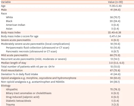
The median LOS was 3 days (Q1=2 days, Q3=6 days) (Table 1).
Analysis of study cohort
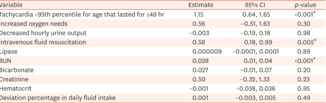
Patients with tachycardia above the 95th percentile for age that lasted for ≥48 hours had on average a 1.15 log (days) longer LOS than those who did not (p<0.001), controlling for oxygen needs, scaled hourly urine output, age, sex, and race (Table 2).
We utilized the laboratory model to analyze the effect of serum BUN level on admission and other laboratory markers on hospital LOS. Although the majority of the cohort had a serum BUN within high normal level (normal range 7–20 mg/dL), the elevation of serum BUN on admission was found to be significantly associated with prolonged LOS (p<0.001)
Table 1. Patient demographics and clinical characteristics
Variable Value (n=92)
Age 11.95±5.40
Male 41 (44.6)
Race
White 58 (70.7)
Black 20 (24.4)
American Indian 2 (2.4)
Asian 2 (2.4)
Body mass index 22.40±8.38
Body mass index z-score for age 0.47±1.54
Severe acute pancreatitis 6 (6.5)
Moderately severe acute pancreatitis (local complications) 18 (19.6) Peripancreatic fluid collection (ultrasound or CT scan) 10 (10.9)
Pancreatic necrosis (ultrasound or CT scan) 8 (8.7)
Mild acute pancreatitis 68 (73.9)
Recurrent acute pancreatitis (mild, moderate or severe) 13 (14.1)
Median length of stay 3.0 (2.0, 6.0)
Total number of patients with nil per os >24 hr 12 (13.0)
Fluid resuscitation 67 (72.8)
Deviation % in daily fluid intake 41 (44.6)
Opioid analgesia e.g. morphine, oxycodone and hydromorphone 58 (63.0)
Non-opioid analgesia e.g. acetaminophen and NSAIDs 24 (26.1)
Etiology
Idiopathic 72 (78.3)
Biliary tract anomalies or cholelithiasis 6 (6.5)
Drug induced (valproic acid) 6 (6.5)
Diabetic ketoacidosis 5 (5.4)
Trauma 3 (3.3)
Values are presented as mean±standard deviation, number (%), or median (1st quartile, 3rd quartile).
CT: computerized tomography, NSAID: non-steroidal anti-inflammatory drug.
as illustrated in Fig. 2. In contrast, serum creatinine, bicarbonate, lipase and hematocrit levels were not found to have a significant effect on LOS, after controlling for all other laboratory markers, age, sex, and race. Similarly, the interaction between serum BUN level on presentation and peak serum lipase during admission did not significantly affect the primary outcome. Table 2 demonstrates the effect of laboratory markers completed during admission on hospital LOS.
Patients who received fluid resuscitation during admission had on average a 0.58 log (days) longer LOS than those who did not (p=0.005), controlling for deviation percentage from maintenance fluid, age, sex and race. There was no association between the percentage deviation of fluid intake from maintenance and increased hospital LOS in our patient population (p=0.49). This finding is illustrated in Table 2.
In the complete regression model that included clinical signs of organ dysfunction, laboratory markers, fluid resuscitation and total fluid intake volume during hospital
admission, we demonstrated that tachycardia above the 95th percentile for age that lasted for
≥48 hours was the leading variable to be associated with an average of 0.99 log (days) longer hospital LOS. This result is illustrated in Table 3.
Table 2. Linear regression model analysis showing the effect of clinical, laboratory and fluid therapy variables on hospital length of stay in log (days)
Variable Estimate 95% CI p-value
Tachycardia >95th percentile for age that lasted for ≥48 hr 1.15 0.64, 1.65 <0.001*
Increased oxygen needs 0.56 −0.51, 1.63 0.30
Decreased hourly urine output −0.003 −0.19, 0.18 0.98
Intravenous fluid resuscitation 0.58 0.18, 0.99 0.005*
Lipase 0.000009 −0.0001, 0.0001 0.89
BUN 0.028 0.01, 0.04 <0.001*
Bicarbonate 0.027 −0.01, 0.07 0.20
Creatinine 0.50 −0.32, 1.33 0.23
Hematocrit −0.001 −0.038, 0.036 0.95
Deviation percentage in daily fluid intake 0.001 −0.003, 0.005 0.49
CI: confidence interval, BUN: blood urea nitrogen.
*Statistically significant.
0 6
4
2
100
Log(lengthofstay)
BUN on admission (mg/dL) 0
Fit
95% perdiction limits
20 40 60 80
Fig. 2. Fit plot curve showing the effect of serum BUN level at admission on length of stay in log (days).
BUN: blood urea nitrogen.
DISCUSSION
Much has been written about the underlying pathophysiology of AP and its complications, however management remains a clinical challenge and is primarily based on supportive therapy [13-15]. Early correction of intravascular volume depletion caused by pancreatitis appears to be widely accepted as the cornerstone intervention in this condition, yet there is little evidence about the value and range of its clinical application. In addition, there is no current standardized or validated protocol for the management of AP in pediatric patients [16-18].
Our study shows that pediatric patients without complex medical co-morbidities, who were admitted with AP, tachycardia above the 95th percentile for age that lasted for ≥48 hours, elevated BUN on admission and need for fluid resuscitation had longer hospital LOS. The amount of fluid intake above recommended maintenance volumes during hospital stay did not affect the LOS in our patient population.
Unlike observations reported from adult studies, there is limited data regarding the outcomes of pediatric AP [19]. Earlier efforts to build a prognostic system for AP in children have not been widely generalizable, as studies were based on major medical institutions, predominantly quaternary referral hospitals with a greater proportion of patients who had prior co-morbidities and more severe disease.
Our organ failure model was developed to examine the effect of clinical signs of cardiac, pulmonary and renal dysfunction on LOS. In this model, elevated heart rate above 95th percentile for age that lasted for ≥48 hours was found to be associated with increased LOS.
Only three of our patients with SAP had decreased SBP and MBP so these blood pressure variables were not included. The finding that most patients are not hypotensive may be due to the limited number of patients with SAP in our cohort or the physiologic delay in development of hypotension in pediatric hypovolemic shock [19]. Similarly, decreased hourly scaled urine output and increased oxygen needs did not have a statistically significant effect on LOS as they appear to be a delayed sign of organ failure in pediatric population [20].
Analysis of the laboratory markers model showed that elevated serum BUN on admission was associated with increased LOS. The majority of BUN values were in the higher normal level for pediatric age (20 mg/dL). In a prior study that included 671 adult patients with AP, authors Table 3. Complete linear regression model analysis showing the effect of variables on length of stay in log (days)
Variable Estimate 95% CI p-value
Tachycardia >95th percentile for age that lasted for ≥48 hr 0.99 0.45, 1.53 <0.001*
Increased oxygen needs −0.05 −2.75, 2.61 0.97
Decreased hourly urine output −0.02 −0.22, 0.18 0.83
Intravenous fluid resuscitation 0.33 −0.15, 0.81 0.17
Deviation percentage in daily fluid intake −0.001 −0.006, 0.004 0.69
Serum Lipase −0.000006 −0.0001, 0.0001 0.93
Serum BUN 0.02 −0.01, 0.05 0.20
Serum bicarbonate 0.02 −0.02, 0.07 0.28
Serum creatinine 0.28 −0.59, 1.16 0.52
Blood hematocrit 0.004 −0.037, 0.045 0.83
Z-score of body mass index −0.09 −0.24, 0.05 0.20
CI: confidence interval, BUN: blood urea nitrogen.
*Statistically significant.
concluded that the best timing for BUN measurement to predict in-hospital mortality related to AP was at admission and with a value of 37.3 mg/dL for optimal response [21]. This cut-off value in pediatric patients would be different as serum BUN levels are physiologically lower than those of adults due to their anabolic growth state and higher glomerular reserve [22].
A retrospective study of 202 pediatric patients with AP showed that an increase in BUN at 48 hours was associated with more severe disease outcome based on comparable scores to Ranson and modified Glasgow systems. However, the authors did not suggest a cut-off value [23,24].
In our study, elevated serum lipase was not associated with increased LOS. This is
comparable to widely accepted adult pancreatitis severity systems, including APACHE II and Ranson's criteria, that showed serum lipase was not found to correlate with disease outcome [25]. In a different pediatric study published by Szabo et al, the authors concluded that serum lipase greater than or equal seven times the ULN predicted severity of AP, defined as organ failure, local pancreatic complications, need for surgery or death, with 85% sensitivity and 56% specificity [26]. This finding was not validated in other studies.
Most of adult severity predictive systems include blood hematocrit on admission as a predictor of severe disease and marker of hemoconcentration [27-29]. Based upon this, we hypothesized that increased blood hematocrit in pediatric patients on admission would correlate with increased LOS [30,31]. However, our results did not support this hypothesis.
Perhaps the higher prevalence of anemia and age-related variation of hemoglobin level in the pediatric population may reduce the sensitivity of hematocrit as a marker of intravascular hypovolemia [32]. In our study cohort, we suspect that could be the reason why elevated blood hematocrit was not found to be associated with increased hospital LOS. We must also note that no cases of hemorrhagic pancreatitis were diagnosed in our study group.
The finding that fluid resuscitation was associated with longer LOS could be related to the fact that patients who needed fluid boluses had a relatively more severe course. Fluid hydration provides circulatory support to prevent the cascade of events leading to pancreatic necrosis and complications [33-35]. In addition, fluid resuscitation improves hypovolemia resulting from reduced oral intake, vomiting and third space extravasation. Our patient cohort received fluid resuscitation with either NS or LR using volume a of 20 mg/kg/bolus with a maximum of 3 boluses.
Previous guidelines published by the INSPPIRE group recommended that pediatric patients with AP receive 1.5–2.0 times daily calculated maintenance fluids [8,36]. This finding could be validated by a future prospective observational study or randomized controlled clinical trial.
As a retrospective cohort design, our study has limitations related to identifying causality and selection bias. The potential source of selection bias is due to management decisions being left to the discretion of each patient's individual treating physician. To reduce this bias, we considered all demographic and clinical variables in the regression models. Although this is the largest sample size of pediatric patients with AP who did not have other severity confounding co-morbidities, we had a limited number of patients with severe AP.
In conclusion we found that an elevated serum BUN on admission, tachycardia that lasted for
≥48 hours and the need for fluid resuscitation were associated with increase LOS in pediatric AP. We did not find evidence that daily total fluid intake above recommended maintenance was associated with reduced hospital LOS as a proxy marker for severity. Our study highlights the importance of tachycardia and elevated BUN as simple and universal markers for identifying
higher severity cases of pancreatitis. In addition, it provides insight for future research aiming to better determine the severity of AP and the parameters for fluid therapy in pediatric AP.
REFERENCES
1. Morinville VD, Barmada MM, Lowe ME. Increasing incidence of acute pancreatitis at an American pediatric tertiary care center: is greater awareness among physicians responsible? Pancreas 2010;39:5-8.
PUBMED | CROSSREF
2. Nydegger A, Heine RG, Ranuh R, Gegati-Levy R, Crameri J, Oliver MR. Changing incidence of acute pancreatitis: 10-year experience at the Royal Children's Hospital, Melbourne. J Gastroenterol Hepatol 2007;22:1313-6.
PUBMED | CROSSREF
3. Park A, Latif SU, Shah AU, Tian J, Werlin S, Hsiao A, et al. Changing referral trends of acute pancreatitis in children: a 12-year single-center analysis. J Pediatr Gastroenterol Nutr 2009;49:316-22.
PUBMED | CROSSREF
4. Pant C, Deshpande A, Olyaee M, Anderson MP, Bitar A, Steele MI, et al. Epidemiology of acute pancreatitis in hospitalized children in the United States from 2000-2009. PLoS One 2014;9:e95552.
PUBMED | CROSSREF
5. Abu-El-Haija M, El-Dika S, Hinton A, Conwell DL. Acute pancreatitis admission trends: a national estimate through the Kids' Inpatient Database. J Pediatr 2018;194:147-51.e1.
PUBMED | CROSSREF
6. Kumar S, Ooi CY, Werlin S, Abu-El-Haija M, Barth B, Bellin MD, et al. Risk factors associated with pediatric acute recurrent and chronic pancreatitis: lessons from INSPPIRE. JAMA Pediatr 2016;170:562-9.
PUBMED | CROSSREF
7. Morinville VD, Husain SZ, Bai H, Barth B, Alhosh R, Durie PR, et al. Definitions of pediatric pancreatitis and survey of present clinical practices. J Pediatr Gastroenterol Nutr 2012;55:261-5.
PUBMED | CROSSREF
8. Abu-El-Haija M, Lin TK, Palermo J. Update to the management of pediatric acute pancreatitis:
highlighting areas in need of research. J Pediatr Gastroenterol Nutr 2014;58:689-93.
PUBMED | CROSSREF
9. Abu-El-Haija M, Kumar S, Szabo F, Werlin S, Conwell D, Banks P, et al. Classification of acute pancreatitis in the pediatric population: clinical report from the NASPGHAN pancreas committee. J Pediatr
Gastroenterol Nutr 2017;64:984-90.
PUBMED | CROSSREF
10. Bai HX, Lowe ME, Husain SZ. What have we learned about acute pancreatitis in children? J Pediatr Gastroenterol Nutr 2011;52:262-70.
PUBMED | CROSSREF
11. Pant C, Sferra TJ, Lee BR, Cocjin JT, Olyaee M. Acute recurrent pancreatitis in children: a study from the Pediatric Health Information System. J Pediatr Gastroenterol Nutr 2016;62:450-2.
PUBMED | CROSSREF
12. Gay AC, Barreto NB, Schrager SM, Russell CJ. Factors associated with length of stay and 30-day revisits in pediatric acute pancreatitis. J Pediatr Gastroenterol Nutr 2018;67:e30-5.
PUBMED | CROSSREF
13. Aggarwal A, Manrai M, Kochhar R. Fluid resuscitation in acute pancreatitis. World J Gastroenterol 2014;20:18092-103.
PUBMED | CROSSREF
14. Mayer J, Rau B, Gansauge F, Beger HG. Inflammatory mediators in human acute pancreatitis: clinical and pathophysiological implications. Gut 2000;47:546-52.
PUBMED | CROSSREF
15. Hack CE, Zeerleder S. The endothelium in sepsis: source of and a target for inflammation. Crit Care Med 2001;29(7 Suppl):S21-7.
PUBMED | CROSSREF
16. de-Madaria E, Soler-Sala G, Sánchez-Payá J, Lopez-Font I, Martínez J, Gómez-Escolar L, et al. Influence of fluid therapy on the prognosis of acute pancreatitis: a prospective cohort study. Am J Gastroenterol 2011;106:1843-50.
PUBMED | CROSSREF
17. Eckerwall G, Olin H, Andersson B, Andersson R. Fluid resuscitation and nutritional support during severe acute pancreatitis in the past: what have we learned and how can we do better? Clin Nutr 2006;25:497-504.
PUBMED | CROSSREF
18. Mao EQ, Tang YQ, Li L, Qin S, Wu J, Liu W, et al. [Strategy of controlling fluid resuscitation for severe acute pancreatitis in acute phase]. Zhonghua Wai Ke Za Zhi 2007;45:1331-4. Chinese.
PUBMED
19. Ranson JH, Rifkind KM, Roses DF, Fink SD, Eng K, Spencer FC. Prognostic signs and the role of operative management in acute pancreatitis. Surg Gynecol Obstet 1974;139:69-81.
PUBMED
20. Ceneviva G, Paschall JA, Maffei F, Carcillo JA. Hemodynamic support in fluid-refractory pediatric septic shock. Pediatrics 1998;102:e19.
PUBMED | CROSSREF
21. Goldstein B, Giroir B, Randolph A; International Consensus Conference on Pediatric Sepsis.
International pediatric sepsis consensus conference: definitions for sepsis and organ dysfunction in pediatrics. Pediatr Crit Care Med 2005;6:2-8.
PUBMED | CROSSREF
22. Lin S, Hong W, Basharat Z, Wang Q, Pan J, Zhou M. Blood urea nitrogen as a predictor of severe acute pancreatitis based on the revised Atlanta criteria: timing of measurement and cutoff points. Can J Gastroenterol Hepatol 2017;2017:9592831.
PUBMED | CROSSREF
23. Granger J, Remick D. Acute pancreatitis: models, markers, and mediators. Shock 2005;24 Suppl 1:45-51.
PUBMED | CROSSREF
24. Vitale DS, Hornung L, Lin TK, Nathan JD, Prasad S, Thompson T, et al. Blood urea nitrogen elevation is a marker for pediatric severe acute pancreatitis. Pancreas 2019;48:363-6.
PUBMED | CROSSREF
25. DeBanto JR, Goday PS, Pedroso MR, Iftikhar R, Fazel A, Nayyar S, et al. Acute pancreatitis in children.
Am J Gastroenterol 2002;97:1726-31.
PUBMED | CROSSREF
26. Szabo FK, Hornung L, Oparaji JA, Alhosh R, Husain SZ, Liu QY, et al. A prognostic tool to predict severe acute pancreatitis in pediatrics. Pancreatology 2016;16:358-64.
PUBMED | CROSSREF
27. Banks PA. Practice guidelines in acute pancreatitis. Am J Gastroenterol 1997;92:377-86.
PUBMED
28. Nathens AB, Curtis JR, Beale RJ, Cook DJ, Moreno RP, Romand JA, et al. Management of the critically ill patient with severe acute pancreatitis. Crit Care Med 2004;32:2524-36.
PUBMED | CROSSREF
29. Martínez J, Sánchez-Payá J, Palazón JM, Suazo-Barahona J, Robles-Díaz G, Pérez-Mateo M. Is obesity a risk factor in acute pancreatitis? A meta-analysis. Pancreatology 2004;4:42-8.
PUBMED | CROSSREF
30. Johnson CD, Toh SK, Campbell MJ. Combination of APACHE-II score and an obesity score (APACHE-O) for the prediction of severe acute pancreatitis. Pancreatology 2004;4:1-6.
PUBMED | CROSSREF
31. Gloor B, Müller CA, Worni M, Martignoni ME, Uhl W, Büchler MW. Late mortality in patients with severe acute pancreatitis. Br J Surg 2001;88:975-9.
PUBMED | CROSSREF
32. Halonen KI, Leppaniemi AK, Puolakkainen PA, Lundin JE, Kemppainen EA, Hietaranta AJ, et al. Severe acute pancreatitis: prognostic factors in 270 consecutive patients. Pancreas 2000;21:266-71.
PUBMED | CROSSREF
33. Brotanek JM, Gosz J, Weitzman M, Flores G. Iron deficiency in early childhood in the United States: risk factors and racial/ethnic disparities. Pediatrics 2007;120:568-75.
PUBMED | CROSSREF
34. Perel P, Roberts I, Ker K. Colloids versus crystalloids for fluid resuscitation in critically ill patients.
Cochrane Database Syst Rev 2013;(2):CD000567.
PUBMED | CROSSREF
35. Quiros JA, Marcin JP, Kuppermann N, Nasrollahzadeh F, Rewers A, DiCarlo J, et al. Elevated serum amylase and lipase in pediatric diabetic ketoacidosis. Pediatr Crit Care Med 2008;9:418-22.
PUBMED | CROSSREF
36. Abu-El-Haija M, Kumar S, Quiros JA, Balakrishnan K, Barth B, Bitton S, et al. Management of acute pancreatitis in the pediatric population: a clinical report from the North American Society for Pediatric Gastroenterology, Hepatology and Nutrition Pancreas committee. J Pediatr Gastroenterol Nutr 2018;66:159-76.
PUBMED | CROSSREF