서론
치아 결손 부위의 수복을 원하는 환자들에게 임플란트를 이용 한 수복 치료가 보편화되고 있으며, 임상연구를 통하여 임플란 트 수복은 기능적, 심미적으로 높은 성공률을 보인다는 결과가 보고되고 있다.1 또한 수복된 임플란트 보철물의 생존률도 높은 수준으로 보고되고 있어 이를 기반으로 임플란트 수복의 임상 적용은 그 범위가 더욱 넓어지고 있다.2,3
임플란트 수복의 적용 범위가 넓어짐에 따라 환자들의 요구 사항도 다양해지고 있다. 특히 임플란트 식립 후 즉시 수복되는 것을 요구하는 경향이 증가하고 있다. 임플란트 즉시 하중은 이 러한 요구에 맞춰 임상가들 사이에서 확산되었다. 임플란트 즉
시 하중의 임상적인 적용 이외에도 1990년, Schnitman 등4이 임 플란트 즉시 하중에 대한 첫 종적인 연구를 보고한 이후로 즉시 하중에 대한 여러 임상적 연구들이 시행되었고, 임플란트 생존 율에 관하여 긍정적인 결과들이 보고되었다.5
통상적인 임플란트의 수복 과정은 Brånemark 등6에 의해 제 안되었는데, 임플란트 식립 후 하악에서 3 - 4개월, 상악에서 5 - 6개월의 회복기간을 가진 후 임플란트에 하중을 가하게 된다.
2013년 Esposito 등5은 즉시 하중의 개념을 임플란트 식립 후 1 주일 이내에 보철물을 수복하는 것으로 정의하였으며, 식립 후 1 주일에서 2개월 사이에 수복하는 경우를 조기 하중, 3개월 이상 이 지난 뒤에 수복하는 경우를 통상적인, 또는 지연 하중이라 정 의하였다.
https://doi.org/10.4047/jkap.2018.56.2.114 ORIGINAL ARTICLE
즉시 하중 임플란트에서 변연골 흡수량에 관한 후향적 연구
이성훈 정지혜 이진한 조혜원*
원광대학교 치과대학 치과보철학교실
Retrospective study on marginal bone resorption around immediately loaded implants
Sung-Hoon Lee, Ji-Hye Jung, Jin-Han Lee, Hye-Won Cho*
Department of Prosthodontics, College of Dentistry, Wonkwang University, Iksan, Republic of Korea
Purpose: Patients who treated implant immediate loading within a week after implant placement at Wonkwang University Dental Hospital Implant Center were evaluated marginal bone resorption. These retrospective analyses are intended to reinforce the clinical evidence for the implant immediate loading. Materials and methods: Medical history and radiographic data were investigated, which were the patients’ who treated implant immediate loading and restoration with provisional prostheses between January 2005 and June 2016, at Wonkwang University Dental Hospital Implant Center. Total number of implants was 70, marginal bone resorption was measured according to implant length, diameter and connection type. To measure marginal bone resorption, periapical radiographs were taken when the implants were placed and after 6 month.
Statistical analysis was done in Mann-whitney U test and Kruskal-wallis test with SPSS 22.0 software (P<.05). Results: Mean marginal bone resorption around immediately loaded implants according to implant connection type was shown 1.24 ± 0.72 mm in internal hexagon connection type and 1.73 ± 1.27 mm in external hexagon connection type. There was no statically significant difference in marginal bone resorption with implant length and diameter. Conclusion: Implants with immediated loading in internal hexagon connection type showed less marginal bone resorption significantly than in external hexagon connection type. (J Korean Acad Prosthodont 2018;56:114-9) Keywords: Immediate dental implant loading; Bone resorption
*Corresponding Author: Hye-Won Cho
Department of Dentistry, Graduate School, Wonkwang University, 460 Iksan-Daero, Iksan 54538, Republic of Korea
+82 (0)2 63 859 2938: e-mail, [email protected]
Article history: Received January 31, 2018 / Last Revision March 6, 2018 / Accepted March 15, 2018
2018 The Korean Academy of Prosthodontics
This is an Open Access article distributed under the terms of the Creative Commons Attribution Non-Commercial License (http://creativecommons.org/
licenses/by-nc/3.0) which permits unrestricted non-commercial use, distribution, and reproduction in any medium, provided the original work is properly cited.
c cc
※This study was supported by Wonkwang University 2018.
임플란트 식립 후 즉시 하중을 가하는 경우, 보철물을 수복하 기 위해 필요한 기간이 감소하여 환자들에게 치아 상실로 인한 심리적 충격과 스트레스를 줄여줄 수 있다는 장점이 있다.7 또한 임플란트 수술 횟수가 1회로 감소하며, 술 후 임시의치를 제작할 필요가 없어진다. Chaushu 등8은 임플란트 보철물이 즉시 수복 되는 경우 임플란트 주위의 치간 유두를 포함한 연조직의 높이 와 골밀도가 유지되어 추후 최종 보철물 수복 시 심미성이 증가 된다는 보고를 하였다.
임플란트 주위 변연골의 방사선학적 분석은 임플란트 식립 위 치의 적절성의 평가와 임플란트 식립 후의 변연골의 변화를 진단 하는 데에 사용되고 있다. 특히 치근단 방사선 사진은 변연골 높 이 변화에 대한 평가와 골병소를 발견하는 데에 주로 사용되고 있다. 그러나 기존의 방사선학적 기법은 임플란트 방사선사진의 기하학적 투영을 정형화하기 위하여 발전이 되었지만, 미세한 골 변화를 측정하는 데에는 명도의 한계가 있다.9 이를 보완하기 위 하여 디지털 공제술이 도입되었다. 디지털 공제술은 치아나 골 과 같은 경조직의 미세한 변화를 관찰하기에 유용한 술식으로, Queiroz 등10은 두 장의 방사선 사진의 기하학적인 차이를 일치 시킨다면, 디지털 공제술을 이용한 골변화 관측의 정확도는 매 우 높아질 것이라고 보고하였다.
본 연구는 2005년에서 2016년 사이 원광대학교 치과병원 임 플란트 센터에 내원하여 임플란트를 식립한 후 즉시 임시보철물 을 수복하고 6개월 이상의 관찰이 이루어진 환자를 대상으로 임 플란트의 길이 및 직경, 임플란트 제조 회사, 임플란트 체결 방식 을 조사하였다. 그 후 각 임플란트에서 임플란트의 길이 및 직경, 임플란트 제조 회사, 임플란트 체결 방식에 따른 변연골 흡수량 을 디지털 공제술을 이용하여 비교 분석을 시행하였다. 이를 통 하여 임플란트 식립 후 즉시 하중을 가할 시의 임상적 근거를 강 화하고, 추후 보철물의 예후를 보다 높이는 데에 도움이 되고자 한다.
대상 및 방법
2005년 1월 1일부터 2016년 6월 31일 사이에 원광대학교 치과 병원 임플란트센터에서 임플란트를 식립한 후 1주일 내에 임시 보철물을 수복한 환자들 중에서 최소 6개월 이상의 관찰이 이루 어진 환자를 선정하였으며, 환자들의 진료기록부를 조사하였다.
이 중 조절되지 않는 고혈압 환자와 당뇨 환자, 하루에 한 갑 이 상의 담배를 흡연하는 흡연자 및 구강 악습관을 지닌 환자, 임플 란트 실패 후 재식을 시행한 환자는 연구대상에서 제외하였다.
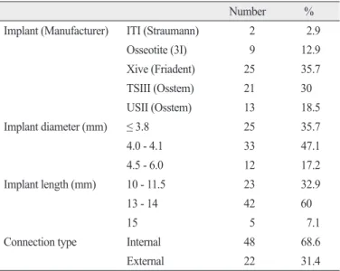
환자들의 진료기록부를 조사한 후, 실험군에서 식립된 임플란 트의 제조사, 직경, 길이, 임플란트 연결 방식을 기준으로 분류하 였으며, 표본 수는 표와 같다 (Table 1).
변연골의 흡수량을 측정하기 위하여 임플란트 식립 직후와 6 개월 후의 치근단 방사선 사진을 변연골 흡수량 측정에 사용하 였다.
방사선 사진은 구강내 디지털 치근단 방사선 사진 촬영기인
KODAK 2200 Intraoral X-ray System (Kodak-Trophy, Paris, France)을 사용하여 70kVp, 7mA로 조건을 설정하여 촬영되 었으며, Kodak RVG 6100 Digital Radiography System (Car- estream Dental, Atlanta, GA, USA)라는 CCD 형태의 센서를 사용하여 영상 정보를 획득하였다. 획득한 영상 정보를 Infinitt PACS (Infinitt, Seoul, Korea)를 통하여 DICOM 영상으로 전환, 이를 Tiff 파일로 변환하여 개인 컴퓨터에 저장하였다.
변연골의 높이를 비교하기 위하여 Emago advanced v5.6 (Oral diagnostic systems, Amsterdam, Netherlands) 소프트웨어를 사 용하였다. 임플란트 식립 직후와 6개월 후에 촬영된 치근단 방사 선 사진을 100% 확대한 영상에서 동일한 위치에 각각 10개의 참 고점을 설정하였으며 (Fig. 1), 두 사진에 설정된 참고점을 기준 으로 기하학적 표준화를 시킨 영상을 획득하였다. 획득한 영상 의 흑화도와 대조도를 임플란트 식립 직후에 촬영된 사진의 조 건과 일치시킨 영상을 얻은 후, 이 영상과 임플란트 식립 직후 촬 영된 영상을 공제하여 디지털 공제 영상을 획득하였다. 최종적으
Table 1. Features of implants investigated in this study
Number %
Implant (Manufacturer) ITI (Straumann) 2 2.9
Osseotite (3I) 9 12.9
Xive (Friadent) 25 35.7
TSIII (Osstem) 21 30
USII (Osstem) 13 18.5
Implant diameter (mm) ≤ 3.8 25 35.7
4.0 - 4.1 33 47.1
4.5 - 6.0 12 17.2
Implant length (mm) 10 - 11.5 23 32.9
13 - 14 42 60
15 5 7.1
Connection type Internal 48 68.6
External 22 31.4
Fig. 1. White arrows indicate 10 reference points with 100% blowed up peri- apical images.
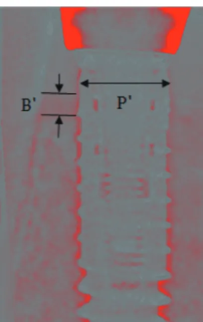
로 획득한 디지털 공제 영상에서 임플란트 플랫폼의 폭(P')을 측 정하고, 임플란트 주변의 흡수된 변연골 높이 중 가장 낮은 지점 을 지정하여 수직적인 변연골 흡수량(B')을 측정한다. 각 임플란 트의 제조사가 제시한 실제 임플란트 플랫폼의 폭(P)을 구하여 기록하였다. 그 후 아래의 비례식을 통하여 실제 임플란트의 변 연골 흡수량을 계산하였다 (Fig. 2).
SPSS ver. 22.0 (SPSS Inc., Chicago, IL, USA) 소프트웨어를 이용하여 모든 통계 처리를 시행하였다. 즉시 하중을 가한 임플 란트 중에서 지대주 연결방식에 따른 변연골 흡수량을 분석하 기 위하여 Mann-whitney U test를 시행하였으며, 임플란트 제조 회사, 직경, 길이에 따른 골 흡수량을 알아보기 위하여 Kruskal- wallis test를 시행하였다.
결과
내부 육각 구조 임플란트의 경우 변연골 흡수량은 평균 1.24 ± 0.72 mm, 외부 육각 구조 임플란트의 경우 1.73 ± 1.27 mm로 나 타나 외부 육각 구조 임플란트에서 더 많은 변연골 흡수가 일어 났으며, 통계적으로 유의한 차이를 보였다 (P < .05) (Table 2).
임플란트 제조회사의 차이에 따른 임플란트 주위 변연골 흡수 량을 조사하였으며, 각 집단 간의 변연골 흡수량의 차이에는 통 계적으로 유의한 차이가 나타나지 않았다 (P > .05) (Table 3).
임플란트의 길이는 규격이 일정치 않아 10 - 11.5 mm, 13 - 14 mm, 15 mm의 범위로 분류하였고, 각 범위 간의 변연골 흡수량 은 유의한 차이가 보이지 않았다 (P > .05). 임플란트의 직경은 각 회사마다 다양하여 3.8 mm 이하, 4.0 - 4.1 mm, 4.5 - 6.0 mm 로 집단을 나누었으며, 각 집단 간의 변연골 흡수량에 유의한 차 이는 나타나지 않았다 (P > .05) (Table 4).
고찰
구강 내 치아 상실의 원인으로는 치아 우식, 치주질환, 외상 등 이 있으며, 보철 수복을 위한 전략적 발거로 인하여 부분 무치악 상태가 될 수 있다. 이런 무치악 부위를 임플란트로 수복하는 방 법은 보편화 되었다. 상악 전치부 상실 환자 또는 상하악 구치부 상실 환자는 치아 상실 후, 또는 임플란트 식립 후 빠른 시일 내 에 심미적 혹은 기능적으로 회복이 되길 요구한다. 이러한 상황 에서 임플란트 즉시 하중은 좋은 치료법이 될 수 있다.
Fig. 2. References used to measure the actual marginal bone resorption.
B': marginal bone resorption on digital subtraction image. P': width of im- plant platform on digital subtraction image.
B = P × B' / P'
P: actual width of implant platform (mm)
P': width of implant platform on digital subtraction image (mm) B: actual marginal bone resorption (mm)
B': marginal bone resorption on digital subtraction image (mm)
Table 2. Marginal bone resorption around immediate loading implants according to implant connection type
Implant connection type Mean ± SD (mm) P
Internal hex 1.24 ± 0.72
External hex 1.73 ± 1.27 .04*
*significantly different (P < .05)
Table 3. Marginal bone resorption around immediate loading implants according to implant manufacturer
Manufacturer Mean ± SD (mm) P
ITI 1.68 ± 1.90
.15
3I 1.24 ± 0.50
XIVE 1.16 ± 0.68
TS III 1.30 ± 0.67
US II 2.07 ± 1.54
Table 4. Marginal bone resorption around immediate loading implants according to implant length and implant diameter
Group Mean ± SD (mm) P
Implant length (mm) 10 - 11.5 1.73 ± 1.35 .27 13 - 14 1.25 ± 0.66
15 1.11 ± 0.28
Implant diameter (mm) ≤ 3.8 1.26 ± 0.59 .91
4.0 - 4.1 1.25 ± 0.75 4.5 - 6.0 1.16 ± 0.77
Misch 등11에 따르면, 임플란트의 완벽한 성공 기준은 동요가 없어야 하며, 방사선 사진 상 임플란트 주위의 방사선 투과상이 관찰되지 않아야 한다. 또한 임플란트를 식립한 첫 해에는 수직 적 골 흡수량이 0.2 mm 미만이어야 하며, 각각의 임플란트에서 동통, 감염, 신경통, 마비 등의 증상이 나타나지 않아야 한다. 만 족스러울 정도로 생존했다는 기준으로는 수직적 골 흡수량이 술 후 2 - 4 mm 정도이어야 하며 나머지 기준들은 완벽히 성공했을 때와 같다고 보고하였다.
임플란트와 지대주 사이의 체결 방식에 따른 평균 변연골 흡 수량은 내부 육각 연결 구조에서 1.24 ± 0.72 mm, 외부 육각 연 결 구조에서 1.73 ± 1.27 mm로 나타나 내부 육각 연결 구조에서 통계적으로 유의하게 더 적은 변연골 흡수량을 보였다. Galindo- Moreno 등12은 내부 육각 연결 구조 임플란트와 외부 육각 연결 구조 임플란트의 변연골 흡수량을 비교한 연구에서 내부 육각 연결 구조 임플란트의 변연골 흡수량은 평균 0.40 ± 0.03 mm, 외부 육각 연결 구조 임플란트의 변연골 흡수량은 평균 0.67 ± 0.03 mm로 나타나 외부 육각 연결 구조에서의 변연골 흡수량이 많이 나타났다고 보고하였다.Tsouknidas 등13과 Chun 등14은 임 플란트 체결 방식에 따른 응력 분포에 관한 연구에서 외부 육각 연결 구조에서 받는 응력의 크기가 내부 육각 연결 구조에서 받 는 응력의 크기보다 더 크다고 보고하였다.이를 통하여 외부 육 각 구조에서의 임플란트 변연골 흡수량이 더 많이 나타날 것이 라고 유추할 수 있다.
통상적인 임플란트 식립 과정에서 임플란트 길이가 변연골 흡 수량에 미치는 영향에 관한 연구 논문들에서는 임플란트 길이는 임플란트의 생존율과 변연골 흡수에 영향을 적게 미친다고 보고 하고 있다.15-18 2003년 Pierrisnard 등19은 임플란트 변연골에 전 달되는 최대 응력은 임플란트 길이에 관계없이 비슷하다고 보고 하였으며, 이를 임플란트 길이와 변연골 흡수량에는 관계가 없 다는 것으로 유추할 수 있다. Degidi 등20은 임플란트에 즉시 하 중을 가하는 경우, 임플란트의 길이는 적어도 10 mm 이상이 되 어야 한다고 보고하였다. 본 연구에서는 각 군마다 변연골 흡수 량에 차이는 있었으나, 통계적으로 유의한 차이는 나타나지 않 아 앞서 언급한 보고들의 결과들과 일치함을 알 수 있었다.
Ding 등21은 임플란트 직경에 따른 응력 분포에 관한 연구를 진행하였는데, 임플란트의 직경이 증가함에 따라 임플란트 변연 골에 가해지는 응력이 감소한다고 보고하였다.22
2004년 Himmlová 등23은 임플란트 직경이 증가할수록 임플 란트 변연골에 가해지는 응력이 감소한다고 보고하였다. 본 연 구에서도 통계적으로 유의한 차이가 나지는 않았지만, 직경이 증가할수록 변연골 흡수량이 감소하는 경향을 보였다.
현재까지 임플란트 즉시 하중의 성공률에 관한 연구들은 많이 시행되었지만, 변연골 흡수량에 관한 연구는 충분치 못한 상황 이었다. 이에 본 연구에서는 임플란트 즉시 하중에 관하여 임플 란트의 여러 가지 조건에 따른 변연골 흡수량을 디지털 공제술 을 통하여 비교 분석하였으며 이를 통해 임플란트의 체결 방식 을 고려하여 임플란트 즉시 하중을 시행할 때, 임플란트 즉시 하
중 술식의 성공률이 높아질 것이라 결론지었다. 그러나 임플란 트 보철물을 수복한 술자의 불일치, 보철물에 가해지는 기능력, 보철물의 형태 등을 파악하지 못하였다는 한계점이 있었다. 추 후에는 이러한 한계점을 보완하여 추가적인 연구가 이루어져야 할 것이다.
결론
내부 연결형 임플란트에서의 변연골 흡수량이 외부 연결형 임 플란트에서의 변연골 흡수량보다 적게 나타났다. 임플란트의 제 조회사에 따른 변연골 흡수량에는 유의한 차이가 없었다. 임플 란트의 길이 및 직경에 따른 변연골 흡수량에는 유의한 차이가 없었다.
References
1. Cochran DL. The evidence for immediate loading of im- plants. J Evid Based Dent Pract 2006;6:155-63.
2. Bergman B. Evaluation of the results of treatment with osseo- integrated implants by the Swedish National Board of Health and Welfare. J Prosthet Dent 1983;50:114-5.
3. Buser D, Mericske-Stern R, Dula K, Lang NP. Clinical expe- rience with one-stage, non-submerged dental implants. Adv Dent Res 1999;13:153-61.
4. Schnitman PA, Wohrle PS, Rubenstein JE. Immediate fixed interim prostheses supported by two-stage threaded implants:
methodology and results. J Oral Implantol 1990;16:96-105.
5. Esposito M, Grusovin MG, Maghaireh H, Worthington HV.
Interventions for replacing missing teeth: different times for loading dental implants. Cochrane Database Syst Rev 2013;(3):CD003878.
6. Brånemark PI, Hansson BO, Adell R, Breine U, Lindström J, Hallén O, Ohman A. Osseointegrated implants in the treat- ment of the edentulous jaw. Experience from a 10-year pe- riod. Scand J Plast Reconstr Surg Suppl 1977;16:1-132.
7. Malchiodi L, Cucchi A, Ghensi P, Nocini PF. Evaluation of the esthetic results of 64 nonfunctional immediately loaded postextraction implants in the maxilla: correlation between in- terproximal alveolar crest and soft tissues at 3 years of follow- up. Clin Implant Dent Relat Res 2013;15:130-42.
8. Chaushu G, Chaushu S, Tzohar A, Dayan D. Immediate load- ing of single-tooth implants: immediate versus non-immediate implantation. A clinical report. Int J Oral Maxillofac Implants 2001;16:267-72.
9. Brägger U, Bürgin W, Lang NP, Buser D. Digital subtraction radiography for the assessment of changes in peri-implant bone density. Int J Oral Maxillofac Implants 1991;6:160-6.
10. Queiroz PM, Oliveira ML, Tanaka JL, Soares MG, Haiter- Neto F, Ono E. Assessment of three methods of geometric image reconstruction for digital subtraction radiography. Den- tomaxillofac Radiol 2016;45:20160120.
11. Misch CE, Perel ML, Wang HL, Sammartino G, Galindo- Moreno P, Trisi P, Steigmann M, Rebaudi A, Palti A, Pikos MA, Schwartz-Arad D, Choukroun J, Gutierrez-Perez JL, Marenzi G, Valavanis DK. Implant success, survival, and fail- ure: the International Congress of Oral Implantologists (ICOI) Pisa Consensus Conference. Implant Dent 2008;17:5-15.
12. Galindo-Moreno P, Fernández-Jiménez A, O’Valle F, Monje A, Silvestre FJ, Juodzbalys G, Sánchez-Fernández E, Catena A. Influence of the crown-implant connection on the pres- ervation of peri-implant bone: a retrospective multifactorial analysis. Int J Oral Maxillofac Implants 2015;30:384-90.
13. Tsouknidas A, Lympoudi E, Michalakis K, Giannopoulos D, Michailidis N, Pissiotis A, Fytanidis D, Kugiumtzis D.
Influence of alveolar bone loss and different alloys on the biomechanical behavior of internal-and external-connection implants: A three-dimensional finite element analysis. Int J Oral Maxillofac Implants 2015;30:e30-42.
14. Chun HJ, Shin HS, Han CH, Lee SH. Influence of implant abutment type on stress distribution in bone under various loading conditions using finite element analysis. Int J Oral Maxillofac Implants 2006;21:195-202.
15. Buser D, Mericske-Stern R, Bernard JP, Behneke A, Behneke N, Hirt HP, Belser UC, Lang NP. Long-term evaluation of non-submerged ITI implants. Part 1: 8-year life table analysis of a prospective multi-center study with 2359 implants. Clin Oral Implants Res 1997;8:161-72.
16. Ellegaard B, Kølsen-Petersen J, Baelum V. Implant therapy
involving maxillary sinus lift in periodontally compromised patients. Clin Oral Implants Res 1997;8:305-15.
17. Feldman S, Boitel N, Weng D, Kohles SS, Stach RM. Five- year survival distributions of short-length (10 mm or less) machined-surfaced and Osseotite implants. Clin Implant Dent Relat Res 2004;6:16-23.
18. Lemmerman KJ, Lemmerman NE. Osseointegrated dental implants in private practice: a long-term case series study. J Periodontol 2005;76:310-9.
19. Pierrisnard L, Renouard F, Renault P, Barquins M. Influence of implant length and bicortical anchorage on implant stress distribution. Clin Implant Dent Relat Res 2003;5:254-62.
20. Degidi M, Piattelli A. Immediate functional and non-func- tional loading of dental implants: a 2- to 60-month follow-up study of 646 titanium implants. J Periodontol 2003;74:225- 21. Ding X, Zhu XH, Liao SH, Zhang XH, Chen H. Implant-bone 41.
interface stress distribution in immediately loaded implants of different diameters: a three-dimensional finite element analy- sis. J Prosthodont 2009;18:393-402.
22. Ding X, Liao SH, Zhu XH, Zhang XH, Zhang L. Effect of diameter and length on stress distribution of the alveolar crest around immediate loading implants. Clin Implant Dent Relat Res 2009;11:279-87.
23. Himmlová L, Dostálová T, Kácovský A, Konvicková S. Influ- ence of implant length and diameter on stress distribution: a finite element analysis. J Prosthet Dent 2004;91:20-5.
즉시 하중 임플란트에서 변연골 흡수량에 관한 후향적 연구
이성훈 정지혜 이진한 조혜원*
원광대학교 치과대학 치과보철학교실
목적: 원광대학교 치과대학병원 임플란트 센터에서 임플란트를 식립한 후 1주일 내에 임시보철물을 수복한 다음, 즉시 하중을 가한 환자들을 대상으 로 골 흡수량을 측정하고자 하였다. 이러한 후향적 분석을 통하여 임플란트에 대한 즉시 하중의 임상적 근거를 보강하고 주의해야 할 사항 등을 파악 하고자 하였다.
대상 및 방법: 2005년 1월 1일부터 2016년 6월 31일 사이에 원광대학교 치과대학병원 임플란트 센터에서 임플란트를 식립한 후 1주일 내에 임시보철
물을 수복한 환자들을 대상으로 하여 진료기록부와 방사선 사진을 조사하였다. 총 임플란트의 수는 70개였으며, 임플란트의 제조회사, 길이, 직경, 체 결 방식에 따른 변연골의 흡수량을 측정하였다. 변연골의 흡수량을 측정하기 위해 임플란트 식립 직후와 6개월 이후의 치근단 방사선 사진을 비교 분석 하였다. 통계 프로그램인 SPSS 22.0을 이용하여 Mann-whitney U test와 Kruskal-wallis test를 시행하였다.
결과: 즉시 하중 임플란트 군에서 임플란트와 지대주의 연결 구조에 따른 변연골 흡수량을 비교한 결과 내부 육각 연결 구조 임플란트에서의 변연골 흡수량은 평균 1.24 ± 0.72 mm, 외부 육각 연결 구조 임플란트에서 1.73 ± 1.27 mm로 나타났으며 통계적으로 유의한 차이를 보였다. 임플란트 제조회 사, 길이, 직경에 따른 변연골 흡수량의 차이는 통계적으로 유의한 차이를 보이지 않았다.
결론: 즉시 하중 임플란트에서 내부 육각 연결 구조 임플란트에서의 변연골 흡수량은 외부 육각 연결 구조 임플란트에서의 변연골 흡수량보다 적게 나 타났으며, 임플란트의 다른 조건들에서는 변연골 흡수량에 차이를 보이지 않았다. (대한치과보철학회지 2018;56:114-9)
주요단어: 즉시 하중 임플란트; 변연골 흡수
*교신저자: 조혜원
54538 전북 익산시 익산대로 460 원광대학교 치과대학 치과보철학교실 063 859 2938: e-mail, [email protected]
원고접수일: 2018년 1월 31일 / 원고최종수정일: 2018년 3월 6일 / 원고채택일: 2018년 3월 15일
2018 대한치과보철학회
이 글은 크리에이티브 커먼즈 코리아 저작자표시-비영리 3.0 대한민국 라이선스에 따라 이용하실 수 있습니다.
c cc
※이 논문은 2018학년도 원광대학교 교비지원에 의해서 발표되었음.