Received: March 6 2020, Revised: March 11 2020, Accepted: April 10 2020
Corresponding author: Na-Kyoung Hwang, Rehabilitation Medical Center, Seoul North Municipal Hospital, 38 Yangwonyeok-ro, Jungnang-gu, Seoul 02062, Korea
Tel: +82-2-2036-0273, Fax: +82-2-2036-0278 E-mail: [email protected]
Copyrights ⓒ The Korean Dysphagia Society, 2020.
Effects of Head Lift Exercise on Oropharyngeal Swallowing Function and Dropout Rate According to Reclining Angle in Patients with Dysphagia
after Stroke: A Randomized Trial
Joo-Young Lee, M.D.
1, Dong-Goo Kim, M.D.
1, Hyo-Jeong Lee, B.H.Sc.
2, Ji-Su Park, Ph.D.
3, Tae-Hyung Yoon, Ph.D.
4, Young-Jin Jung, Ph.D.
5, Jun-Yong Hong, R.T.
6, Na-Kyoung Hwang, M.S.
11Rehabilitation Medical Center, Seoul North Municipal Hospital, Seoul, 2Department of Occupational Therapy, Busan National University Yangsan Hospital, Yangsan, 3Advanced Human Resource Development Project Group for Health Care in Aging Friendly Industry, Dongseo University, Busan, 4Department of Occupational Therapy, Division of Health Sciences, Dongseo University, Busan, 5Department of Radiological Science, Health Sciences Division, Dongseo University, Busan, 6Department of Multidisciplinary Radiological Science, Graduate School, Dongseo University, Busan, Korea
Objective: Although the effects of head lift exercise (HLE) in the reclining position have been reported, there is in- sufficient clinical evidence of the effects. This study compared the effects of HLE in the 0° supine position and 45°
reclining position on the swallowing function and the compliance of patients with dysphagia after stroke after both exercises.
Methods: This was a randomized, assessor-blinded clinical trial. Thirty-five patients with stroke and dysphagia were assigned randomly to HLE in the 0° supine group (n=18) or HLE in the 45° reclining group (n=17). Patients in both groups performed HLE five days a week for four weeks and received the same conventional dysphagia therapy. The videofluoroscopic dy sphagia scale (VDS) was used to evaluate the swallowing function. The dropout rate and sub- jective feedback related to compliance with the two exercises were monitored.
Results: No significant differences in the baseline characteristics were observed between the two groups. Patients in both groups showed significant improvement in the oral and pharyngeal phases of VDS (P<0.05). After the inter- vention, no significant differences were observed between the groups (P>0.05). Dropout rates of 22% and 6% owing to neck discomfort or fatigue were observed in the HLE in 0° supine group and the HLE in 45° reclining group, re- spectively.
Conclusion: HLE in the 45° reclining position has a similar effect on the swallowing function in patients with dys- phagia after stroke to that of HLE in the 0° supine position and is associated with better exercise compliance.
(JKDS 2020;10:159-166)
Keywords: Dysphagia, Head lift exercise, Compliance, Reclining position, Videofluoroscopic dysphagia scale
INTRODUCTION
Head lift exercise (HLE), also called Shaker exer- cise, is a method of improving the opening of the pharyngo-esophageal segment by increasing the st- rength of the muscle groups involved in the opening of the upper esophageal sphincter. During swallow- ing, the su prahyoid mu scles su ch as mylohyoid, geniohyoid, and anterior digastric muscles, help the anterior movement of the hyoid bone and induce the anterosu perior movement of the larynx. Improved strength and endurance of the weakened suprahyoid muscles facilitate the opening of the upper esoph- ageal sphincter1,2. This exercise is performed by sus- taining three head lifts held (isometric) and repetitive head lifts of constant velocity without holding (iso- kinetic) while in the 0° supine position3.
However, despite these positive effects of HLE, it can be a physically demanding exercise. Easterling et al.3 reported a high dropout rate in patients perform- ing the exercise because of muscle discomfort and compliance difficu lty. Other stu dies reported neck soreness, fatigue, dizziness, and discontinuation in non-dysphagic elderly participants3,4. Thus, completion of HLE may be a seriou s challenge, especially in patients with dysphagia along with chronic cough, pneumonia, and malnutrition5. Thus, it is clinically important to investigate an exercise method that is modified for the limitations of HLE and is easier to apply.
To compensate for the disadvantages of this con- ventional HLE, some stu dies tried HLE at a 30°-45°
reclining position. As a result, the reclining HLE was demonstrated to indu ce mu scle activation of the suprahyoid muscles similar to when performing HLE in the 0° supine position4,6. In addition, the reclining position helps to relieve neck pain and improves head and neck posture and range of motion in healthy adults and therefore is often used in clinical prac- tice7. By using surface electromyography (sEMG), Koshi et al.6 reported that HLE is easier and more ap- propriate for reclining the backrest at the 30° than at the 0° position.
Thu s, HLE in a reclining position (approximately 30°-45°) is less strenuous than HLE with high physical demands and may be a new exercise that can be applied to patients with dysphagia. However, pre- vious studies measured only muscle activation using sEMG4,6. Therefore, the effects of HLE in a reclining position on the actu al swallowing fu nction are u n- known. This study aimed to compare the effects of HLE at 0° supine position and that of HLE in a reclining position on swallowing function and the compliance of patients with stroke and dysphagia to these exercises, to identify the possibility of HLE in a reclining position as an alternative to HLE.
MATERIALS AND METHODS 1. Participants
We conducted a randomized, assessor-blinded cli- nical trial. In total, 35 patients with dysphagia after a stroke were recruited from a rehabilitation center (Seoul North Municipal Hospital) in the republic of Korea. Participants were randomly allocated to the 0°
HLE group (n=18) or the 45° HLE group (n=17) by using a random allocation software program by a blinded occupational therapist. Block randomization with block size 4 was performed for 1:1 allocation.
Inclusion criteria were as follows: (1) oropharyn- geal dysphagia after stroke confirmed by a video- flu oroscopic swallowing stu dy (VFSS), (2) observed aspiration or penetration in VFSS, (3) no significant cognitive deficits (Mini-Mental State Examination score
>20), (4) above fair grade obtained on muscle testing of the neck, (5) symmetric postu re of the neck, (6) able to perform HLE in a supine position, and (7) ability to swallow voluntarily. Exclusion criteria were as follows: (1) neck pain or discomfort, (2) poor gen- eral condition precluding further participation in the experiment, (3) communication problems (aphasia, apraxia), (4) unstable medical condition, (5) presence of a tracheostomy tube, and (6) cricopharyngeal dys- function.
We explained the objectives and requirements of our study to all participants, and they voluntarily sign-
Fig. 1. Performing the HLE accord- ing to reclining angle (0° and 45°).
ed the informed consent form. The institutional re- view board of Seoul Medical Center approved the study (SEOUL 2017-03-016-002, approval notice date:
10/5/2017).
2. Sample size estimation
The G-Power 3.1 software (University of Dusseldorf, Dusseldorf, Germany) was used to calculate the sam- ple size. A power of 0.80 was adopted considering a level of significance of 0.05 and an effect size of 0.88.
According to a previous analysis, each group required at least 18 participants.
3. Procedures and intervention
The 0° HLE group performed HLE in a lying posi- tion on a flat surface. Conversely, the 45° HLE group performed HLE in the lying position while maintain- ing a 45° reclining position.(Fig. 1) The angle was controlled by a goniometer. Both groups performed HLE with the same frequency and type of exercise.
Exercise was performed with isotonic and isometric contractions. Participants sustained three head lifts held for 1 min without movement in the supine posi- tion, and a 60-s rest was allowed between the lifts.
Then, the participants performed 30 repetitive head lifts without holding in the same supine position. The participants lifted their head high enough to observe their toes without raising their shoulders. HLE was performed three times per day, 5 days a week, for 4 weeks.
Both the grou ps received additional conventional dysphagia treatment (CDT). CDT was performed for 4 weeks, 5 times a week, for 30 minu tes per day, which included thermal tactile stimulation, orofacial
muscle exercise, and various compensatory maneuvers.
An occu pational therapist with 7 years of clinical experience in treating dysphagia performed CDT in all the participants.
4. Outcome measurement
The primary outcome of this study was to measure the swallowing function using videofluoroscopic dys- phagia scale (VDS) based on the VFSS before and 4 weeks after the intervention. The VDS is a functional evaluation scale that reflects overall swallowing func- tion in stroke survivors based on VFSS findings. The VDS is divided into an oral stage (lip closure, bolus formation, tongue-to-palate contact, mastication, aprax- ia, premature bolus loss, and oral transit time) and pharyngeal stage (pharyngeal triggering, vallecular residues, pyriform sinus residues, laryngeal elevation, pharyngeal wall coating, pharyngeal transit time, and aspiration)8. The VDS findings were evaluated and interpreted by a physician specialized in rehabili- tation medicine.
The secondary outcome of this study was to mea- su re dropou t rate and su bjective feedback to com- pare patient compliance of the HLE at the 0° supine position and at the 45° reclining position. The drop- out rate due to discomfort, pain, and fatigue in the neck and abdomen was monitored until the end of the intervention; dropout rate was not monitored for reasons such as discharge or transfer to another hos- pital. After the intervention was completed, the pa- tients were briefly interviewed about their subjective feedback, such as difficulty and discomfort while per- forming the exercises.
Fig. 2. CONSORT diagram of par- ticipant recruitment.
Table 1. Baseline characteristics of participants.
Characteristics 0° HLE group
(n=12) 45° HLE group (n=13) Age (year), mean±SD 63.00±10.55 63.40±6.65
Gender (male/female) 6/6 7/6
Type of stroke
(Hemorrhage/Infarction) 7/5 7/6
Stroke lesion
(Cortical/Subcortical/Brain stem) 8/4/0 9/4/0
Side of stroke (Right/Left) 4/8 8/5
Time since onset of stroke
months, mean±SD 3.00±1.18 3.90±1.28
SD: standard deviation.
5. Statistical analysis
Participant characteristics were analyzed using a statistical software program (SPSS Statistics 20), and descriptive statistics are presented as mean±SD.
Shapiro-Wilk test was used to check normality of the outcome variables. To evaluate the intervention ef- fects, Wilcoxon signed-rank test was used to compare pre- and post-intervention measures in each group.
Mann-Whitney U test was used to compare inter- group changes in outcome measures. Significance level was set at P<0.05.
RESULTS
1. Patient characteristics and baseline information
Thirty-five participants were included in this study.
Ten participants dropped out before the follow-up becau se of transferring to another hospital (0° HLE group [n=2] and 45° HLE group [n=3]) and giving up due to neck muscle discomfort, pain, and fatigue (0°
HLE group [n=4] and 45° HLE group [n=1]).(Fig. 2) A summary of the baseline information features of the
participants (n=25) is shown in Table 1. The homo- geneity test of each measurement item (VDS) showed no significant differences in baseline characteristics between the groups (oral and pharyngeal phase in liquid type of VDS evaluation; P=0.153 and 0.145, oral and pharyngeal phase in semisolid type of VDS eval- uation; P=0.934 and 0.125).
2. Swallowing function
In this study, the swallowing function of the parti- cipants in the reclining position is compared with those in the supine position. The 0° HLE group show-
Table 3. Subjective feedback of participants on performing two exercises.
0° HLE group
Participant 1 It seems to be a burden on the neck muscles. Appropriate preparation such as sufficient stretching before exercise is necessary
Participant 2 Some pain occurred in the neck, but it was fine soon. Overall, it was a tough exercise
Participant 3 A lot of effort of the abdominal muscles was required, and it was not easy to perform the exercise Participant 4 It was a very difficult exercise, especially when I had to keep my head lifted up
Participant 5 Temporary muscle pain was experienced in the abdominal muscles, but it was relieved after a few days Participant 6 Neck and abdominal pain was intense, and required a lot of effort. Hence, I did not want to do this exercise Participant 7 It felt like a burden on the neck and shoulders. After the exercise, the shoulders and neck were stiff
Participant 8 I think it was an extremely tedious schedule, as the exercise had to be performed for four weeks. It is a physically difficult exercise, especially for the neck and shoulders
45° HLE group
Participant 1 The neck feels uncomfortable
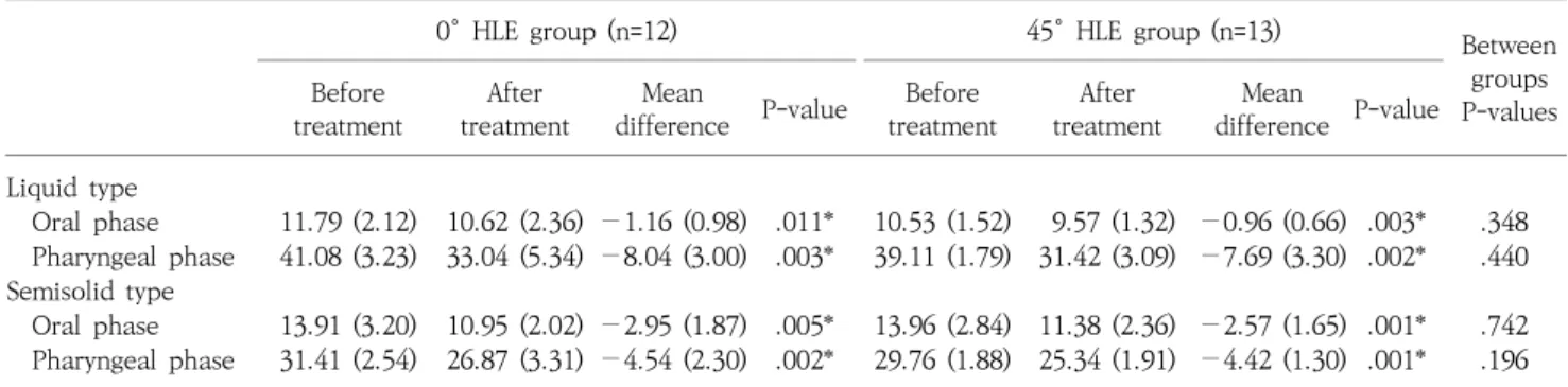
Participant 2 The neck and abdominal muscles required considerable effort and were difficult to perform Table 2. Comparison of swallowing function within and between two groups.
0° HLE group (n=12) 45° HLE group (n=13) Between
groups P-values Before
treatment After
treatment Mean
difference P-value Before
treatment After
treatment Mean
difference P-value Liquid type
Oral phase 11.79 (2.12) 10.62 (2.36) −1.16 (0.98) .011* 10.53 (1.52) 9.57 (1.32) −0.96 (0.66) .003* .348 Pharyngeal phase 41.08 (3.23) 33.04 (5.34) −8.04 (3.00) .003* 39.11 (1.79) 31.42 (3.09) −7.69 (3.30) .002* .440 Semisolid type
Oral phase 13.91 (3.20) 10.95 (2.02) −2.95 (1.87) .005* 13.96 (2.84) 11.38 (2.36) −2.57 (1.65) .001* .742 Pharyngeal phase 31.41 (2.54) 26.87 (3.31) −4.54 (2.30) .002* 29.76 (1.88) 25.34 (1.91) −4.42 (1.30) .001* .196 The values are mean±standard deviation.
HLE: head lift exercise, VDS: videofluoroscopy dysphagia scale.
*P<0.05 by Wilcoxon signed-rank test.
ed significant improvement in the oral and pharyn- geal phase of VDS (liqu id type; P=0.011 and 0.003, semisolid type; P=0.005 and 0.002). The 45° HLE group also showed significant improvement in the oral and pharyngeal phase of VDS (liqu id type; P=0.003 and 0.002, semisolid type; P=0.001 and 0.001). A compari- son of both the groups after the intervention showed no significant differences (all, P>0.05).(Table 2)
3. Dropout-rate related compliance
Four out of 18 patients in the 0° HLE group drop- ped out, indicating a dropout rate of 22%. In the 45°
HLE group, 1 out of 17 patients dropped out, indi- cating a dropou t rate of 6%. The reasons for the dropouts were temporary pain in the neck, muscle weakness, and lack of endurance. There were no side effects such as prolonged muscle pain, discomfort,
and severe fatigue after the intervention.
4. Subjective feedback of participants based on brief interview
Eight patients included in the 0° HLE group re- ported difficulty in performing the exercise because it was very challenging and bothersome to the neck and required significant abdominal effort. Two pa- tients in the 45° HLE group reported abdominal and neck discomfort. All the other patients admitted that the neck and abdominal exercises requ ired some effort, but the effort did not preclude performance of the exercise. Table 3 shows the subjective feedback related to HLE performance of 10 patients who re- ported relative difficulty in performance after the intervention.
DISCUSSION
Both exercises led to significant improvements in the swallowing function; however, there was no sig- nificant difference between the two groups. This sug- gested that HLE in a reclining position and regular HLE have similar effects on stroke patients with dys- phagia. In addition, HLE in a reclining position had better exercise compliance, as evident from the lower dropout rates and subjective feedback.
The present study confirmed the effectiveness of the two exercise methods in the improvement of the oral phase of VDS in patients with dysphagia after stroke. Oral phase items of VDS are mainly related to tongue function (e.g., bolus formation, tongue to palate contact). Both Exercises are head-raising workouts against gravity from head lifting to the 45° reclining and 0° supine positions that requires tongue stability4. Previous studies have also shown that HLE positively affects functional movement of the tongue in the oral phase and induces extensive activation of extrinsic muscles such as the hyoglossus9,10. Mishra et al.4 re- ported that the HLE in a reclining position as well as in a su pine position showed an increase in tongu e strength. These previous studies support the results of this study.
The present study showed improvement of the pharyngeal phase of VDS in both groups. There was no difference between the two grou ps after inter- vention. HLE has been proven in many research studies to indu ce the activation of the su prahyoid mu scles, and recent research has shown that both the exercise methods induce similar high levels of muscle acti- vation4,6. This supports the results of the present study.
Sufficient muscle activation means considerable recruit- ment of motor units and can be expected to improve muscle strength when repeatedly performed. The im- provement of muscular strength of the suprahyoid muscles suggests that it may contribute to normal swal- lowing mechanisms in the pharyngeal phase such as airway protection (reduced aspiration) and UES open- ing by inducing sufficient anterior-superior move- ment of the hyoid bone11-13.
This stu dy confirmed that both exercises have similar effects on the oropharyngeal swallowing. Never- theless, the efficiency aspect is also important for the effective use of exercise, and efficient aspects of treatment are directly related to patient compliance.
Several stu dies have reported a large nu mber of dropouts among participants during HLE2,3. Twenty- five percent of the HLE grou p in ou r stu dy did not perform to completion (75% of patients performed to completion), whereas only 7% of the patients who performed HLE in the reclining position refused and 93% completed the treatment. The dropouts refused to perform the exercise because of neck pain, muscle fatigue, lack of endurance, and discomfort. Sluijs et al.14 reported that discomfort is an important factor in a patient’s attitu de toward exercising; pain and fatigue due to exercise are correlated with compli- ance to exercise and physical therapy. In their study, some participants reported that the exercise was diffi- cult, painful, or tiring.
Compliance is an important factor in rehabilitative approaches in dysphagia treatment, and HLE in a reclining position has the potential to increase com- pliance in patients who experience inconvenience or discomfort when performing head lifts in a supine position. Previous sEMG studies have reported neck muscle (such as the sternocleidomastoid muscles) fatigue during HLE15-17, which results in noncompli- ance and ultimately makes it difficult to reach ther- apeutic goals. Thus, HLE in a reclining position is relatively easy to perform because sternocleidomas- toid fatigue can be reduced with decreased loading during head lifting.
Chin tuck against resistance (CTAR) exercise5,16, jaw-opening exercise18,19, and jaw-opening-against- resistance (JOAR) exercise20 have been introduced as therapeutic exercise methods that complement the limitations of HLE. A comparative study on HLE re- ported the effectiveness of swallowing function and compliance; however, no study has yet compared the effectiveness of swallowing function with 45° HLE and the above mentioned exercises. Further research in this regard is needed. CTAR, JOE, and JOAR are ap-
plicable to patients who can understand and perform the correct exercise to achieve therapeutic effects, and are limited to patients with severe cognitive de- cline or those unable to maintain a sitting position21. Therefore, 45° HLE can be a clinically useful method in that it is less stringent with regard to the subject criteria and can strengthen the suprahyoid muscles while alleviating neck pain and limitation of the range of motion.
This study demonstrated that the effect of reclining HLE is as effective as HLE on swallowing function in patients with dysphagic stroke. Thus, reclining HLE can be provided for patients with dysphagia as an alternative and less strenuous exercise. Further, HLE in a reclining position can be an alternative method for patients who cannot perform HLE in a su pine position because of problems of posture and pain, or complain of muscle fatigue during HLE.
There are some limitations of our study. The sam- ple size was small, and the findings are difficu lt to generalize. Activation of the suprahyoid muscles by u sing EMG was not evalu ated; thu s, the association between hyoid movement and increasing strength of the su prahyoid mu scles cou ld not be confirmed. It was difficult to compare only the effects of two HLEs excluding CDT because the participants of this study were inpatients with dysphagia. Finally, this study was not objective, because the patients were followed through interviews only to investigate compliance to the HLE.
This study used a goniometer to control the re- clining angle at the supine position; however, the angle of the head lift slightly differed among the partici- pants because the angle of head lift from the bed was not strictly controlled. Nevertheless, a merit of this study is that an alternative to regular HLE was at- tempted to demonstrate exercise compliance and to evaluate the improvement in the swallowing function.
Further studies with a larger sample size are needed to determine the long-term therapeu tic benefits of the two exercises.
CONCLUSION
HLE in a reclining position not only has an effect similar to that of regular HLE on swallowing function in patients with dysphagia after stroke but also has better exercise compliance. Therefore, HLE in a re- clining position can be recommended as an alter- native to regular HLE.
CONFLICT OF INTEREST
The authors declare no conflicts of interest.
ACKNOWLEDGEMENTS
This work was supported by the 2017 Seoul Medical Center Medical Research Institu te grant and BB21+
project in 2020.
REFERENCES
1. Easterling C. Does an exercise aimed at improving swal- low function have an effect on vocal function in the healthy elderly? Dysphagia. 2008;23:317-326.
2. Park JS, Hwang NK, Oh DH, Chang MY. Effect of head lift exercise on kinematic motion of the hyolaryngeal com- plex and aspiration in patients with dysphagic stroke. J Oral Rehabil. 2017;44:385-391.
3. Easterling C, Grande B, Kern M, Sears K, Shaker R. At- taining and maintaining isometric and isokinetic goals of the Shaker exercise. Dysphagia. 2005;20:133-138.
4. Mishra A, Rajappa A, Tipton E, Malandraki GA. The Re- cline Exercise: Comparisons with the Head Lift Exercise in Healthy Adults. Dysphagia. 2015;30:730-737.
5. Yoon WL, Khoo JK, Rickard Liow SJ. Chin tuck against resistance (CTAR): new method for enhancing supra- hyoid muscle activity using a Shaker-type exercise. Dys- phagia. 2014;29:243-248.
6. Koshi N, Matsumoto H, Hiramatsu T, Shimizu Y, Hagino H. Influence of backrest angle on swallowing muscu- lature activity and physical strain during the head lift exercise in elderly women compared with young women.
J Oral Rehabil. 2018;45:532-538.
7. Pearson ND, Walmsley RP. Trial into the effects of re- peated neck retractions in normal subjects. Spine. 1995;
20:1245-1250.
8. Han TR, Paik NJ, Park JW, Kwon BS. The prediction of persistent dy sphagia bey ond six months after stroke.
Dysphagia. 2008;23:59-64.
9. Forsberg CM, Hellsing E, Linder-Aronson S, Sheikholeslam A. EMG activity in neck and masticatory muscles in rela- tion to extension and flexion of the head. Eur J Orthod.
1985;7:177-184.
10. Palmer PM, Jaffe DM, McCulloch TM, Finnegan EM, Van Daele DJ, Luschei ES. Quantitative contributions of the muscles of the tongue, floor-of-mouth, jaw, and velum to tongue-to-palate pressure generation. J Speech Lang Hear Res. 2008;51:828-835.
11. Ertekin C, Aydogdu I. Neurophysiology of swallowing.
Clin Neurophysiol. 2003;114:2226-2244.
12. Matsuo K, Palmer JB. Anatomy and physiology of feed- ing and swallowing: normal and abnormal. Phys Med Rehabil Clin N Am. 2008;19:691-707.
13. Sommerich CM, Joines SM, Hermans V, Moon SD. Use of surface electromyography to estimate neck muscle acti- vity. J Electromyogr Kinesiol. 2000;10:377-398.
14. Sluijs EM, Kok GJ, van der Zee J. Correlates of exercise compliance in physical therapy. Phys Ther. 1993;73:771- 782.
15. Ferdjallah M, Wertsch JJ, Shaker R. Spectral analy sis of surface electromyography (EMG) of upper esophageal sphincter-opening muscles during head lift exercise. J Rehabil Res Dev. 2000;37:335-340.
16. Sze WP, Yoon WL, Escoffier N, Rickard Liow SJ. Evaluating the Training Effects of Two Swallowing Rehabilitation Therapies Using Surface Electromyography--Chin Tuck Against Resistance (CTAR) Exercise and the Shaker Exer- cise. Dysphagia. 2016;31:195-205.
17. White KT, Easterling C, Roberts N, Wertsch J, Shaker R.
Fatigue analysis before and after shaker exercise: phys- iologic tool for exercise design. Dysphagia. 2008;23:385- 391.
18. Hughes T, Watts CR. Effects of 2 resistive exercises on electrophysiological measures of submandibular muscle activity. Archives of physical medicine and rehabilitation.
2016;97:1552-1557.
19. Wada S, Tohara H, Iida T, Inoue M, Sato M, Ueda K.
Jaw-opening exercise for insufficient opening of upper esophageal sphincter. Archives of physical medicine and rehabilitation. 2012;93:1995-1999.
20. Watts CR. Measurement of hyolaryngeal muscle activa- tion using surface electromyography for comparison of two rehabilitative dysphagia exercises. Archives of phys- ical medicine and rehabilitation. 2013;94:2542-2548.
21. Park JS, Hwang NK, Oh DH, Chang MY. Therapeutic Exercises for Strengthening Suprahyoid Muscles. Journal of the Korean Dysphagia Society. 2018;8:8-14.