pISSN 2288-9272 eISSN 2383-8493 J Oral Med Pain 2016;41(4):169-179 https://doi.org/10.14476/jomp.2016.41.4.169
Conservative Treatment with Occlusal Appliance for Temporomandibular Disorder Patients with Rheumatoid Arthritis
Young-Ae Kim 1,2 , Kyung-Hee Kim 3 , Soo-Min Ok 1,2 , Yong-Woo Ahn 1,2 , Sung-Hee Jeong 1,2
1 Department of Oral Medicine, School of Dentistry, Pusan National University, Yangsan, Korea
2 Department of Oral Medicine and Dental Research Institute, Pusan National University Dental Hospital, Yangsan, Korea
3 Department of Oral Medicine, Busan Paik Hospital, Inje University College of Medicine, Busan, Korea
Received November 21, 2016 Revised December 10, 2016 Accepted December 10, 2016
Purpose:
Purpose: This study is designed to analyse etiology and bone pattern at the first visit using cone-beam computed tomography (CBCT) and to evaluate the treatment outcome of conserva- tive treatment in temporomandibular disorder (TMD) patients with rheumatoid arthritis (RA).
Methods:
Methods: One hundred condyles in 50 subjects with RA were chosen among the patients who presented to the Department of Oral Medicine of Pusan National University Dental Hospital, di- agnosed as TMD. Condylar bone changes were classified by normal, erosive bony change, pro- liferative bony change and combined group (erosive bony change+proliferative bony change).
They were treated conservatively with physical therapy, medication, behavioral therapy and/or occlusal stabilizing splint therapy. After 3 months on average, patients were re-evaluated with regards to subjective symptoms and the clinical findings were investigated.
Results:
Results: TMD patients with RA have behavioral contributing factors such as parafunctional habit. The results that analyse bone pattern at the first visit using CBCT proliferative bony changes group (32.6%) were more common than erosive bony changes group (15.2%). In com- parison between unilateral and bilateral bony change in temporomandibular joint, the ratio showed no significant differences. After 3 months of conservative treatments, pain, noise, limi- tation of motion (LOM) were markedly improved regardless of occlusal splint therapy. However only LOM was significantly improved through occlusal splint therapy during 3 months.
Conclusions:
Conclusions: TMD patients with RA had similar behavioral contributing factors and character- istics of CBCT images shown in general TMD patients and also similar response to conservative treatment so it is difficult to differentiate. Therefore when TMD patients show symptoms cor- responding to clinical diagnostic criteria of RA at the first visit, serological testing should be conducted and through this, early diagnosis and treatment of RA should be initiated.
Key Words:
Key Words: Arthritis, rheumatoid; Cone-beam computed tomography; Conservative treatment;
Temporomandibular joint disorders
Correspondence to:
Sung-Hee Jeong
Department of Oral Medicine, School of Dentistry, Pusan National University, 49 Busandaehak-ro, Mulgeum-eup, Yangsan 50612, Korea Tel: +82-55-360-5242
Fax: +82-55-360-5234 E-mail: [email protected] This work was supported by a 2-year Research Grant of Pusan National University.
JOMP Journal of Oral Medicine and Pain
Copyright Ⓒ 2016 Korean Academy of Orofacial Pain and Oral Medicine. All rights reserved.
CC
This is an open-access article distributed under the terms of the Creative Commons Attribution Non-Commercial License (http://creativecommons.org/licenses/by-nc/4.0/), which permits unrestricted non-commercial use, distribution, and reproduction in any medium, provided the original work is properly cited.
INTRODUCTION
Rheumatoid arthritis (RA) is a chronic, systemic, inflam- matory autoimmune disease that leads to symmetrical polyarthritis of large and small joints. It typically has an onset in patients aged between 30 to 50 years old 1) with women being affected more than men by a factor of two
or three times. 2) This difference in the prevalence of RA
between the genders suggests the influence of reproduc-
tive and hormonal factors but this notion is still controver-
sial. 3-5) Genetic factors definitely play an important role in
RA pathogenesis 6) and environmental risk factors such as
smoking, alcohol intake, vitamin D status, use of oral con-
traceptives and low socioeconomic status also can influence
on development of RA. 7) The temporomandibular joint (TMJ) also can be influenced by RA with morbidity rates vary- ing from 5% to 86%, according to the study population, di- agnostic criteria and evaluation methods. 8-10) Patients with TMJ affected by RA show various signs and symptoms such as bilateral pain, tenderness to palpation and limitation of jaw opening. 11) Behavioral factors such as parafunctional habits play a role in causing and perpetuating TMJ disor- ders. However, it has not yet been fully elucidated whether patients with RA of the TMJ have these factors. Goupille et al. 12) reported that computed tomography (CT) findings of TMJs in patients with RA included erosion of condylar and glenoid fossa, subcondral cysts, flattening of articular emi- nence and joint space narrowing. Kretapirom et al. 13) have indicated that magnetic resonance imaging (MRI) images of RA patients with TMJ involvement showed more frequent joint effusion and synovial proliferation than temporoman- dibular disorder (TMD) patients. In recent times, cone-beam CT (CBCT) has been used for assessment of TMJs because it is more economical and has a lower radiation exposure than conventional CT. 14) Other studies have proposed that CBCT images are superior to corrected angle linear tomog- raphy, panoramic projections 15) and MRI. 16) Despite the ad- vantages of CBCT in diagnosing osseous change of TMJs, the study evaluating CBCT TMJ images of RA patients has not been performed yet. Conservative treatments such as patient education, cognitive behavioral intervention, medi- cation, physical therapy, and oral appliances have been supported initial care of TMD patients, because they are re- versible and most of patients experience symptom relief. 17) Indeed, the efficacy of conservative treatments in general TMD patients has been well documented by various stud- ies; however, there is no study to date which has adequate- ly assessed the efficacy on conservative treatment options in patients with RA in the TMJ. Therefore, the aim of this study was to assess the efficacy of conservative treatments including oral appliances for TMD patients with RA. The study also analysed condylar bone pattern of CBCT images at the first visit in TMD patients with RA.
MATERIALS AND METHODS
1. Subjects
Among patients who had visited the Department of Oral Medicine, Pusan National University Dental Hospital (Yangsan, Korea) between 2003 and 2013 with the chief complaint of TMD, 50 patients (8 male, 42 female) who were diagnosed with RA by the Rheumatology Clinic or the Department of Orthopedic Surgery were designated as the experimental group. The control group was comprised of 43 TMD patients (2 male, 41 female) who were not diagnosed with RA as confirmed by laboratory results. Patients with other rheumatic diseases except RA and degenerative joint disease were excluded while patients with systemic diseas- es such as diabetes and hypertension were included in the study. This study was approved by the Institutional Review Board of Pusan National University Dental Hospital (IRB No. PNUDH-2013-030).
2. Methods
1) Investigation procedure
(1) The Questionnaire for TMD patients (Appendix 1) and etiological factors from chart review were compared and in- vestigated between the experimental and control group.
(2) Out of the 50 patients in the experimental group, 23 patients underwent CBCT scan at the first visit. Forty- six condylar bone CBCT images were analyzed based on readings by an oral and maxillofacial radiologist in Pusan National University Dental Hospital.
(3) Out of the 50 patients in the experimental group,
18 patients who were treated with conservative treatment
(medication, physical therapy, behavioral therapy, and oc-
clusal stabilization splint therapy) for more than 3 months
were selected. Pain, noise, limitation of motion (LOM) on
numerical rating scale (NRS; range 0-10) and maximum
comfortable opening (MCO) in millimeter were measured for
each patient during their first visit and again at 3 months
after initiation of conservative treatment. These results
were used to determine efficacy of the treatments. Among
these, 15 patients who were treated with occlusal stabili-
zation splint were selected for further investigation. The
same measurements were taken just prior to the fitting of
the splint and then again after 3 months of splint therapy.
These results were used to evaluate the efficacy of occlusal stabilization splint therapy.
2) Radiographic examination and analysis method CBCT scans (PaX-Zenith 3D; VATEC, Hwaseong, Korea) were performed on patients showing suspicious condylar bone changes on X-ray images taken during the first vis- it, at the Department of Oral and Maxillofacial Radiology, Pusan National University Dental Hospital. The scan set- tings were 85 kVp tube voltage, 5 mA tube current. By re- ferring to the study of Zhao et al. 18) condylar bone change in CBCT images were classified as follows: Group I, nor- mal group; Group II, erosive bony change group; Group III, proliferative bony change group; and Group IV, combined group including both erosive and proliferative bony change groups.
3) Statistical analysis
IBM SPSS Statistics version 20.0 (IBM Co., Armonk, NY, USA) was used for statistical analysis. Normality test was done and if data followed normal distribution, independent t-test, paired t-test were used and if not, Mann-Whitney U test, Wilcoxon signed rank test, Kruskal-Wallis test were used. Chi-square test (χ 2 test) and linear by linear associa- tion were used for nominal variables. The significance level was set to 5% (p≤0.05).
RESULTS
1. The Comparison of TMD Questionnaire and Etiological Factors
1) Subjects analysis
The experimental group consisted of 8 male patients (40.50±15.72 years old) and 42 female patients (46.21±12.28 years old). Both male and female patients had an average
age in the forties but the female patients outnumbered the male patients by a ratio of more than 5 to 1. The average age of control group was 38.50±12.02 years old for male patients and 46.15±16.03 years old for female patients (Table 1). A majority of the patients (94.0%) in the experi- mental group were taking regular medication to treat RA.
Of the total patients, 12 patients (24.0%) were taking medi- cation to treat systemic diseases (such as diabetes and hy- pertension), other than rheumatic disease excluding RA and degenerative joint disease. The average duration for a pa- tient suffering from RA and TMD were 43.55±67.52 months and 13.26±18.07 months, respectively.
2) Analysis of the questionnaires for TMD patients In part 1, more patients in the experimental group re- sponded yes to question 2 (Do you have difficulty when opening your mouth because your jaws aren’t moving well?) and 6 (Is it hard to open your mouth as wide as you want?), asking about limitation of motion, and question 3 (Do you have pain when opening your mouth widely or chewing?) and 4 (Do you have pain in ear or preauricular area?), asking about pain compared to the patients in the control group but it was statistically insignificant (Table 2).
In part 2, significantly more patients in the control group responded yes to question 1 (Have you felt bruxing during night time?) than patients in the experimental group. More patients in the experimental group responded yes to ques- tion 4 (Are you chewing food on one side only?) than the patients in the control group but it was statistically insig- nificant (Table 2). In part 3, more patients in the experimen- tal group responded yes to most all questions except ques- tion 5 (Do you feel frustration or depression because of pain or discomfort?) compared to patients in the control group but it was statistically insignificant. In part 4, significantly more patients in the experimental group responded yes to
Table 1.
Table 1. Demographics of control and experimental groups
Sex Control group Experimental group
No. of patients Age (y) No. of patients Age (y)
Male 2 (4.65) 38.50±12.02 8 (16.00) 40.50±15.72
Female 41 (95.35) 46.15±16.03 42 (84.00) 46.21±12.28
Total 43 (100) 42.33±14.03 50 (100) 43.36±14.00
Values are presented as number (%) or mean±standard deviation.
Table 2.
Table 2. Comparison of temporomandibular disorder questionnaire between control and experimental group
Part Question Control group Experimental group
χ
2p-value
No Yes No Yes
Part 1 Question 1 16 (37.2) 27 (62.8) 26 (53.1) 23 (46.9) 2.320 0.128
Question 2 25 (58.1) 18 (41.9) 20 (40.8) 29 (59.2) 2.750 0.097
Question 3 10 (23.3) 33 (76.7) 8 (16.3) 41 (83.7) 0.699 0.403
Question 4 14 (32.6) 29 (67.4) 9 (18.4) 40 (81.6) 2.460 0.117
Question 5 16 (37.2) 27 (62.8) 19 (38.8) 30 (61.2) 0.024 0.877
Question 6 19 (44.2) 24 (55.8) 15 (30.6) 34 (69.4) 1.811 0.178
Question 7 25 (58.1) 18 (41.9) 34 (69.4) 15 (30.6) 1.260 0.262
Question 8 17 (39.5) 26 (60.5) 20 (40.8) 29 (59.2) 0.016 0.900
Question 9 21 (48.8) 22 (51.2) 32 (65.3) 17 (34.7) 2.544 0.111
Part 2 Question 1 31 (72.1) 12 (27.9) 45 (91.8) 4 (8.2) 6.214 0.013*
Question 2 26 (60.5) 17 (39.5) 33 (67.3) 16 (32.7) 0.472 0.492
Question 3 23 (53.5) 20 (46.5) 35 (71.4) 14 (28.6) 3.164 0.075
Question 4 22 (51.2) 21 (48.8) 24 (49.0) 45 (51.0) 0.044 0.834
Question 5 38 (88.4) 5 (11.6) 46 (93.9) 3 (6.1) 0.874 0.466
Question 6 41 (95.3) 2 (4.7) 45 (91.8) 4 (8.2) 0.463 0.681
Part 3 Question 1 28 (65.1) 15 (34.9) 30 (62.5) 18 (37.5) 0.067 0.796
Question 2 27 (62.8) 16 (37.2) 28 (57.1) 21 (42.9) 0.304 0.581
Question 3 32 (74.4) 11 (25.6) 35 (71.4) 14 (28.6) 0.103 0.748
Question 4 30 (69.8) 13 (30.2) 28 (57.1) 21 (42.9) 1.567 0.211
Question 5 27 (62.8) 16 (37.2) 31 (63.3) 18 (36.7) 0.002 0.962
Part 4 Question 1 26 (60.5) 17 (39.5) 3 (6.1) 46 (93.9) 31.333 <0.001***
Question 2 28 (65.1) 15 (34.9) 33 (67.3) 16 (32.7) 0.051 0.821
Question 3 28 (65.1) 15 (34.9) 35 (71.4) 14 (28.6) 0.423 0.516
Question 4 15 (34.9) 28 (65.1) 21 (42.9) 28 (57.1) 0.611 0.434
Question 5 37 (86.0) 6 (14.0) 38 (77.6) 11 (22.4) 1.097 0.295
Question 6 27 (62.8) 16 (37.2) 34 (69.4) 15 (30.6) 0.446 0.504
Values are presented as number (%).
Each question is to be presented in Appendix 1.
By χ
2-test.
*p<0.05, ***p<0.001.
Table 3.
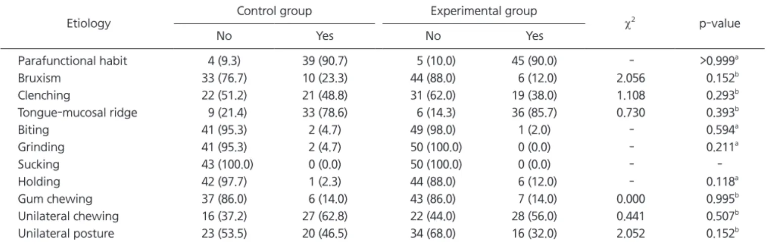
Table 3. Etiology of temporomandibular disorders between control and experimental group
Etiology Control group Experimental group
χ
2p-value
No Yes No Yes
Parafunctional habit 4 (9.3) 39 (90.7) 5 (10.0) 45 (90.0) - >0.999
aBruxism 33 (76.7) 10 (23.3) 44 (88.0) 6 (12.0) 2.056 0.152
bClenching 22 (51.2) 21 (48.8) 31 (62.0) 19 (38.0) 1.108 0.293
bTongue-mucosal ridge 9 (21.4) 33 (78.6) 6 (14.3) 36 (85.7) 0.730 0.393
bBiting 41 (95.3) 2 (4.7) 49 (98.0) 1 (2.0) - 0.594
aGrinding 41 (95.3) 2 (4.7) 50 (100.0) 0 (0.0) - 0.211
aSucking 43 (100.0) 0 (0.0) 50 (100.0) 0 (0.0) - -
Holding 42 (97.7) 1 (2.3) 44 (88.0) 6 (12.0) - 0.118
aGum chewing 37 (86.0) 6 (14.0) 43 (86.0) 7 (14.0) 0.000 0.995
bUnilateral chewing 16 (37.2) 27 (62.8) 22 (44.0) 28 (56.0) 0.441 0.507
bUnilateral posture 23 (53.5) 20 (46.5) 34 (68.0) 16 (32.0) 2.052 0.152
bValues are presented as number (%).
a
By Fisher’ s exact test.
bBy χ
2-test.
question 1 (Are you suffering from inflammation or pain in other joints?) than the patients in the control group. Over 50% of the patients in both the experimental and control groups responded yes to question 4 (Table 2).
3) Etiology analysis
In comparison of parafunctional habits between the ex- perimental and control groups, over 90% of the patients in both groups responded that they had at least one para- functional habit. Fifty six percents of the patients in the ex- perimental group and 62.8% of the patients in the control group had a unilateral chewing habit. In the case of tougue- mucosal ridge, 85.7% of the patients in the experimental group and 78.6% of the patients in the control group were observed. Over 50% of the patients in both groups had a
unilateral chewing habit and tongue-mucosal ridging but there was no significant difference in statistics (Table 3).
2. Analysis of CBCT Images in the Experimental Group 1) Based on the analysis of CBCT images of condylar bone during their first visit, 41.3% of the patients were as- signed to the normal group and 32.6% of patients were as- signed to the proliferative bony change group (Table 4).
2) From the result of analysis on the distribution of bony pattern during the patient’s first visit in relation to dura- tion of TMD, 85.7% of erosive bony change group were acute TMD patients and 66.7% of proliferative bony change group were chronic TMD patients but it was statistically in- significant (p=0.893; Table 5).
Table 4.
Table 4. Cone-beam computed tomography findings of condylar bony changes at the first visit in experimental groups
Group No. of patients (%)
Group I 19 (41.3)
Group II 7 (15.2)
Group III 15 (32.6)
Group IV 5 (10.9)
Total 46 (100)
Group I, normal; Group II, erosive bony changes; Group III, pro- liferative bony changes; Group IV, combined group=erosive bony changes+proliferative bony changes.
Table 5.
Table 5. Distribution of bony patterns in acute and chronic group of temporomandibular disorders
Bone pattern Acute Chronic Total
Group I 7 (36.8) 12 (63.2) 19 (100)
Group II 6 (85.7) 1 (14.3) 7 (100)
Group III 5 (33.3) 10 (66.7) 15 (100)
Group IV 2 (40.0) 3 (60.0) 5 (100)
Group I, normal; Group II, erosive bony changes; Group III, pro- liferative bony changes; Group IV, combined group=erosive bony change+roliferative bony changes.
Values are presented as number (%).
Statistical methods were linear by linear association (p=0.893).
Table 6.
Table 6. Bone pattern distribution by duration of rheumatoid arthritis
Bone pattern <5 y 5-9 y 10-15 y >15 y
Group I 13/28 (46.4) 4/12 (33.3) 1/2 (50.0) 1/4 (25.0)
Group II 6/28 (21.4) 0 1/2 (50.0) 0
Group III 9/28 (32.1) 4/12 (33.3) 0 2/4 (50.0)
Group IV 0 4/12 (33.3) 0 1/4 (25.0)
Total 28/46 (60.9) 12/46 (26.1) 2/46 (4.3) 4/46 (8.7)
Group I, normal; Group II, erosive bony changes; Group III, proliferative bony changes; Group IV, combined group=erosive bony change+
roliferative bony changes.
Values are presented as number (%).
Statistical methods were linear by linear association (p=0.106).
Table 7.
Table 7. The comparison between unilateral and bilateral bony change in temporomandibular joint
Comparison item Unilateral Bilateral Normal Total p-value
No. of patients 11 (47.8) 8 (34.8) 4 (17.4) 23 (100) 0.200
aRA duration (mo) 53.41±75.69 83.28±113.93 26.00±28.25 59.29±85.69 0.455
bRA, rheumatoid arthritis.
Values are presented as number (%) or mean±standard deviation.
a
By one sample chi-square test.
bBy Kruskal-Wallis test.
From the result of analysis on the bone pattern distribu- tion in relation to duration of RA, patients suffering for less than 5 years of RA were the most frequently seen in all groups except the combined group but it was statistically insignificant (p=0.106; Table 6).
3) From the result of an analysis of bony change in TMJ by one sample chi-square test (p=0.200), there was no dif- ference in ratio between unilaterally and bilaterally affected patients. More patients had unilateral bony change (47.8%) than bilateral bony change (34.8%). The patients with bilat- eral bony change had a longer duration of suffering RA but this was statistically insignificant (Table 7).
3. The Analysis of Efficacy of Conservative Treatment and Occlusal Stabilization Splint Therapy
1) The results of conservative treatment for 3 months (1) There was no significant difference in MCO measure- ment between the first visit and the visit 3 months later.
However, pain (p<0.001), noise (p=0.021), LOM (p<0.001) score in NRS improved significantly after 3 months (Table 8).
(2) In comparison between conservative treatment with- out occlusal stabilization splint (physical therapy, medica- tion, and behavioral therapy) and conservative treatment with occlusal stabilization splint (physical therapy, medi- cation, behaviour therapy, and occlusal stabilization splint therapy), both methods showed symptom improvement but there was no significant difference according to treatment method (Table 9).
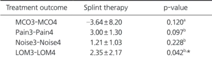
2) Efficacy of occlusal stabilization splint therapy for 3 months
There was no significant difference in MCO measure- ment, pain, noise score between the first visit and the visit 3 months later, but only LOM score improved significantly (Table 10).
DISCUSSION
In a joint affected by RA, synovial cells are differentiated into fibroblast and fibroblast-like cells and inflammatory destruction of cartilage arises and leads to bone erosion, typically at the junction of bone and cartilage. 19) This bone erosion begins within 2-3 years after disease onset and pro- gresses rapidly. 20,21) On the other hand, osteoarthritis on TMJ occurs from the destruction of joint tissue when overload- ing exceeds adaptive capacity of the joint and progresses slowly. A few biochemical materials such as interleukin 1β,
Table 8.
Table 8. Difference of MCO and NRS between the first visit and 3 months visit for pain, LOM by conservative treatment
Treatment outcome Conservative treatment p-value
MCO1-MCO2 –2.61±8.58 0.214
aPain1-Pain2 3.36±1.80 <0.001
b,***
Noise1-Noise2 1.11±1.85 0.021
b,*
LOM1-LOM2 2.36±1.92 <0.001
b,***
MCO, maximum comfortable opening; NRS, numerical rating scale (range 0-10); LOM, limitation of motion.
MCO1: MCO (mm) at the first visit; MCO2: MCO (mm) after 3 months treatment; Pain1, Noise1, and LOM1: NRS at the first visit; Pain2, Noise2, and LOM2: NRS after 3 months conservative treatment.
Values are presented as mean±standard deviation.
a
By paired t-test.
bBy Wilcoxon signed rank test.
*p<0.05, ***p<0.001.
Table 9.
Table 9. Difference of MCO and NRS between the first visit and 3 months visit according to different treatment modalities
Treatment outcome PT/M (n=5) PT/M+SP (n=13) p-value MCO1-MCO2 –3.20±7.98 –2.38±9.10 0.863
aPain1-Pain2 3.20±2.17 3.42±1.73 0.822
aNoise1-Noise2 0.70±2.39 1.27±1.69 0.468
bLOM1-LOM2 2.60±2.79 2.27±1.62 0.755
aMCO, maximum comfortable opening; NRS, numerical rating scale (range 0-10); PT/M, physical therapy+medication; PT/M+SP, physical therapy+medication+splint therapy; LOM, limitation of motion.
MCO1: MCO (mm) at the first visit; MCO2: MCO (mm) after 3 months treatment; Pain1, Noise1, and LOM1: NRS at the first visit; Pain2, Noise2, and LOM2: NRS after 3 months treatment.
Values are presented as mean±standard deviation.
a
By independent t-test.
bBy Mann-Whitney U test.
Table 10
Table 10. Difference of MCO and NRS between the first visit and 3 months visit for pain, LOM by splint treatment
Treatment outcome Splint therapy p-value
MCO3-MCO4 –3.64±8.20 0.120
aPain3-Pain4 3.00±1.30 0.097
bNoise3-Noise4 1.21±1.03 0.228
bLOM3-LOM4 2.35±2.17 0.042
b,*
MCO, maximum comfortable opening; NRS, numerical rating scale (range 0-10); LOM, limitation of motion.
MCO3: MCO (mm) at the splint delivery; MCO4: MCO (mm) after 3 months splint treatment; Pain3, Noise3, and LOM3: NRS at the splint delivery; Pain4, Noise4, and LOM4: NRS after 3 months splint treatment.
Values are presented as mean±standard deviation.
a