Dysesthesia after Tooth Extraction and Implant Surgery Reported by Dentists
Ji-Won Ryu
1, D.D.S.,M.S.D., Jeong-Seung Kwon
2, D.D.S.,M.S.D.
Department of Oral Medicine, College of Dentistry, Chosun University1 Department of Oral Medicine, College of Dentistry, Yonsei University2
The purpose of this study was to analyze the nerve damage after tooth extraction and implant surgery, and to establish a predictive model for assessment and management of dysesthesia.
In this questionnaire study, the subjects chosen for this study were 276 dentists who answered the questionnaire about dysesthesia after tooth extraction and implant surgery. The analysis of the results consist of the sex and age distribution, affected site, associated symptoms, rate and duration of the recovery.
The results are summarized as follows. :
1. There were no significant difference between the sex and the dysesthesia.
2. The most common affected site was the mandibular region. In the group of the implant surgery, 100% affected the mandibular site. The tooth extraction group was 93.2% affected.
3. Pain was one of the most associated symptom with dysesthesia-46.5% of the tooth extraction and 44.8% of the implant surgery.
4. The recovery ratio was 72.3% in the tooth extraction, 71.8% in the implant surgery. Most of them, they recovered in 1~6 months.
In conclusion, most of dysesthesia may be recovered within 1 year. However, the possibility of persistent dysesthesia should not be neglected. Therefore, practitioners must discuss the possibility of nerve injury with their patients, and include this possibility in the consent forms. Various methods of monitoring recovery of sensation should be considered for objective assessment of prognosis. In addition, immediate referral to orofacial pain specialists can offer the patients an opportunity for more effective and noninvasive treatments.
Key Words : Dysesthesia, Implant surgery, Nerve injury, Tooth extraction
Corresponding Author : Jeong-Seung Kwon
Department of Oral Medicine, College of Dentistry, Yonsei University, 134 Shinchon-Dong, Seodaemun-Ku, Seoul 120-752, Korea
Tel: 82-2-2228-8880 FAX: 82-2-393-5673 E-mail: jskwon@yuhs.ac Received: 2007-06-02 Accepted: 2007-08-10
I. INTRODUCTION
Nerve damage can affect a single nerve or several
nerves and result in sensory, motor, autonomic
deficits in the affected region. Causes of cranial
nerve damage can be classified as intracranial or
extracranial. Intracranial causes include stroke,
transient ischemic attack and tumors. Extracranial
causes can include trauma, malignancy and
infection. In addition, there may be iatrogenic causes
such as radiotherapy, chemotherapy, mandibular surgery and dental treatment.
Reported dental causes of dysesthesia include third molar extractions, surgical molar/premolar extractions, implant surgery, needle trauma following block anesthesia, periapical inflammation, denture compression of the nerve and neural injury following endodontic treatment.
Dysesthesia is defined as an unpleasant abnormal sensation, whether spontaneous or evoked. A dysesthesia should always be unpleasant and a paresthesia not, although difficulties can emerge when a patient must decide whether a sensation is pleasant or unpleasant. This altered perception may become manifest by the impairment of sensation of the nerve. Sometimes just the sensation of pain is disturbed(hyperalgesia, hypoalgesia or analgesia), while in other cases the tactile and temperature senses are affected simultaneously. All these changes can be transient or persistent depending on the degree of the irritation of the nerve.
1)Injury of the lingual or the inferior alveolar nerves sustained during removal of lower third molars in among the most common causes of litigation in dentistry,
2)because the lingual nerve and inferior alveolar nerve(IAN) are at risk because of close anatomic relationships.
3-5)However, advances in dental implant technology and studies documenting predictable long-term success have led to an interest in this tooth-replacement option over the last 10 years.
Reports in the literature on the incidence of such complications have been received with deep concern.
6-9)In Ellies LG and Hawker PB study,
7)the prevalence of dysesthesia following mandibular implant surgery did not significantly differ from that found in the Toronto study,
6)suggesting that nerve injuries during implant placement occur at a similar frequency regardless of the type of implant being placed or of the operating surgeon, and of the type of initial incision line.
There was a study about the incidence of
al.
10)studied the type of medical accident, and the questionnaire study subjected to 1,133 dentists who had been registered in the Seoul Dental association in 1997. In the medical accident cases, the number of the accidents associated with tooth extraction was 704(15.76%), and 107 cases(15.20% of 704 cases) was the experience of dysesthesia after wisdom tooth removal.
Therefore, the number of malpractice suits related to implant surgery and tooth extraction has increased significantly awards among the highest in dentistry. Dysesthesia following implant surgery or tooth extraction may result in liability claims. As a result, it seems prudent to review these cases to better understand the causes and characterization of such actions to prevent complications and reduce future litigation.
The purposes of this study were to analyze the nerve damage after tooth extraction or implant surgery evaluated by dentists, through questionnaire, to establish a predictive model for assessment and management of dysesthesia.
Ⅱ. MATERIALS AND METHODS 1. Subjects
The subjects for the study were 276 dentists who attended implant seminars from June to August, 2004.
2. Method
Questionnaires were delivered to dentists who attended implant seminars directly and returned at that time. Returned questionnaires were 276 pieces.
The questionnaire consisted of 16 question(Table 1),
which questions related to tooth extraction and
implant surgery were 8 each, and they were
answered independently. The respondents were
grouped according to the following 2 yes/no type
questions : 1) "Do/Did you have patients with
dysesthesia after tooth extraction?" If yes, they
1. Have you ever experienced any patient presenting numbness or dysesthesia after tooth extraction ?
① Yes ② No
2. If yes, which tooth was it?
(It is possible to answer the questions plurally) ① Upper anterior tooth
② Lower anterior tooth ③ Upper posterior tooth ④ Lower posterior tooth ⑤ Lower lip
⑥ Chin area (symphysis) ⑦ the others
3. Have you ever experienced any symptom associated with the dysesthesia?
① Yes ② No
4. If yes, can you describe the symptom(s)?
(For example, tingling on touch, unpleasant feeling, spilling food while eating)
( )
5. Was the numbness or dysesthesia recovered?
① Yes ② No
6. If yes, how long did it take to recover?
( ) months
7. Have you performed any medication or treatment for the dysesthesia?
① Yes ② No
If yes, please describe the medication or the treatment(s) you had performed specifically
( )
8. Please write down the sex and age of the patient(s) presenting the above symptom.
(It is possible to answer the questions plurally) (Male, Female) ( ) year-old.
1. Have you ever experienced any patient presenting numbness or dysesthesia after implant surgery ?
① Yes ② No
2. If yes, which tooth was it?
(It is possible to answer the questions plurally) ① Upper anterior tooth
② Lower anterior tooth ③ Upper posterior tooth ④ Lower posterior tooth ⑤ Lower lip
⑥ Chin area (symphysis) ⑦ the others
3. Have you ever experienced any symptom associated with the dysesthesia?
① Yes ② No
4. If yes, can you describe the symptom(s)?
(For example, tingling on touch, unpleasant feeling, spilling food while eating)
( )
5. Was the numbness or dysesthesia recovered?
① Yes ② No
6. If yes, how long did it take to recover?
( ) months
7. Have you performed any medication or treatment for the dysesthesia?
① Yes ② No
If yes, please describe the medication or the treatment(s) you had performed specifically
( )
8. Please write down the sex and age of the patient(s) presenting the above symptom.
(It is possible to answer the questions plurally) (Male, Female) ( ) year-old.
How many years have you been in dental practice? ( ) years
Thank you for your cooperation!
Table 1. The questionnaire about trigeminal dysesthesia after tooth extraction and implant surgery
2) "Do/Did you have patients with dysesthesia after implant surgery?". If yes, they were grouped as dysesthesia after implant surgery. The combination of the two groups might be possible. In this study, only "yes" group were analysed, except for one question about dentists' clinical experience.
Ⅲ. RESULTS 1. The analysis of the respondents
276 dentists answered the questionnaire, among them, 141(51.1%) responded to the question of the years of clinical experience(Table 2). The mean year was 10.48(SD=2.80, range=2-33 years). 135 respondents who did not answer to this question were excluded from the analysis of the respondents.
From this questionnaire, 108 dentists(39.1%) had dysesthetic patients after tooth extraction, and 68(24.6%) dentists answered that they had dysesthetic patients after implant surgery.
2. The analysis of dysesthesia after tooth extraction
1) Sex distribution of the patients
Out of 115 answers(including plural answers), there were 23 males and 38 females. The plural answers were 7. There were 54 unknown group which respondent did not describe their patients' information. Male:Female ratio was about 3:5.
Clinical experience(year) Number of dentists (%) 0~ 5
6~10 11~15 16~20 21~25 26~30 31~35
41 (29.1) 35 (24.8) 34 (24.1) 18 (12.8) 8 (5.7) 4 (2.8) 1 (0.7) Table 2. Distribution of clinical experience in questionnaire
respondents
2) Age distribution of the patients
Out of 116 answers(including plural answers), the mean age was 37.92(SD=10.53, range=15-65 years).
The 51 unknown cases in which did not dictate patients' age were excluded.
3) Site distribution of dysesthesia
Out of the 117 answers(including plural answers), the major site of dysesthesia was mandibular molar region, 82 cases(70.1%) out of 117. Lower lip was affected in 20 cases(17.1%), tongue was 2(1.7%), and symphysis was 1(0.9%). The unknown group in which the respondents did not describe the affected site was 5(4.2%)(Table 3).
4) Associated symptoms
Out of 108 cases , there were 58 cases(53.7%) that showed associated symptom. Table 4 shows various types of associated symptoms after tooth extraction.
The major symptom was pain, 33 cases(46.5%) out of 71(including plural answers). 20 cases (28.2%) were reported that discomfort was accompanied with dysesthesia. Loss of taste was 1 case(1.4%).
Motor dysfunction, (for example, frequent biting of lower lip or tongue, drooling, having pieces of foods remaining on the lip without feeling them, difficulty in pronouncing certain words) were reported in 17 cases(23.9%).
5) Rate and duration of recovery
Out of the 112 answers(including plural answers),
Site Number of patients (%) Maxillary anterior
Maxillary molar Mandibular anterior Mandibular molar Lower lip Tongue Symphysis
1 (0.9) 2 (1.7) 4 (3.4) 82 (70.1) 20 (17.1) 2 (1.7) 1 (0.9) Table 3. Affected oral site distribution of dysesthesia after tooth
extraction
81(72.3%) cases had recovered. No recovery was reported in 16 cases(14.3%). The mean duration of the recovery was 3.6 months(SD=2.51, range=0.5-11 months). In the recovery group, most cases recovered within 1-6 months, 42 cases(90.1%) out of 81(Table 5).
6) Recommended treatment
Out of 108 cases, 39(36.1%) dentists tried some treatment for their patients, whereas 60(55.5%) dentists did not give any treatment. Table 6 shows various type
Symptom Number of patients (%) Pain
Discomfort Loss of taste Motor dysfunction
33 (46.5) 20 (28.2) 1 (1.4) 17 (23.9) Table 4. Associated symptoms
Recovery Duration(month) Number of patients
Yes
0-1 1-3 4-6 7-9 10-12
12-
6 42 25 4 4 -
No recovery 16
Unknown 15
Table 5. Rate and duration of recovery
Prescription or treatment Number of patients
Yes
medication physical therapy refer to another clinics no answer
37 12 2 5
No 60
Unknown 9
Table 6. Recommended treatment
of treatments which the respondent offered to their dysesthetic patients. Out of 125 answers(including plural answers), the most recommended treatment was medication, 37 cases. Out of 37 cases, steroid was recommended in 14 cases(37.8%), and vitamin was recommended in 16 cases(43.2%). In 39 cases which were grouped as "treatment" group, 5 cases with no answer were excluded.
3. The analysis of dysesthesia after implant surgery
1) Sex distribution of the patients
Out of 71 answers(including plural answers), there were 14 males and 14 females. The plural answers were 3. There were 43 unknown group which respondent did not describe their patients' information. Male:Female ratio was about 1:1.
2) Age distribution of the patients
Out of 71 answers(including plural answers), the mean age was 50.31(SD=17.50, range=25-65 years).
The 39 unknown group in which did not dictate patients' age was excluded.
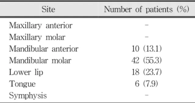
3) Site of dysesthesia
Of the 76 answers(including plural answers), the major site of dysesthesia was mandibular molar region, 42 cases(55.3%) out of 76 cases. Lower lip was affected in 18 cases(23.7%), mandibular anterior tooth region was 10 case(13.1%) and tongue was 6 cases(7.9%)(Table 7).
Site Number of patients (%) Maxillary anterior
Maxillary molar Mandibular anterior Mandibular molar Lower lip Tongue Symphysis
- - 10 (13.1) 42 (55.3) 18 (23.7) 6 (7.9)
-
Table 7. Affected Oral site distribution of dysesthesia after implant surgery
4) Associated symptoms
Out of 68 cases, there were 46(67.6%) cases that showed associated symptom. The major symptom was pain, 30 cases(44.8%) out of 67(including plural answers). 19 cases(28.3%) were reported that discomfort was accompanied with dysesthesia. In 17 cases(25,4%), motor dysfunction was reported.
Unknown 1 cases in which did not answer the question was excluded(Table 8).
5) Rate and duration of recovery
Out of the 71 answers(including plural answers), 51 cases(71.8%) had recovered. No recovery was reported in 18 cases(25.4%). The mean duration of the recovery was 4.01 months(SD=2.87, range=
0.5-12 months). In the recovery group, most cases recovered within 1-6 months, 45 cases(88.2%) out of 51(Table 9).
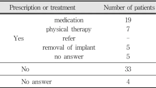
6) Recommended treatment
Out of 68 cases, 31(45.6%) dentists tried some treatment for their patients, however, 33(48.5%) dentists did not give any treatment. Table 10 shows
Symptom Number of patients (%) Pain
Discomfort motor dysfunction Unknown
30(44.8) 19(28.3) 17(25.4) 1(1.5) Table 8. Associated symptoms
Recovery Duration(month) Number of patients
Yes
0-1 1-3 4-6 7-9 10-12
12-
4 29 12 2 3 1
No 18
Table 9. Rate and duration of recovery
the various type of treatments which the respondents offered to their dysesthetic patients.
Out of the 73 answers(including plural answers), the most recommended treatment was medication, 19 cases. Out of 19 cases, anticonvulsant(gabapentin) were recommended in 5 cases(26.3%), and steroid was in 4 cases(21.1%), and vitamin was in 5 cases(26.3%). In the 31 cases which was grouped as
"treatment" group, 5 cases with no answer were excluded.
Ⅳ. DISCUSSION
Dysesthesia is well known complication of dental and maxillofacial surgery and has been well documented in the long term evaluation of patients after maxillofacial trauma, third molar and orthognathic surgery, vestibuloplasty, and ridge augmentation.
11-15)The cause of dysesthesia in dentistry can be systemic or local factors. Systemic factors include demyelinating diseases(eg, multiple sclerosis), viral infections, metabolic disorders(eg, diabetes and hypothyroidism), hypovitaminosis, Paget's disease, syphilis, transitory ischemic attack, cerebral strokes, neural neoplasia, metastatic malignancies, and some systemic drug reactions.
16-22)Local factors include traumatic injuries such as mandibular fractures, expanding compressive lesions(benign or malignant neoplasia and cysts), impacted teeth, local infections(osteomyelitis, periapical, and peri-implant infections), iatrogenic
Prescription or treatment Number of patients
Yes
medication physical therapy
refer removal of implant
no answer
19 7 - 5 5
No 33
Table 10. Recommended treatment
lesions after tooth extraction, anesthetic injection, endodontic therapy (overfilling and apical surgery), operative treatment,
23)implant surgery, orthodontic surgery, and prosthetic surgery.
Thus, various dental treatments can be a cause of dysesthesia, however, a lot of literature have shown that dysesthesia after tooth extraction and implant surgery is quite frequent.
17,18,20-22,24)In the aspects of dysesthesia after tooth extraction, a lot of studies had focused on the relationship between third molar extraction and inferior alveolar and lingual nerve damage. The incidence of inferior alveolar nerve damage after lower third molar extraction ranges from 0.5%
25)to 8%,
26,27)approximately; the usual rate is about 5%.
13,25,28,29)Van Steenberghe et al.
30)reported a multicenter study of partially edentulous patients who were treated with implants, in which 17% of the patients experienced an altered sensation of the lower lip after implant surgery. Kiyak et al.
31)reported that preoperatively 4% of the patients expect some form of sensory disturbance. Ellies and Hawker
7)reported on a retrospective analysis of a multicenter study which took place in Toronto(Canada) and Adelaide(Australia). Two weeks after implant surgery they found altered sensation of the inferior alveolar nerve in 37% and 36% of patients respectively. In both centers, these complaints were persistent in 13% of the patients.
In this study, the incidence of dysesthesia after tooth extraction was 39.1% and the incidence of the dysesthesia after implant surgery was 24.6%. In addition, the incidence of permanent dysesthesia out of the dysesthetic cases was 14.3% after tooth extraction and 25.4% after implant surgery, respectively. Compared with the reviewed study above, the result of our study may be interesting values. To discuss with true incidence, the study should be directed to the patients, but the subjects of our study were dentists. Therefore, the incidence of our study was deduced indirectly from the dental practitioners. For that reason, the revised study should be needed.
The male:female sex ratio was about 3:5, in the cases of tooth extraction, and 1:1, in the cases of implant surgery. From the ratio of the tooth extraction, Similar sex ratios have been found in some studies: 2:3,
32)2:5,
33)and 19:35.
13)In this connection, it is of interest that Howe and Poyton found that the impacted third molar was frequently closer to the mandibular canal on radiographs in women than in men in their study of 1,355 third molar extraction cases. The prediliction of female can be explained by the anatomical gap and sex hormones.
34)However, many studies shows that there were no significant difference between the sex and the dysesthesia.
In most of the cases, between the tooth extraction and implant surgery, the affected site was mandibular region: the innervation of inferior alveolar nerve. In the group of the implant surgery, 100% affected the mandibular site. The extraction group was also 93.2% affected. This result was explained by close anatomic relationships.
3-5)In both of them, Pain was one of the most associated symptom with dysesthesia: 46.5% in the tooth extraction and 44.8% in the implant surgery.
This result was in accordance with that of the Sandstedt and Sörensen.
2)It is well established that the oral and perioral regions are among the most sensitive areas in the human body.
35)Considering this fact, it is not difficult to understand why minor oral nerve damage can be a major handicap for the individual. For many practitioners, it is commonly believed that nerve damage will be spontaneously recovered over time. However, complications and delayed recovery can cause demoralizing and disappointing, leading to loss of confidence in the clinician even if there would be happened only in a minority of the patients. Occasionally, a patient interprets them as negligence and malpractice with subsequent filing of a legal claim against the treating dentist.
36)The number of malpractice suits related to
implants has increased significantly, with awards
that are among the largest in dentistry.
37)Some
patients may even claim damage where there is
none, purely for financial gain because symptoms of neurosensory loss are largely subjective and based on what the patient states.
Therefore, practitioners must discuss the possibility of nerve injury with their patients and include this possibility in the consent forms. If dysethesia occurs after the procedure, the clinician must monitor recovery of sensation with various methods including not only a simple assessment but also light touch sensation, 2-point discrimination, brush stroke direction, thermal stimulation, pin prick/pain sensation, and pain-pressure threshold,
12,38-42)
for objective assesment of prognosis.
Ⅴ. CONCLUSION
Most of dysesthesia may be recovered within 1 year. However, the possibility of persistent dysesthesia should not be neglected. Therefore, practitioners must discuss the possibility of nerve injury with their patients, and include this possibility in the consent forms. Various methods of monitoring recovery of sensation should be considered for objective assessment of prognosis. In addition, immediate referral to orofacial pain specialists can offer the patients an opportunity for more effective and noninvasive treatments.
REFERENCES
1. Merrill RG. Prevention, treatment, and prognosis for nerve injury related to the difficult impaction. Dent Clin North Am 1979;23:471-88.
2. Sandstedt P, Sörensen S. Neurosensory disturbances of the trigeminal nerve: a long-term follow-up of traumatic injuries. J Oral Maxillofac Surg 1995;53:
498-505.
3. Kiesselbach J, Chamberlain J. Clinical and anatomic observations on the relationship of the lingual nerve to the mandibular third molar region. J Oral Maxillofac Surg 1984;42:565-567.
4. Pogrel MA, Renaut A, Schmidt B, Ammar A. The relationship of the lingual nerve to the mandibular third molar region: an anatomic study. J Oral Maxillofac Surg 1995;53:1178-1181.
Assessment of the lingual nerve in the third molar region using magnetic resonance imaging. J Oral Maxillofac Surg 1997;55:134-137.
6. Ellies LG. Altered sensation following mandibular implant surgery: a retrospective study. J Prosthet Dent 1992;68:664-671.
7. Ellies LG, Hawker PB. The prevalence of altered sensation associated with implant surgery. Int J Oral Maxillofac Implants 1993;8:674-679.
8. Wismeijer D, van Waas MA, Vermeeren JI, Kalk W.
Patients' perception of sensory disturbances of the mental nerve before and after implant surgery: a prospective study of 110 patients. Br J Oral Maxillofac Surg 1997;35:254-259.
9. Bartling R, Freeman K, Kraut RA. The incidence of altered sensation of the mental nerve after mandibular implant placement. J Oral Maxillofac Surg 1999;57:
1408-1412.
10. Kim JH, Choi JH, Kim CY. A study on the types of the medical accidents and the counterplan of the dentists in Seoul. Korean J Oral Med 1998;23:157-191.
11. De Koomen HA. A prosthetic view on vestibuloplasty with free mucosal graft. Int J Oral Surg 1977;6:
388-411.
12. Baily PH, Bays RA. Evaluation of long term sensory changes following mandibular augmentation proce- dures. J Oral Maxillofac Surg 1984;42:722-727.
13. Kipp DP, Gildstein BH, Weiss WW. Dysesthesia after mandibular third molar surgery. A retrospective study and analysis of 1377 surgical procedures. J Am Dent Assoc 1980;100:185-192.
14. Simpson HE. Injuries to the inferior dental and mental nerves. J Oral Surg 1958;16:300-305.
15. Nickel JJ. A retrospective study of paraesthesia of the dental alveolar nerves. Anesth Prog 1990;37:42-45.
16. Morse DR. Infection-related mental and inferior alveolar nerve paresthesia: literature review and presentation of two cases. J Endod 1997;23:457-460.
17. Antrim DD, Linda L. Paresthesia of the inferior alveolar nerve caused by periapical pathology. J Endod 1978;4:220-221.
18. Cohenca N, Rotstein I. Mental nerve paresthesia associated with a non-vital tooth. Endod Dent Traumatol 1996;12:298-300.
19. Joubert JJ, Farman AG, Nortjé CJ. Lip paresthesia of dental origin. J Oral Med 1979;34:26-27.
20. Gilbert BO, Dickerson AW II. Paresthesia of the mental nerve after an acute exacerbation of chronic
588-590.
21. Glassmann GD. Flare-up with associated paresthesia of a mandibular second premolar with three root canals. Oral Surg Oral Med Oral Pathol 1987;64:
110-113.
22. Lambrianidis T, Molyvdas J. Paresthesia of the inferior alveolar nerve caused by periodontal-endodontic pathosis. Oral Surg Oral Med Oral Pathol 1987;63:
90-92.
23. Osvaldo Zmener. Mental Nerve Paresthesia Associated with an adhesive resin restoration-a case report. J of Endodontics 2004;30:117-119.
24. Morse DR. Endodontic-related inferior alveolar nerve and mental foramen paresthesia. Comp Cont Ed Dent 1997;18:963-987.
25. Sisk AL, Hammer WB, Shelton DW, Joy ED Jr.
Complications following removal of impacted third molars: the role of the experience of the surgeon. J Oral Maxillofac Surg 1986;44:855-859.
26. Rood JP. Permanent damage to inferior alveolar and lingual nerves during the removal of impacted mandibular third molars. Comparison of two methods of bone removal. Br Dent J 1992;172:108-110.
27. Bruce RA, Frederickson GC, Small GS. Age of patients and morbidity associated with mandibular third molar surgery. J Am Dent Assoc 1980;101:240-245.
28. Carmichael FA, McGowan DA. Incidence of nerve damage following third molar removal. A West of Scotland Oral Surgery Research Group study. Br J Oral Maxillofac Surg 1992;30:78-82.
29. Rood JP. Lingual split technique. Damage to inferior alveolar and lingual nerves during removal of impacted mandibular third molars. Br Dent J 1983;154:402-403.
30. Van Steenberghe D, Lekhol U, Bolender C et al..
Applicability of osseointegrated oral implants in the rehabilitation of partial edentulism: a prospective multicenter study on 558 fixtures. Int J Oral Maxillofac Implants 1990;5:272-281.
31. Kiyak HA, Beach BH, Worthington P, Taylor T, Bolender C, Evans J. Psychological impact of osseointegrated dental implants. Int J Oral Maxillofac Implants 1990;5:61-69.
32. Howe GL, Poyton HG. Prevention of damage to the inferior dental nerve during the extraction of mandibular third molars. Br Dent J 1960;109:335-336.
33. Ferdousi AM, MacGregor AJ. The response of the peripheral branches of the trigeminal nerve to trauma.
Int J Oral Surg 1985;14:41-46.
34. Graff-Radford SB, Evans RW. Lingual nerve injury.
Headache 2003;43:975-983.
35. Waiters H. Reducing lingual nerve damage in third molar surgery: A clinical audit of 1350 cases. Br Dent J 1995;178:140-144.
36. Smith RS. Long-term complications of osseointegrated implants. In Kaban BL, Pogrel MA, Perrot HD(Eds).
Complications in Oral and Maxillofacial Surgery.
Philadelphia. 1997, Saunders, pp. 319-358.
37. Palat M. Assessing legal responsibility for implant failure. Compendium 1991;12:295-296.
38. Campbell RL, Shamaskin RG, Harkins SW.
Assessment of recovery from injury to inferior alveolar and mental nerves. Oral Surg Oral Med Oral Pathol 1987;64:519-526.
39. Van Sickels JE, Zysset M, Nishioka GJ, Thrash WJ. A comparative study of normal sensibility of the inferior alveolar nerve and the infraorbital nerve. Oral Surg Oral Med Oral Pathol 1989;67:255-257.
40. Omer GE. Methods of assessment of injury and recovery of peripheral nerves. Surg Clin North Am 1981;61:303-319.
41. Robinson PP, Smith KG, Johnson FP, Coppins DA.
Equipment and methods for simple sensory testing. Br J Oral Maxillofac Surg 1992;30:387-389.
42. Davenport JC. Pressure-pain thresholds in the oral cavity in man. Arch Oral Biol 1969;14:1267-1274.
국문요약
치과의사에 의해 보고된 발치 및 임프란트 수술 후 지각이상에 대한 분석
조선대학교 치과대학 구강내과학교실1, 연세대학교 치과대학 구강내과학교실2