soft tissue sealing; an in vitro and in vivo study
Hyo-Jung Lee1,†, Jaden Lee2,8,†, Jung-Tae Lee1,3, Ji-Soo Hong4,9, Bum-Soon Lim5,9, Hee-Jung Park6, Young-Kwang Kim7, Tae-Il Kim8,9*
1Department of Periodontology, Section of Dentistry, Seoul National University Bundang Hospital, Seongnam, Korea
2Department of Oral Health Sciences, Medical University of South Carolina College of Dental Medicine, Charleston, SC, USA
3Department of Periodontics, Dankook University College of Dentistry Jukjeon Dental Hospital, Yongin, Korea
4Department of Oral Pathology, Seoul National University School of Dentistry, Seoul, Korea
5Department of Dental Biomaterials, Seoul National University School of Dentistry, Seoul, Korea.
6Department of Public Health Sciences, Korea University, Seoul, Korea
7Department of General Dentistry, Boston University School of Dental Medicine, Boston, MA, USA
8Department of Periodontology, Seoul National University School of Dentistry, Seoul, Korea
9Dental Research Institute, Seoul National University School of Dentistry, Seoul, Korea
Research Article
J Periodontal Implant Sci 2015;45:120-126 http://dx.doi.org/10.5051/jpis.2015.45.3.120
Purpose: With the significance of stable adhesion of alveolar bone and peri-implant soft tissue on the surface of titanium for successful dental implantation procedure, the purpose of this study was to apply microgrooves on the titanium surface and investigate their ef- fects on peri-implant cells and tissues.
Methods: Three types of commercially pure titanium discs were prepared; machined-sur- face discs (A), sandblasted, large-grit, acid-etched (SLA)-treated discs (B), SLA and micro- groove-formed discs (C). After surface topography of the discs was examined by confocal laser scanning electron microscopy, water contact angle and surface energy were mea- sured. Human gingival fibroblasts (hGFs) and murine osteoblastic cells (MC3T3-E1) were seeded onto the titanium discs for immunofluorescence assay of adhesion proteins. Com- mercially pure titanium implants with microgrooves on the coronal microthreads design were inserted into the edentulous mandible of beagle dogs. After 2 weeks and 6 weeks of implant insertion, the animal subjects were euthanized to confirm peri-implant tissue heal- ing pattern in histologic specimens.
Results: Group C presented the lowest water contact angle (62.89±5.66 θ), highest surface energy (45±1.2 mN/m), and highest surface roughness (Ra=22.351±2.766 μm). The ex- pression of adhesion molecules of hGFs and MC3T30E1 cells was prominent in group C. Ti- tanium implants with microgrooves on the coronal portion showed firm adhesion to peri- implant soft tissue.
Conclusions: Microgrooves on the titanium surface promoted the adhesion of gingival fi- broblasts and osteoblastic cells, as well as favorable peri-implant soft tissue sealing.
Keywords: Cell adhesion, Dental implants, Titanium, Wound healing.
Received: Mar. 20, 2015 Accepted: May 25, 2015
*Correspondence:
Tae-Il Kim
Department of Periodontology, Seoul National University School of Dentistry, 101 Daehak-ro, Jongno-gu, Seoul 110-749, Korea
Email: [email protected] Tel: +82-2-2072-2642 Fax: +82-2-744-1349
†Hyo-Jung Lee and Jaden Lee contributed equally to this study.
INTRODUCTION
Titanium is used as a reliable biomaterial through dental implant procedure to treat eden- tulous areas. Osseointegration, a direct structural and functional binding reaction between bone and titanium, ensures stable dental implant fixation in the alveolar bone [1]. Crestal bone loss around titanium implant, however, has been reported with the presence of an in- flammatory infiltrate in the titanium implant-surrounding gingival soft tissue [2]. Since
This is an Open Access article distributed under the terms of the Creative Commons Attribution Non-Commercial License (http://creativecommons.org/licenses/by-nc/3.0/).
peri-implant soft tissue does not attach to the surface of titanium, it is vulnerable to bacterial invasion which often leads to peri-im- plant disease [3]. Therefore, tight soft tissue sealing around titanium implant is also regarded as important as osseointegration.
Several studies have been performed to promote peri-implant soft tissue attachment by physical and chemical modification of titanium surface. It was demonstrated that the soft tissue attachment was not influenced by the roughness of the titanium surface [4]. Howev- er, concave transmucosal-profiled titanium implants may promote connective tissue attachment around implants during the early healing phase [5]. In those studies, researchers modified the abut- ment or transmucosal area of titanium implants to investigate peri- implant soft tissue reactions.
As the actual morphology of alveolar ridge is not flat, it often oc- curs that not all surfaces of titanium implant fixtures are covered with the recipient bone in dental implant fixture installation proce- dure. In this context, successful outcome of dental implant treat- ment requires stable hard/soft tissue attachment to the titanium implant fixtures, while little has been known of the peri-implant soft tissue reaction on non-submerged coronal part of titanium implant fixtures.
In this study, we applied microgrooves on the titanium surface and investigated their effects on peri-implant soft tissue cells and tissues by in vitro and in vivo experiments.
MATERIALS AND METHODS
Titanium discs preparation
Commercially pure, grade 4 titanium discs (Dentium Co., Seoul, Korea) with 10 mm diameter and 2 mm thickness were divided into three groups as follows; machined-surface discs (A), sandblasted, large-grit, acid-etched (SLA)-treated discs (B), SLA-treated and mi- crogroove (width×depth, 200 μm×100 μm; distance between mi- crogrooves, 50 μm)-formed discs (C). All samples were washed in an ultrasonic bath by using distilled water and stored in 100% ethanol at room temperature.
Surface characterization
Surface roughness was estimated using laser scanning confocal microscope (Zeiss LSM 5 Pascal, Carl Zeiss Inc., Oberkochen, Germa- ny) with imaging software (Zeiss LSM Image Examiner, Carl Zeiss Inc.). The multi-argon laser emits light with a wavelength of 633 nm. This allows the calculation of the arithmetic mean of surface roughness from a mean plane in the sampling area (900×900×350 µm). To evaluate surface wettability, water contact angle was mea- sured by sessile drop method at room temperature. A video camera with an image analyzer (Phoenix 300, Surface Electro Optics, Seoul, Korea) visualized the shape of the drop and provided the contact angle. Three probe liquids of different polarities were used: 1-bro- monaphthalene, formamide, and deionized water. The volume of liquid drops was controlled using an instrument with a computer- ized interface and pictures of the drops were taken as soon as they
landed on the surfaces using a video camera. Right and left contact angles of each drop were automatically averaged to give one con- tact angle per drop. Then, the surface free energy was calculated using three contact angle values from three different probe liquids according to the van Oss model. Each experiment was repeated 5 times for 3 specimens of each experimental group. The surface en- ergy was calculated using the Owens–Wendt equation [6].
Immunofluorescence analysis
Human gingival fibroblasts (hGFs), purchased from ScienCell (Carlsbad, CA, USA), and murine osteoblastic MC3T3-E1 cells were seeded at 1×10⁴ cells/well in a 24-well plate. After 24 hours cell culture, the cells were washed with PBS and fixed using 4% para- formaldehyde solution. The fixed cells were permeabilized with buffered 0.3% Triton X-100 at room temperature. The samples were blocked with 1% bovine serum albumin (BSA) for 1 hour. Actin (Abcam, Cambridge, MA, USA) was applied as the primary antibody and then reacted with fluorescein isothiocyanate (FITC)-conjugated secondary antibodies (Sigma, St. Louis, MO, USA). The samples were then reacted with 4,6-diamidino-2-phenylindole (DAPI) (Sigma) for visualizing the nuclei. All the labeled cells were examined using the Confocal Laser Scanning Microscope (FV300, Olympus, Center Val- ley, PA, USA) equipped with FITC - and DAPI - channel filter systems (Olympus).
Animal surgery
Four adult male beagle dogs (average age, 2 years; average weight, 13 kg) received dental prophylaxis for sustaining healthy periodontal conditions. Animal experiments were carried out in ac- cordance with the Guidelines of the National Institute of Health (NIH) regarding the care and use of animals for experimental pro- cedures and with the protocols approved by the Institutional Ani- mal Care and Use Committee of Seoul National University (14-016- BA1402-147-007-01). Under general anesthesia by 2% xylazine hy- drochloride (Rumpun, Bayer Korea, Seoul, Korea) with tiletamine hydro-chloride (Zoletil, Virbac, Ltd., Carros, France) and local anes- thesia using 2% lidocaine hydrochloride with 1:100,000 epineph- rine (Lidocaine, Huons, Seoul, Korea), mandibular 2nd, 3rd, and 4th premolars on both sides were extracted. After 12 weeks of healing under the same anesthetic conditions as those of the tooth extrac- tion procedure, SLA-treated titanium implants (diameter×length;
3.1 mm×9mm), with microgrooves (width×depth, 200 μm×100 μm; distance between microgrooves, 50 μm) in the coronal portion, were installed according to the guideline of manufacturer (Den- tium, co, Seoul, Korea). The experiment was performed with a bal- anced block design in which different groups of implants were ran- domly placed in each extraction socket according to the Latin block experimental design; group A, implants installed with 1 mm expo- sure of coronal microgroove portion; group B, implants installed with 2 mm exposure of coronal microgroove portion (Fig. 1). After connecting healing abutments and suturing, antibiotics (Cefazolin, 30 mg/kg, Chong Kun Dang Pharmaceutical Co., Seoul, Korea) were
intramuscularly injected with soft diet feeding. After 2 and 6 weeks, the animals were euthanized for mandibular block sections.
Histologic specimen preparation
In preparation for histologic specimens, the mandibular blocks were dehydrated in 70-100% ethanol, embedded in methacrylate (Technovit 7200 VCL, Kulzer, Wehrheim, Germany), and sectioned in the mesio-distal plane by using a diamond saw (Exakt, Apparatebau, Norderstedt, Germany). From all the block sections, the central four sections were reduced to a final thickness of 50 μm and stained by hematoxylin and eosin. The specimens were histologically analyzed under an optical microscope (Leica DM 2500, Leica Microsystems, Wetzlar, Germany).
Histometric analysis
Digital images were captured and a computer-based image anal- ysis system (Image-Pro Plus, Media Cybernetic, Silver Spring, MD, USA) was used to quantify the findings. The soft tissue contact ratio
was measured along the continuous line of soft tissue attachment within 3 cycles of micro-grooved pitch from the coronal part of fixture.
Statistical analysis
All statistical analyses were conducted using the statistical pro- gram (SPSS Inc., Chicago, IL, USA). One-way analysis of variance (ANOVA) was used to compare the mean values of the contact an- gle and surface energy. Mann–Whitney U test was used to compare the mean values from the results of histometry. The threshold for statistical significance was set at P<0.05.
RESULTS
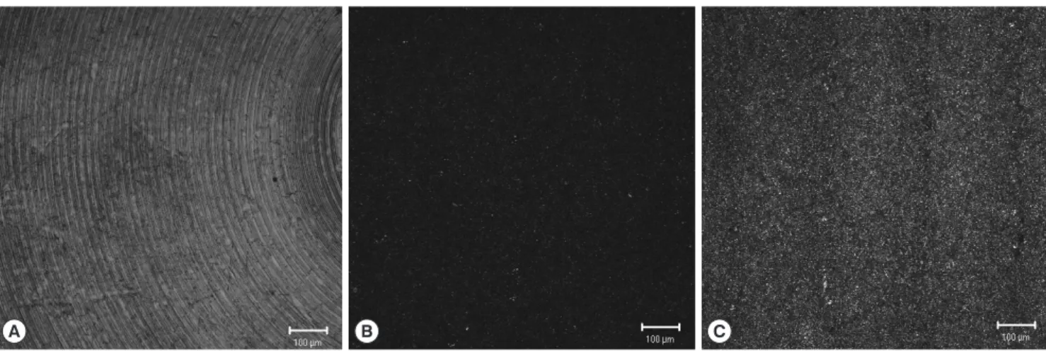
Each group of titanium discs showed unique surface characteris- tics (Fig. 2). The machined-surface presented the lowest value of av- erage roughness (Ra=0.537±0.051 μm), root mean square rough- ness (Rq=0.681±0.059 μm) and valley depth (Rz=2.169±0.239
A B
Figure 1. Sandblasted, large-grit, acid-etched (SLA)-treated titanium implants with microgrooves in the coronal portion were installed in the edentulous man- dible of experimental animal (A), and gingival flaps were sutured (B).
A B C
Figure 2. Laser scanning confocal microscopic images of titanium discs. A, machined-surface discs; B, sandblasted, large-grit, acid-etched (SLA)-treated discs; C, SLA-treated and microgroove-formed discs. Bar=100 µm.
μm), while the SLA-treated group showed increased values (Ra=
1.285±0.025 μm, Rq=1.759±0.037 μm, Rz=6.652±0.797 μm). The SLA-treated and microgroove-formed discs showed the highest val-
ues in the average roughness, root mean square roughness, and val- ley depth, which were 22.351±2.766 μm, 25.202±2.472 μm, and 46.161±4.904 μm, respectively (Table 1).
Multiple-comparison results of water contact angle analysis re- vealed that microgrooves and subsequent SLA treatment signifi- cantly increased hydrophilicity of titanium comparing to single SLA treatment procedure (P<0.05) (Fig. 3). Moreover, The SLA-treated and microgroove-formed discs showed the highest surface energy value, while there were no significant differences compared to SLA- treated group (Fig. 4).
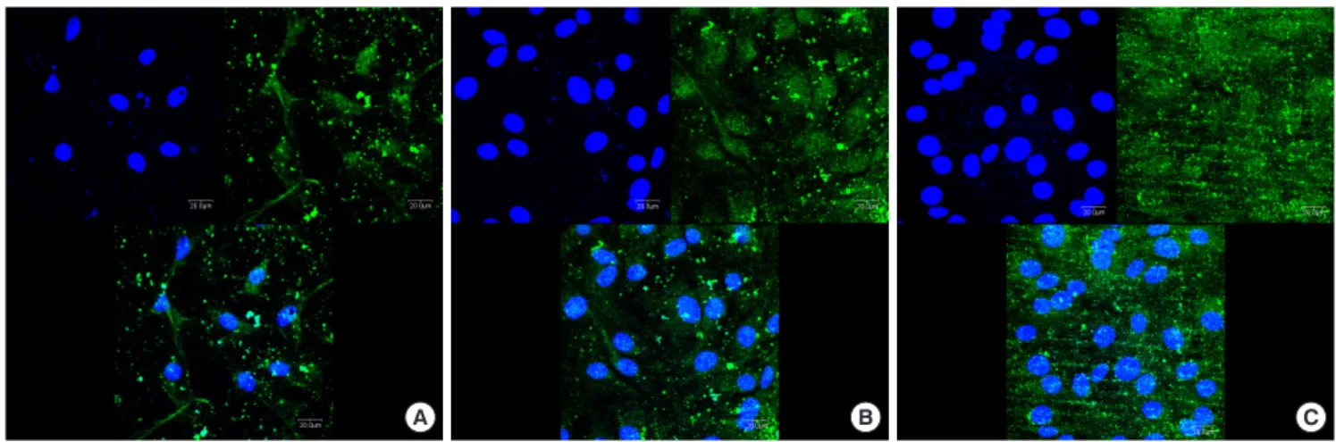
Immunofluorescence analysis for hGFs and MC3T3-E1 cells ad- hesion revealed prominent expression of actin filaments and nuclei on the surface of the SLA-treated and microgroove-formed discs (Figs. 5 and 6).
Table 1. Surface roughness parameters of titanium discs using laser scanning confocal microscopy.
A B C
Ra (μm) 0.537±0.051 1.285±0.025 22.351±2.766
Rq (μm) 0.681±0.059 1.759±0.037 25.202±2.472
Rz (μm) 2.169±0.239 6.652±0.797 46.161±4.904
A, machined-surface discs; B, sandblasted, large-grit, acid-etched (SLA)- treated discs; C, SLA-treated and microgroove-formed discs.
Ra, roughness average; Rq, root mean square roughness; Rz, valley depth.
100
80
60
40
20
0
Water contact angle (θ)
a)
a)
A B C
Figure 3. Water contact angle determination on titanium discs. A, machined- surface discs; B, sandblasted, large-grit, acid-etched (SLA)-treated discs; C, SLA- treated and microgroove-formed discs. a)P<0.05, using the One-way ANOVA.
80
60
40
20
0
Surface energy (mN/m)
a) a)
A B C
Figure 4. Surface energy determination on titanium discs. A, machined-surface discs; B, sandblasted, large-grit, acid-etched (SLA)-treated discs; C, SLA-treated and microgroove-formed discs. a)P<0.05, using the One-way ANOVA.
A B C
Figure 5. Immunofluorescence images showing actin filaments (green) and nuclei (blue) of human gingival fibroblasts on titanium discs for 24 hours. Promi- nent cell adhesion was induced in group C. A, machined-surface discs; B, sandblasted, large-grit, acid-etched (SLA)-treated discs; C, SLA-treated and micro- groove-formed discs. Bar=20 µm.
None of the implants, installed in the mandible of experimental animals, failed without any serious complications. There were no
A B C
Figure 6. Immunofluorescence images showing actin filaments (green) and nuclei (blue) of murine osteoblastic cell line (MC3T3-E1) on titanium discs for 24 hours. Prominent cell adhesion was induced in group C. A, machined-surface discs; B, sandblasted, large-grit, acid-etched (SLA)-treated discs; C, SLA-treated and microgroove-formed discs. Bar=20 µm.
signs of abscess, infection, or any wound dehiscence throughout the experimental period. Histological samples showed direct soft tissue contact with the surface of microgroove portion of the im- plants. Under microscopic examination, tight sealing of soft tissue contact was well established as most of the collagen fibers ran par- allel to the surface of microgroove. Collagen fibers showed addi- tional running patterns around microgroove surface, including a circular direction (Fig. 7).
At 2 weeks after implant insertion, the soft tissue contact ratio in group A was lower than that of group B on the area of microgroove Figure 7. Light microscopic photographs at 2 weeks after implant installation.
Peri-implant soft tissue was tightly attached to SLA-treated microgroove por- tion of titanium implant surface. Dense collagen fibers with circular alignment around microgroove surface were found (asterisk). Bar=50 µm.
100
80
60
40
20
0
Soft tissue contact ratio (%)
a)
2 6
Week
A B
Figure 8. Soft tissue contact ratio determination on SLA-treated microgroove portion of titanium implant surface. A, implants installed with 1 mm exposure of coronal microgroove portion; B, implants installed with 2 mm exposure of coronal microgroove portion. a)P<0.05, using the Mann–Whitney U test.
implant surface (Fig. 8). There was no statistically significant dif- ference between two groups at 2 weeks (P>0.05). However, at 6 weeks after implant installation, both groups showed increased soft tissue contact ratio comparing to the values of 2 weeks after surgery. The soft tissue contact ratio of group A was higher than that of group B with a statistically significance (P<0.05)
DISCUSSION
Our present study showed that the SLA-treated and microgroove- formed titanium discs pertained the roughest surface characteristics and maintained the highest hydrophilicity comparing to the ma- chined-surface or SLA-treated groups. Furthermore, the SLA-treated and microgroove-formed titanium surface promoted the adhesion of gingival fibroblasts and osteoblastic cell in vitro, and peri-implant soft tissue healing in vivo to a greater extent.
It has been reported that the roughness and surface energy of ti- tanium surface affect the cellular adhesion [7-11]. In this present study, the dimensions of microgrooves (200 μm width and 100 μm depth) were determined considering the mean size of hGFs (100 μm). Our in vitro study results revealed that microgrooves with sub- sequent SLA treatment on titanium surface increased hydrophilicity significantly and promoted the adhesion of peri-implant soft tissue cells, i.e., gingival fibroblasts and osteoblastic cells. Based on these in vitro results, we performed animal experiments using titanium implants which had microgrooves with subsequent SLA treatment in their coronal portion.
Previous report demonstrated that the alveolar crestal bone level was located up to 2 mm below the implant-abutment junction [2].
In order to simulate this condition, we prepared titanium implants, which have microgrooves with subsequent SLA treatment in the coronal 2 mm portion, and intentionally positioned them with 1 mm or 2 mm coronal exposure to the peri-implant soft tissue. In the present study, connective tissue with abundant fibroblasts ac- companying a few vessels surrounded exposed microgroove por- tion, which corresponds to previous reports on peri-implant soft tissue healing [5,12,13]. While peri-implant soft tissue contact to the microgroove portion was increased in a time-dependent man- ner after implant installation surgery, 2 mm coronal exposure group showed significantly lower soft tissue contact ratio than that of 1 mm coronal exposure group at 6 weeks after surgery. This phenom- enon might be a possible explanation as to why peri-implant soft tissue is vulnerable to bacterial invasion, often leading to peri-im- plant disease, which was addressed previously [3, 14-16]. The soft tissue attachment to dental implants differs from that of natural teeth in the orientation of the connective tissue attachment. In teeth, an attachment apparatus with Sharpey’s fibers is found em- bedded in the cementum and covering the root surface at an oblique angle, while the implants have firm bundles of connective tissue fibers running parallel to the implant surface [17]. Our pres- ent findings were in agreement with this observational report. Con- sidering the present results of soft tissue contact ratio measure-
ment, we can deduct that more coronal exposure of implant fix- tures leads to less soft tissue contact to implant, which may allow inflammatory infiltrate.
From a clinical viewpoint, with alveolar ridge not always being flat, it often occurs that not all coronal surfaces of titanium im- plant fixtures are covered with the recipient alveolar bone in den- tal implant fixture installation procedure. Thus, if the coronal sur- face of implant fixtures facilitates soft tissue contact, it can pro- mote tight soft tissue sealing which is important to resist possible bacterial intrusion.
In this study, as the SLA-treated and microgroove-formed titani- um surface also enhanced osteoblastic adhesion, it would be ex- pected to promote alveolar bone contact if this surface was sub- merged in alveolar bone. Therefore, it is possible that the SLA-treat- ed microgroove in the coronal portion of titanium implant may promote tight peri-implant soft tissue sealing together with osseo- integration which is essential to the maintenance of dental im- plants. The determination of long-term in vivo response of peri-im- plant tissues may provide further evidence for the benefit of SLA- treated microgroove structures on the reliable dental implantation.
CONFLICT OF INTEREST
No potential conflict of interest relevant to this article was re- ported.
ACKNOWLEDGEMENTS
This research was supported by Seoul National University (Grant number: 860-20140001) and the International Research & Devel- opment Program of the National Research Foundation of Korea (NRF) funded by the Ministry of Science, ICT & Future Planning (Grant number: 2014K1A3A1A21001365).
ORCID
Hyo-Jung Lee http://orcid.org/0000-0002-0439-7389 Jaden Lee http://orcid.org/0000-0001-6937-9416 Jung-Tae Lee http://orcid.org/0000-0001-5383-3004 Ji-Soo Hong http://orcid.org/0000-0001-5698-0894 Bum-Soon Lim http://orcid.org/0000-0003-3112-0227 Hee-Jung Park http://orcid.org/0000-0002-6789-9247 Young-Kwang Kim http://orcid.org/0000-0002-1984-206X Tae-Il Kim http://orcid.org/0000-0003-4087-8021
REFERENCES
1. Brånemark PI, Adell R, Albrektsson T, Lekholm U, Lundkvist S, Rockler B. Osseointegrated titanium fixtures in the treatment of edentulousness. Biomaterials 1983;4:25-8.
2. Hermann JS, Buser D, Schenk RK, Higginbottom FL, Cochran DL.
Biologic width around titanium implants. A physiologically formed
and stable dimension over time. Clin Oral Implants Res 2000;11:
1-11.
3. Geurs NC, Vassilopoulos PJ, Reddy MS. Soft tissue considerations in implant site development. Oral Maxillofac Surg Clin North Am 2010;22:387-405, vi-vii.
4. Abrahamsson I, Zitzmann NU, Berglundh T, Linder E, Wennerberg A, Lindhe J. The mucosal attachment to titanium implants with different surface characteristics: an experimental study in dogs.
J Clin Periodontol 2002;29:448-55.
5. Huh JB, Rheu GB, Kim YS, Jeong CM, Lee JY, Shin SW. Influence of Implant transmucosal design on early peri-implant tissue re- sponses in beagle dogs. Clin Oral Implants Res 2014;25:962-8.
6. Owens DK, Wendt RC. Estimation of surface free energy of poly- mers. J Appl Polym Sci 1969;13:1741-7.
7. Bächle M, Kohal RJ. A systematic review of the influence of dif- ferent titanium surfaces on proliferation, differentiation and pro- tein synthesis of osteoblast-like MG63 cells. Clin Oral Implants Res 2004;15:683-92.
8. Cooper LF, Zhou Y, Takebe J, Guo J, Abron A, Holmén A, et al. Flu- oride modification effects on osteoblast behavior and bone for- mation at TiO2 grit-blasted c.p. titanium endosseous implants.
Biomaterials 2006;27:926-36.
9. Kim MJ, Choi MU, Kim CW. Activation of phospholipase D1 by surface roughness of titanium in MG63 osteoblast-like cell. Bio- materials 2006;27:5502-11.
10. Marinucci L, Balloni S, Becchetti E, Belcastro S, Guerra M, Calvitti
M, et al. Effect of titanium surface roughness on human osteo- blast proliferation and gene expression in vitro. Int J Oral Maxil- lofac Implants 2006;21:719-25.
11. Sader MS, Balduino A, Soares GD, Borojevic R. Effect of three dis- tinct treatments of titanium surface on osteoblast attachment, proliferation, and differentiation. Clin Oral Implants Res 2005;16:
667-75.
12. Cochran DL, Hermann JS, Schenk RK, Higginbottom FL, Buser D.
Biologic width around titanium implants. A histometric analysis of the implanto-gingival junction around unloaded and loaded nonsubmerged implants in the canine mandible. J Periodontol 1997;68:186-98.
13. Ruggeri A, Franchi M, Marini N, Trisi P, Piatelli A. Supracrestal cir- cular collagen fiber network around osseointegrated nonsub- merged titanium implants. Clin Oral Implants Res 1992;3:169-75.
14. Broggini N, McManus LM, Hermann JS, Medina R, Schenk RK, Buser D, et al. Peri-implant inflammation defined by the implant- abutment interface. J Dent Res 2006;85:473-8.
15. Chehroudi B, Gould TR, Brunette DM. The role of connective tis- sue in inhibiting epithelial downgrowth on titanium-coated per- cutaneous implants. J Biomed Mater Res 1992;26:493-515.
16. Linkevicius T, Apse P. Biologic width around implants. An evidence- based review. Stomatologija 2008;10:27-35.
17. Gargiulo AW, Wentz FM, Orban B. Dimensions and relations of dentogingival junction in humans. J Periodontol 1961;32:261-7.